Journal of Medical & Surgical Pathology
Open Access
ISSN: 2472-4971
ISSN: 2472-4971
Case Report - (2023)Volume 8, Issue 2
Sternoclavicular joint dislocations represent 3% of all shoulder joint injuries, anterior dislocations are the most common type which carries a high rate of recurrence, it presents a therapeutic challenge, sternoclavicular ligament reconstruction with autograft provides the best stability. The objective of this study is to report the case of a 38-yearold male patient, involved in a road traffic accident and admitted with traumatic left anterior sternoclavicular joint dislocation.
Surgical intervention was indicated due to the significant deformity, which was causing aesthetic concerns for the patient. The treatment involved a ligament reconstruction using an autograft from the ipsilateral palmaris longus tendon. After 6 months, the patient had normal joint range of motion without pain and full muscle strength without any deficits.
Sternoclavicular joint; Allograft reconstruction; Clavicle; Instability; Palmaris longus tendon allograft
Sternoclavicular joint dislocations represent 3% of all shoulder joint injuries. Among which, posterior dislocations mandate urgent/emergent closed reduction due to its high incidence of associated injuries and complications [1].
Anterior dislocations are the most common type which carries a high rate of recurrence. It presents a therapeutic challenge. The main approach involves symptom relief, short-term immobilization, and rehabilitation. Surgical treatment is required in cases of instability or recurrent pain [2].
Sternoclavicular ligament reconstruction with autograft provides the best stability, while other techniques include resection of the proximal end of the clavicle or osteosynthesis with a plate. The use of pins is contraindicated due to a high risk of mediastinal migration [3].
The objective of this study is to report the case of a 38-year-old male patient, involved in a road traffic accident and admitted with traumatic left anterior sternoclavicular joint dislocation. The patient underwent a sternoclavicular ligament reconstruction using the tendon of the palmaris longus muscle.
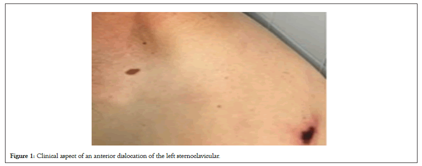
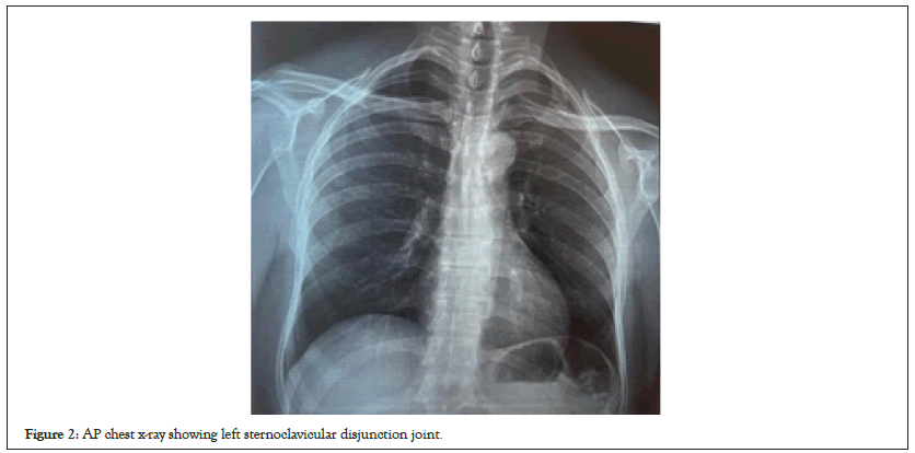
This is a case of a 38-year-old male patient who was admitted to the emergency department following a motor vehicle accident (motorcycle fall). Upon clinical examination, there was pain and swelling over the left sternoclavicular joint with significant deformity. The diagnosis of anterior sternoclavicular joint dislocation was confirmed by an Anterior Posterior (AP) chest X-ray and thoracic Computerized Tomography (CT) scan (Figures 1-3). Surgical intervention was indicated due to the significant deformity, which was causing aesthetic concerns for the patient. The treatment involved a ligament reconstruction using an autograft from the ipsilateral palmaris longus tendon. The procedure was performed under general anesthesia with the patient in a semi-sitting position, with a cushion placed under the thoracic spine to facilitate access to the left sternoclavicular joint. After thorough scrubbing and draping of the left upper limb, a surgical incision was made over the left sternoclavicular joint, first along the superior border of the proximal end of the clavicle and then vertically along the inner edge of the sternal manubrium. The joint was exposed, and the anterior sternoclavicular ligament was found to be torn. Careful periosteal stripping was performed while ensuring the protection of underlying vascular structures.

Figure 1: Clinical aspect of an anterior dislocation of the left sternoclavicular.

Figure 2: AP chest x-ray showing left sternoclavicular disjunction joint.

Figure 3: CT image showing left sternoclavicular disjunction.
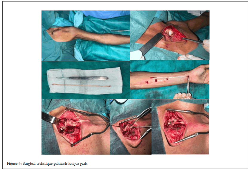
Subsequently, the meniscus was excised, and two tunnels were drilled on each side (proximal end of the clavicle and sternal manubrium) using a 3.2 mm drill. After reduction of the dislocation and temporary stabilization with two Kirschner wires (removed at the end), the harvested palmaris longus graft was passed through the tunnels in a figure of eight configuration, and was sutured end to end (Figure 4).

Figure 4: Surgical technique palmaris longus graft.
In the postoperative period, a follow-up X-ray confirmed the reduction of the dislocation. The patient was immobilized with an arm-to-body sling for a period of 3 weeks. Afterward, passive rehabilitation was initiated, and the immobilization was removed after the sixth week, allowing for active movements.
After 6 months, the patient had normal joint range of motion without pain and full muscle strength without any deficits. A radiograph showed a well-reduced joint. The patient was able to resume normal daily activities.
Road traffic accidents are the leading cause of sternoclavicular dislocations, while sports-related accidents involving falls on the shoulder are less common. In some patients, no specific cause is found. Non-traumatic, voluntary instabilities are a contraindication for surgery and should prompt an investigation for collagen abnormalities or Ehlers-Danlos syndrome [4,5].
When facing an anterior dislocation, it is important to determine whether it is traumatic or non-traumatic in origin. In the case of traumatic anterior dislocations, reduction can be attempted, but recurrence is frequent. For cases of irreducible dislocations, unsatisfactory outcomes with stabilizing treatments shifted the management toward a conservative or functional treatment. However, if the symptoms are severely bothersome and persistent, surgical stabilization may be considered after a thorough discussion with the patient about the potential benefits and risks of such an intervention [6].
Several techniques have been reported in the literature for sternoclavicular joint ligament reconstruction, involving autografts from tendons, with the most commonly used being the semimembranosus tendon. These techniques have been shown to have fewer complications compared to other known techniques such as the use of a plate and screws [7].
Spencer, et al. [8] conducted a cadaveric study comparing the use of the semimembranosus tendon in a figure of eight reconstruction with the subclavius tendon. The first technique demonstrated better resistance and provided greater biomechanical stability.
Bak, et al. [9] conducted a study involving 32 patients, using the palmaris longus tendon in 7 patients and the gracilis tendon in 25 patients. The total Western Ontario Shoulder Instability (WOSI) score improved from 44% to 75% with 2 failures (7.4%), although 40% of the patients reported persistent discomfort. Petri, et al. [10] reported a reconstruction using hamstring tendons in 21 patients, with no reported recurrences and a significant improvement in ASES and QuickDASH scores. In our patient, the choice of graft was based on the criticisms found in the literature regarding the semitendinosus tendon, such as decreased knee flexion strength and residual pain at the donor site. However, another option could be the use of a graft obtained from a tissue bank. Lyons, et al. [11] conducted a study on 21 patients using pins, and the results showed 8 deaths. In all cases, the use of pins is prohibited as they can lead to migration to distant, mediastinal, or even intracardiac locations, with dramatic consequences.
In this article, we present the case of a patient who suffered an anterior sternoclavicular dislocation as a result of a road traffic accident. The patient underwent a figure of eight ligament reconstruction using the palmaris longus tendon with satisfying results at 6 months post-operative. Sternoclavicular joint reconstruction, using a palmaris longus constitutes a good technique for its repair, and allows recovering normal articular amplitude of the shoulder, while avoiding the complications related to the other grafts like the semitendinosus one for example: possible damage to the flexion strength of the knee. However, the discomfort from the palmaris longus is normal transient paraesthesia from the palmar cutaneous branch of the median nerve.
[Crossref] [Google Scholar] [PubMed]
[Crossref] [Google Scholar] [PubMed]
[Crossref] [Google Scholar] [PubMed]
[Crossref] [Google Scholar] [PubMed]
[Google Scholar] [PubMed]
[Crossref] [Google Scholar] [PubMed]
[Crossref] [Google Scholar] [PubMed]
[Crossref] [Google Scholar] [PubMed]
[Crossref] [Google Scholar] [PubMed]
Citation: Sennouni M, Gouri AM, Zitoune A, Zakaria A, Adaoui OE, Fadili M (2023) A Case Report on Sternoclavicular Joint Autograft Reconstruction. J Med Surg Pathol. 08:275.
Received: 26-May-2023, Manuscript No. JMSP-23-24310; Editor assigned: 30-May-2023, Pre QC No. JMSP-23-24310 (PQ); Reviewed: 13-Jun-2023, QC No. JMSP-23-24310; Revised: 20-Jun-2023, Manuscript No. JMSP-23-24310 (R); Published: 27-Jun-2023 , DOI: 10.35248/2472-4971.23.08.275
Copyright: © 2023 Sennouni M, et al. This is an open-access article distributed under the terms of the Creative Commons Attribution License, which permits unrestricted use, distribution, and reproduction in any medium, provided the original author and source are credited.