Journal of Medical & Surgical Pathology
Open Access
ISSN: 2472-4971
ISSN: 2472-4971
Short Communication - (2021)Volume 6, Issue 1
An eccrine spiradenoma is a rare benign tumor most often seen in the head, neck and upper trunk of young adults. It is usually tender and malignant transformation has been reported in long standing cases. Although eccrine spiradenoma has been described in earlier literature in the neck, trunk and proximal limb it has so far not been described in the distal lower limb. Here is a case report of 30-year-old gentleman with eccrine spiradenoma managed at our hospital.
Eccrine spiradenoma; Fever; Swelling; Tumors
30 year old gentleman with nil premorbid illness came with pain and swelling over the left medial malleolus since 5 years, aggravated since 1 week. There was no history of trauma, fever, and discharge from swelling. Examination revealed a solitary 2 × 1 cm tender swelling over the left medial malleolus, firm, mobile and plane of swelling is intradermal (Figure 1). There was no local rise of temperature or punctum. With The diagnosis of sebaceous cyst, excision of the lesion under local anesthesia with an elliptical incision 1 cm in length was done. Grossly the specimen was grayish white with a diameter of 1 cm. Microscopy showed well circumscribed encapsulated tumour composed of 2 cell population of basaloid cell with central cells showing moderate eosinophilic cytoplasm, peripheral smaller cells, arranged in trabecular pattern with peripheral palisading showing minimal pleomorphism and mitosis, interspread by scant fibrous stroma with hyaline material and focal marked edema (Figure 2). Few dilated spaces filled with eosinophilic secretions are seen. Few dilated vascular spaces surrounded by hyalinization are also seen. Thus a final diagnosis of eccrine spiradenoma of lower limb was established (Figure 3).
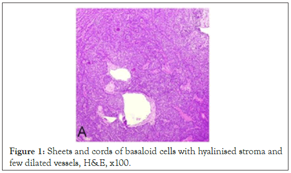
Figure 1: Sheets and cords of basaloid cells with hyalinised stroma and few dilated vessels, H&E, x100.
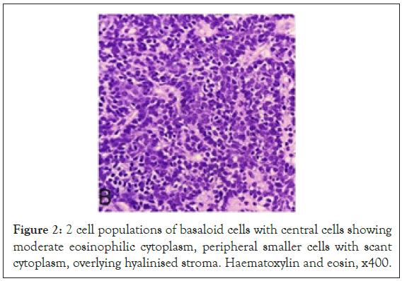
Figure 2: 2 cell populations of basaloid cells with central cells showing moderate eosinophilic cytoplasm, peripheral smaller cells with scant cytoplasm, overlying hyalinised stroma. Haematoxylin and eosin, x400.
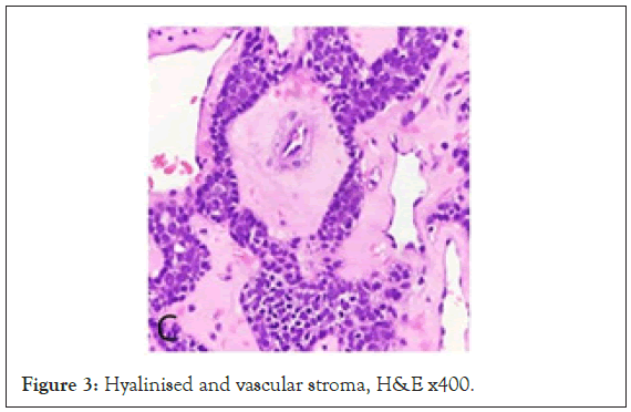
Figure 3: Hyalinised and vascular stroma, H&E x400.
Eccrine spiradenoma is a benign adnexal tumor that appears as a small and bluish nodule typically tender on palpation. This was first reported by Kerstein et al. in 1955. In their study of 134 eccrine spiradenomas, more than 97% of the tumors were solitary blue-red dermal or subcutaneous nodules, ranging from 0.5 to 3 cm in diameter [1]. Eccrine spiradenomas mostly occur in 15-35 years age group and female preponderance. Eccrine spiradenomas can be painful (Figure 4). The rate of malignant transformation is very low. Such transformation to malignancy has been reported to develop spontaneously with a metastasis rate of 50%, which can cause death [2]. Eccrine spiradenoma has been mostly reported in literature as random multiple tumors involving the chest, upper extremities, forehead and scalp. Histopathologically, eccrine spiradenoma consists of one or more large, sharply defined basophilic nodules in the dermis, also described as 'cannon balls' or 'big blue balls' in the dermis (Figure 5). They are not attached to the overlying epidermis and sometimes extend into the subcutis. They consist of groups of cells in cords or islands or sheets. Two types of cells are found in these nodules. There are small, dark, basaloid cells with hyperchromatic nuclei, and cells with large, pale vesicular and ovoid nuclei. Pale cells tend to be at the center of the lesions (Figure 6) [3-5].
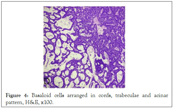
Figure 4: Basaloid cells arranged in cords, trabeculae and acinar pattern, H&E, x100.
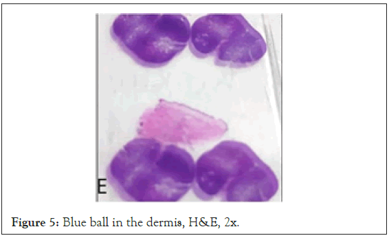
Figure 5: Blue ball in the dermis, H&E, 2x.
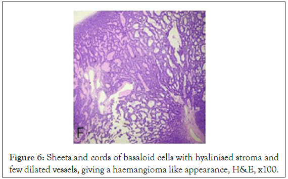
Figure 6: Sheets and cords of basaloid cells with hyalinised stroma and few dilated vessels, giving a haemangioma like appearance, H&E, x100.
Management depends on proper diagnosis and it is based on regional lymph nodes, lungs, brain, and liver involvement (in descending order of frequency). Histopathologically, proliferation of cells with hyper chromatic nuclei, increased mitoses, loss of Periodic Acid-Schiff positive basement membrane cords and invasion of the surrounding tissues characterizes malignant transformation in eccrine spiradenoma. The various treatment modalities include excision, dermabrasion, electrodessication, cryotherapy, and radiotherapy or applying argon and CO2 lasers. It has also been shown that the administration of aspirin and its derivatives can result in the rapid formation of new lesions. Treatments for spiradenoma have not yet been clearly established, but surgical excision is still the gold standard with low rates of recurrence.
Citation: Agarwal K, Anto J, Priya P (2021) A Rare Case of Eccrine Spiradenoma of the Lower Limb. J Med Surg Pathol. 6:194.
Received: 29-Jan-2021 Accepted: 12-Feb-2021 Published: 19-Feb-2021 , DOI: 10.35248/2472-4971.21.6.194
Copyright: © 2021 Agarwal K, et al. This is an open-access article distributed under the terms of the Creative Commons Attribution License, which permits unrestricted use, distribution, and reproduction in any medium, provided the original author and source are credited.