Journal of Women's Health Care
Open Access
ISSN: 2167-0420
ISSN: 2167-0420
Case Report - (2024)Volume 13, Issue 2
Tailgut cyst or retrorectal cystic hamartoma is a congenital lesion located in the retrorectal-presacral space and is a remnant of the embryonic hindgut. It is more common between the ages of 30-60 and in women (f/e=5/1). Although it is usually asymptomatic, it may cause abdominal pain or constipation. Our aim is to highlight this rare cause of pelvic floor pain through 2 cases.
Constipation, Pelvic floor pain, Tailgut cyst
Masses in the retrorectal region are rarely seen and their incidence is reported as 1/40,000–63,000 [1]. Tailgut cyst or retrorectal cystic hamartoma is a rare congenital lesion located in the retrorectalpresacral space and is a remnant of the embryonic hindgut. The tailgut undergoes regression in the 8th week of embryological life; Tailgut cysts form as a result of the regression defect in this period [2]. Tailgut cysts are seen more frequently between the ages of 30-60 and in women (f/e=5/1) [3]. Although it is usually asymptomatic, it may rarely cause abdominal pain and constipation [4].
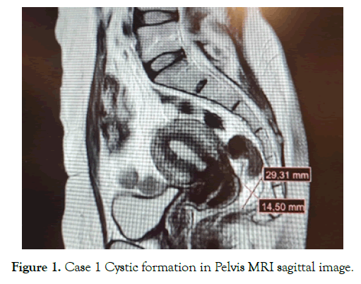
Case 1: A 44-year-old female patient came with hip and groin pain. There was a gastroenterology application due to constipation in her medical history, but no pathology was detected. The musculoskeletal system examination was unremarkable. In the contrast-enhanced pelvic MRI examination, a 29*14*16 mm noncontrast cystic formation in the right posterolateral rectum was reported as a tailgut cyst [Figure 1].

Figure 1. Case 1 Cystic formation in Pelvis MRI sagittal image.
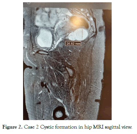
Case 2: A 36-year-old female patient came with left hip pain. On examination, the hip joint was open and there was pain radiating to the perineal area. There was no neurological deficit. In the contrast-enhanced hip magnetic resonance (MR) image, a septate cystic formation of 47*42*27 mm in size, located on the right posterolateral side, deviating the rectum to the left, appearing hyper intense on T2 and hypo intense on T1, was reported in favor of a tailgut cyst [Figure 2].

Figure 2. Case 2 Cystic formation in hip MRI sagittal view.
Both patients signed an informed consent form and were referred to general surgery. Case 1 was lost to follow-up. Case 2 was operated; there was no pain in the postoperative period.
Retrorectal tumors can be divided into 5 categories: congenital, neurogenic, inflammatory, osseous and other tumors. Congenital tumors constitute 55-70% of all retrorectal tumors. Congenital tumors include chordoma (notochord remnant), teratomas, anterior sacral meningocele, and developmental cysts (dermoid, epidermoid, enteric duplication, or tailgut cysts) [5].
Although most of the tailgut cysts, which are developmental cysts, are found in the retrorectal area, they can also be found in the anterior rectal, perianal, perirenal and posterior sacral regions. They are congenital lesions originating from the embryological tailgut, which are generally benign and may rarely show malignant transformation [6,7].
It can be palpated during physical examination during digital rectal examination. Double-contrast colon radiography, transrectal ultrasonography, computed tomography and MRI can be used in the radiological diagnosis of tailgut cysts [8]. MR signal features of tailgut cyst appear as hypointense on T1W sequences and hyper intense on T2W sequences [9].
Although it is generally asymptomatic, it can cause urological conditions such as acute urinary retention due to abdominal pain or pressure on surrounding organs, neurological conditions such as nerve compression such as the sciatic nerve, and constipation and obstipation [4, 10, 11].
In the differential diagnosis, rectal duplication cyst, anterior meningocele, chordoma, teratoma, epidermal cyst, anal gland cyst and cystic lymphangiomas should be kept in mind [12]. The most important complications of tailgut cyst are infection and malignant degeneration of the cyst [13]. The treatment is surgical excision [7].
Tailgut cyst should also be investigated as a rare but possible cause in the differential diagnosis in female patients presenting with perineal pain.
Indexed at, Google Scholar, Cross Ref
Indexed at, Google Scholar, Cross Ref
Indexed at, Google Scholar, Cross Ref
Indexed at, Google Scholar, Cross Ref
Indexed at, Google Scholar, Cross Ref
Indexed at, Google Scholar, Cross Ref
Indexed at, Google Scholar, Cross Ref
Indexed at, Google Scholar, Cross Ref
Citation: Yavas AD (2024) A Rare Cause of Pelvic Pain: Tailgut Cyst. 13(2):710.
Received: 25-Jan-2024, Manuscript No. 29349; Editor assigned: 29-Jan-2024, Pre QC No. 29349; Reviewed: 12-Feb-2024, QC No. 29349; Revised: 16-Feb-2024, Manuscript No. 29349; Published: 24-Feb-2024 , DOI: 10.35248/2167- 0420.24.13.710
Copyright: © 2024 Yavas AD. This is an open-access article distributed under the terms of the Creative Commons Attribution License, which permits unrestricted use, distribution, and reproduction in any medium, provided the original author and source are credited