Journal of Clinical and Experimental Ophthalmology
Open Access
ISSN: 2155-9570
ISSN: 2155-9570
Clinical image - (2021)Volume 12, Issue 5
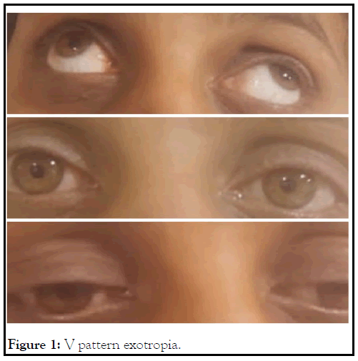
A 28-year-old female visited our department with a history of itching bilateral eyes off and on. No other significant history was given. Her visual aquity was 6/6 in both the eyes while the fundus, color vision, slit lamp examination and intraocular pressure were within normal limits. The only positive finding was a V pattern exotropia (Figure 1) on torch examination about which the patient had no concern and hence further ocular investigations were not done. She was prescribed ocular lubricants plus was told to undergo a Computed Tomography (CT) scan of the brain and orbit for which she agreed. Asymmetric size of the muscle belly of Lateral Rectus (LR) muscle was reported on CT scan (Figure 2). No further intervention was done. Our case highlights an asymmetric size of the muscle belly of lateral rectus muscle which is a very rare finding.

Figure 1: V pattern exotropia.
Figure 2: Asymmetric size of the muscle belly of Lateral Rectus (LR) muscle.
Major anatomical variations seen in the LR muscles include duplication, absence, hypoplasia, bifurcation, posteriorly malinsertion and absence of the LR muscle [1]. V pattern exotropia is a vertical gaze incomitance of more than 15 Prism Dioptres between upgaze and downgaze with eyes diverging more in upgaze. Various etiologies causing this phenomenon include over-action of inferior oblique muscles, over-action or increased innervation of the LR muscle in up gaze or medial rectus muscle in down gaze, displacement of rectus muscle pulleys or its path, ex-cyclorotation of the orbits as in craniofacial anomalies and desagitalisation of the superior oblique muscle in plagiocephaly. The common treatment options available for V pattern strabismus are inferior oblique weakening or vertical offsetting of horizontal recti muscles [2].
Citation: Chauhan A, Sharma KD, Wapa A, Thakur KP, Sharma V(2021) A Rare Finding: Lateral Rectus Anomaly. J Clin Exp Ophthalmol. 12:886.
Received: 08-Oct-2021 Accepted: 22-Oct-2021 Published: 29-Oct-2021 , DOI: 10.35248/2155-9570.21.12.886
Copyright: © 2021 Chauhan A, et al. This is an open-access article distributed under the terms of the Creative Commons Attribution License, which permits unrestricted use, distribution, and reproduction in any medium, provided the original author and source are credited.