Anatomy & Physiology: Current Research
Open Access
ISSN: 2161-0940
ISSN: 2161-0940
Case Report - (2022)Volume 12, Issue 5
Objective: To report and describe a rare variation of the hamstring muscle group known as the tensor fasciae suralis muscle.
Case Presentation: The anomalous muscle was discovered during a routine dissection of a cadaver in the posterior right lower limb. The left lower limb was dissected but displayed no discernable muscle variations.
Results: The variation of the tensor fasciae suralis that was discovered arises from the long head of the biceps femoris, descends superficially across the popliteal region, and inserts on both heads of the gastrocnemius muscle and the overlaying crural fascia.
Conclusion: The tensor fasciae suralis is rarely reported in literature and may have resulted from a disruption in the regulation of muscle and tendon patterning during primary myogenesis in the early development of the lower limb. Although individuals with the tensor fasciae suralis are likely to present clinically with no symptoms, it is of interest due to its proximity to important neurovascular structures of the popliteal fossa, and its potential to show up as a pathological structure on an MRI.
Tensor fasciae suralis; Anatomical variation; Hamstrings; Biceps femoris; Popliteal fossa
The hamstrings refer to a group of three muscles in the posterior compartment of the thigh: semitendinosus, semimembranosus, and the long head of biceps femoris. These biarticular muscles share a common origin, the ischial tuberosity, and are innervated by the tibial portion of the sciatic nerve. Semitendinosus arises from the ischial tuberosity from a conjoined tendon with the long head of the biceps femoris and runs more medially in the posterior thigh. Distally its long, rounded tendon inserts on the upper medial surface of the tibia as part of the pes anserinus tendon, along with gracilis and sartorius. Semimembranosus is located deep to semitendinosus on the medial side of the thigh and has a flattened membranous appearance proximally. Its distal tendon splits into three parts: one part which inserts on the posterior aspect of the medial condyle of the tibia, another that inserts on the popliteal fascia, and a third reflected part that forms the oblique popliteal ligament. The biceps femoris is the most lateral of the three muscles and has two heads of origin, a long head and a short head. The long head is considered to be a true hamstring muscle, while the short head is not. The short head is innervated by the common fibular portion of the sciatic nerve and arises from the linea aspera and lateral supracondylar line of the femur. It combines with the long head distally to form a conjoined tendon that inserts on the head of the fibula. Together the hamstring muscles primarily extend the thigh at the hip joint and flex the leg at the knee joint [1].
Discovering a muscle variation through dissection is not an unusual occurrence. However, variations in the muscles of the posterior thigh are uncommon in literature. A rare hamstring muscle variation is the tensor fasciae suralis (TFS) muscle, also known as the ischioaponeuroticus. It is speculated to be a weak flexor of the knee [2]. This accessory muscle is usually innervated by the tibial portion of the sciatic nerve and has been reported to arise from the distal portion of any of the three hamstring muscles as well as the short head of biceps femoris, and may insert on the proximal gastrocnemius, posterior crural fascia, or calcaneal tendon [3,4]. Although, the TFS can arise from any of the three hamstring muscles, it is reported to arise most often from semitendinosus [3]. Any structural variations of the hamstrings may affect the function of this muscle group, which could predispose a patient to injury, affect gait, compress underlaying structures, or mimic a soft tumor in the popliteal fossa, and are therefore of clinical interest. Documenting and adding to the knowledge of these different hamstring muscle variations can help surgeons and radiologists expand the differential diagnostic possibilities and avoid diagnostic errors. In this report, we show a rare tensor fasciae suralis muscle arising from the long head of the biceps femoris.
During a routine dissection of a of 93-year-old white male cadaver a variation in the hamstring muscle group was discovered in the right posterior thigh. The gluteal region and the posterior thigh of the right lower limb were carefully dissected and an accessory muscle of the biceps femoris muscle was noted. The posterior right leg was dissected as well to show the insertion of this anomalous muscle. Measurements of the length and width of the anomalous muscle were recorded, and images were taken. The left lower limb was dissected and no variations of the hamstrings were detected.
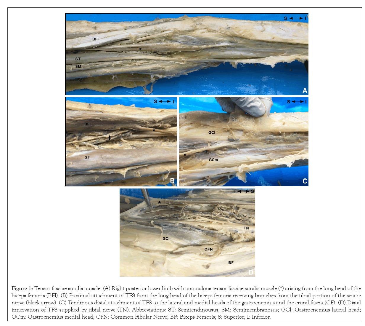
On the right posterior thigh, a long slender muscle was discovered arising from the distal third of the long head of the biceps femoris (Figures 1A-1D). The accessory muscle arose from the long head and descended centrally in the posterior thigh, then traveled superficially across the popliteal region, and between the two heads of the gastrocnemius muscle before inserting on muscles and fascia of the posterior leg (Figure 1A). This muscle crossed the distal portion of the sciatic nerve just before it bifurcated and the proximal part of the tibial nerve in the popliteal fossa. The distal tendon appeared to have three main insertion points, the crural fascia overlaying the proximal gastrocnemius and two other slips that inserted on the medial and lateral heads of the gastrocnemius (Figure 1C). This muscle received branches from the tibial portion of the sciatic nerve and branches from the tibial nerve distally (Figures 1B and 1D). The muscle was 34 cm long from its proximal to its distal attachments and had a minimum width of 0.5 cm and a maximum width of 0.7 cm. Based on these observations, this muscle is consistent with the description for the TFS muscle.

Figure 1: Tensor fasciae suralis muscle. (A) Right posterior lower limb with anomalous tensor fasciae suralis muscle (*) arising from the long head of the biceps femoris (BFl). (B) Proximal attachment of TFS from the long head of the biceps femoris receiving branches from the tibial portion of the sciatic nerve (black arrow). (C) Tendinous distal attachment of TFS to the lateral and medial heads of the gastrocnemius and the crural fascia (CF). (D) Distal innervation of TFS supplied by tibial nerve (TN). Abbreviations: ST: Semitendinousus; SM: Semimembranosus; GCl: Gastrocnemius lateral head; GCm: Gastrocnemius medial head; CFN: Common Fibular Nerve; BF: Biceps Femoris; S: Superior; I: Inferior.
We have identified a rare variation of the hamstring muscles, the tensor fasciae suralis. This muscle may arise from any of the hamstring muscles or the short head of biceps femoris, but in the present case the TFS originates from the long head of the biceps femoris. The prevalence of this muscle in different populations is unknown since incidences of TFS are rarely reported in anatomical, radiological, or surgical findings. Recently, in a study by Bale and Herrin (2019), the authors looked at a sample population of 236 cadavers and found that TFS was present in only three cadavers, resulting in a prevalence rate of 1.3% in this population. Additional data is needed to get a more accurate prevalence rate for the TFS and to see if any sex differences exist. This information is necessary for clinicians as it may help them to avoid misdiagnosis and unnecessary procedures or surgical exploration in the popliteal fossa.
This type of muscle anomaly likely occurred as a result of a disturbance early in the process of limb development. The lower limb buds appear in the lower lumbar and upper sacral region of the embryo at about 28 days [5]. The mesenchymal core of the limb bud gives rise to the bones, ligaments, tendons, and the dermis of the limbs. Myoblasts from the myotome region of adjacent somites migrate into the mesenchymal core of limb buds starting in the 5th week [5]. The myoblasts proliferate, fuse, and differentiate to form dorsal and ventral masses of muscle in each limb bud with the dorsal mass giving rise to the extensor components of the lower limb and the ventral mass giving rise to the flexor components. The muscle masses will then split into subdomains to form the basic pattern for the individual adult muscles in the embryo [6,7]. The muscle precursors migrating into the limb are not predetermined to form a specific muscle, instead it has been demonstrated that local signaling in the muscle connective tissue (MCT) derived from the limb bud mesenchyme participates in the patterning of muscles as well as tendons. Several transcription factors expressed in MCT have been implicated in the regulation of muscle and tendon patterning, including Shox2, Hox11, Tbx3, Tbx4, and Tbx5 [7]. Deletion of genes that control MCT organization early in limb development, such as Tbx4 and Tbx5, have been shown to disrupt adjacent limb muscle and tendon patterning, resulting in muscles that abnormally split and insert at inappropriate locations [6]. In the case of the TFS, a disruption in MCT regulated muscle and tendon patterning may have occurred to result in the development of this accessory muscle of the biceps femoris.
Most muscle variations do not present with clinical problems, but the TFS is of clinical significance as it crosses over the popliteal fossa and runs across several important neurovascular structures. Injury or hypertrophy of the TFS may cause pain or result in swelling in the popliteal fossa, which might be mistaken for a Baker’s cyst, soft tissue tumors, an abscess, or a popliteal artery aneurysm upon examination [2]. An accessory muscle crossing this region can result in compressive neuropathy of the sciatic, tibial, common fibular or sural nerves [2]. Furthermore, this anomalous muscle may also cause an anatomical form of popliteal artery entrapment syndrome (PAES), where there is an abnormal relationship between the artery and the surrounding musculotendinous structures [8]. Although the pathophysiology of the syndrome is unclear, it is believed that anatomical PAES occurs as a result of abnormal embryonic development of the popliteal artery and the medial head of the gastrocnemius muscle [9]. There is a possibility that genetic factors may play a role in the etiology of anatomical PAES as a few cases of siblings with the syndrome have been reported [9-11]. Individuals with anatomical PAES can be asymptomatic, so it is possible that more familial cases exist than are reported; however, further evidence is needed to support the feasibility of familial inheritance of the syndrome [11].
Anatomic variations of the hamstring muscles, especially those involving variation of insertion, may alter the biomechanical properties of the muscle or muscle group and may increase an individual’s risk for hamstring injury or strain [12]. Hamstring injuries can have lengthy recovery periods depending on the severity and location of the strain and the tissue types injured [13]. Mild to moderate hamstring strains often respond to conservative treatment with rest, ice, gentle therapeutic exercise, and a gradual return to activities, and have an average recovery time of 11-25 days. A more severe strain, such as a complete or partial tearing of the muscles, would likely require surgical intervention with extensive post-operative rehabilitation and may take up to months to heal [13,14]. The hamstrings are important for sport activities that involve sprints, jumps, tackling, and kicking. In sports that require high velocity running or repeated sprinting, such as soccer or track, the biceps femoris muscle is the most commonly injured hamstring muscle [15]. During sprinting, the hamstrings are most prone to injury during the late swing phase of the gait cycle when the muscles work eccentrically to decelerate the forward movement of the hip and knee. The biceps femoris muscle is at an increased risk for hamstring injury due to the dual innervation of its long and short heads leading to the possibility of asynchronous stimulation and impaired coordination of the two heads [12]. Moreover, theoretical models have implicated that biceps femoris is required to exert proportionally more force in eccentric contraction than the other hamstring muscles during repeated sprints, increasing its risk to injury during sprinting [15]. Anatomical variations of the biceps femoris like the TFS may further predispose this muscle to risk of strain or injury.
The tensor fasciae suralis is a rare hamstring variation that is clinically significant due to its close relationship to the neurovascular structures of the popliteal fossa and the possibility that its presence may alter the biomechanics of the hamstrings and predispose this group to injury. Though clinically relevant, the prevalence of this muscle in the population is not well known since the TFS is rarely documented; therefore, it is necessary to record the presence of this muscle when it appears. We have found one variation of the TFS, but efforts to document its many different variations should be made.
The authors would like to thank those who donated their bodies to the Anatomical Gift Program at Albany Medical College so that anatomical research could be performed.
The authors declare that they have no conflicts of interest.
[Cross Ref] [Google scholar] [Pub Med]
[Cross Ref] [Google scholar] [Pub Med]
[Cross Ref] [Google scholar] [Pub Med]
[Cross Ref] [Google scholar] [Pub Med]
[Cross Ref] [Google scholar] [Pub Med]
[Cross Ref] [Google scholar] [Pub Med]
[Cross Ref] [Google scholar] [Pub Med]
Citation: Khan AS, Lowry N, Fahl J, Smith MP (2022) A Rare Unilateral Tensor Fasciae Suralis Muscle: Case Report and Clinical Significance. Anat Physiol.12: 400.
Received: 28-Oct-2022, Manuscript No. APCR-22-18311; Editor assigned: 01-Nov-2022, Pre QC No. APCR-22-18311(PQ); Reviewed: 16-Nov-2022, QC No. APCR-22-18311; Revised: 24-Nov-2022, Manuscript No. APCR-22-18311(R); Published: 30-Nov-2022 , DOI: 10.35248/2161-0940.22.12.400
Copyright: © 2022 Khan AS, et al. This is an open-access article distributed under the terms of the Creative Commons Attribution License, which permits unrestricted use, distribution, and reproduction in any medium, provided the original author and source are credited.