Clinical & Experimental Cardiology
Open Access
ISSN: 2155-9880
ISSN: 2155-9880
Research Article - (2023)Volume 14, Issue 1
Aim: The study aimed to report a case of anatomical variation in the mouse iliac artery.
Methods: Totally 75 aortas of male ApoE-/- mice were dissected, including the aortic tree and their distal branches.
Results: Among the 75 arteries, 1 of them showed prominent anatomic variants unexpectedly. The terminal abdominal aorta descended as a trunk and finally extended into the left external iliac artery after divided out several main branches, instead of divided into the left and right iliac arteries communis as reported in previous studies.
Conclusion: The present study identified a rare case of iliac artery anatomical variation in ApoE-/-mice. Mice as vertebrate mammals highly close to human anatomy, the finding might help in the pre-intervention for catheter surgery in human patients and interpreting angiography of the pelvic region by radiologists.
Abdominal aorta terminus; Iliac artery; Angiograms
CIA: Common Iliac Artery; CIIA: Common Internal Iliac Artery; EIA: External Iliac Artery; IAV: Iliac Artery Variant; IIA: Internal Iliac Arteries RA: Rectal Artery
ApoE deficient (ApoE-/-) male mice are often used in cardiovascular studies on diseases based on atherosclerosis because their spontaneous atherosclerosis in a distribution similar to that in human beings [1]. The atherosclerosis can be rapid inducted with a high-fat diet which containing high components of cholesterol to establish animal models [2]. Studies need to collect the aortas and their main branches to compare the composition and the volume of atherosclerotic plaques. When separating the aortas in our study, a rare and interesting Iliac Artery Variant (IAV) was found in a mouse by chance. We would present it herein.
Totally 75 eight-week-old male ApoE-/- mice (C57BL/6J background) were purchased commercially from Cyagen Bioscience Inc (Suzhou, China) and were fed in individual ventilated cages at 25°C in the Laboratory Animal Center of Weifang Medical University.
Experimental mice were randomly grouped into west-diet group fed a high-fat diet and control group fed a standard laboratory diet. Samples were collected 16 weeks later. All mice were anaesthetized with 3% isoflurane in oxygen for two to five minutes until immobile in a closed chamber. Each mouse was then killed with cervical dislocation method after blood extraction. The chests and abdomens were opened and the hearts and vascular trees were perfused with cold saline at a 0.9% concentration. After blood was rinsed out, the hearts and the aortas including the aortic tree and Common Iliac Arteries (CIA) were completely isolated and collected.
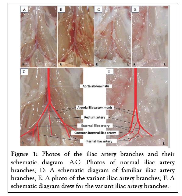
The distal branches of the aorta were dissected. 74 of them were consistent with previous documents [3]. The left and right CIA were two branches of the abdominal aorta terminus. Each CIA descended along the medial lumbar macro muscle as a trunk, and then divided into an External Iliac Artery (EIA) and an Internal Iliac Artery (IIA). Among the 74 mice anatomy, 60 inferior rectal arteries originated from the proximal segment of right CIAs (Figure 1A), 4 rectal artery from the abdominal aorta terminus (Figure 1B), and 10 rectal arteries from left CIAs (Figure 1C). A schematic diagram with A as an example is shown in Figure 1D.
One mouse showed an IAV unexpectedly. The terminal abdominal aorta descended as a trunk and finally extended into the left EIA after divided out several main branches. The first branch was thick and should work as a right CIA, but only extended into right EIC. The second branch was thin and went downward to be a RA. The third branch moved forward the central axis and divided into a left IIA and a right IIA symmetrically. For convenience, we named it as a Common Internal Iliac Artery (CIIA) (Figure 1E) A schematic diagram with E is shown in Figure 1F.

Figure 1: Photos of the iliac artery branches and their schematic diagram. A-C: Photos of normal iliac artery branches; D: A schematic diagram of familiar iliac artery branches; E: A photo of the variant iliac artery branches; F: A schematic diagram drew for the variant iliac artery branches.
Profound knowledge of arteries of the pelvis is essential to interpret and identify all branches of iliac arteries to reduce the intraoperative bleeding and iatrogenic injury. Interesting variations of iliac arteries have long received the concerning of anatomists and surgeons. In the present era of interventional radiology, diagnostic and interventional radiologic procedures on the pelvic arteries are becoming more and more frequent. Attention to the anatomical details helps to guide the interventional procedures [4]. The detailed knowledge of morphology helps in guiding the interventional radiologist in intra-arterial procedures during arterial embolization for hemorrhage, during selective catheterization of the intra-arterial therapy, and embolization of the pelvic tumors [5].
IAVs were uncommon. They were incidentally discovered during autopsy or on imaging in human beings [6]. There are few reports of congenital variations of the caudal aorta and iliac vessels [7]. Only a few branching variations at the abdominal aortic bifurcation have been reported in the literature. Most of these reported variations are related to the length or the abnormalities of level of branching deviations in respect to the pathognostic reference landmarks. Engin Kara, et al. have found that the right external iliac artery arose from the caudal abdominal aorta as a separate branch by angiography [8]. Congenital absence of the iliac arteries is extremely rare and occasionally resulted from postoperative disabling arterial insufficiency. Oduro, et al. reported a case of absence of left common and internal iliac arteries. An abnormal left external iliac artery arose from the left renal artery instead of the end of the abdominal aorta [9]. A case of bilateral congenital absence of internal iliac arteries was reported by Harb, et al. The absent arteries were compensated by lumbar arteries [10]. Koyama, et al. reported a case of absent left external iliac arteries. A welldeveloped left internal iliac artery was continuous with the left common femoral artery to supply blood for left lower limb [11].
Besides, some progresses were also made in the anatomical abnormalities of the iliac arteries branches. A common trunk arising from external iliac which divided into downstream arteries has been observed by Bilgic and Sahin [12]. Aloop of external iliac artery in the lesser pelvis by Moul, et al. [13]. Deep circumflex iliac artery from the external iliac artery has been found by Rusu, et al. [14]. An obturator artery arose from external iliac artery was found by Nayak SB, et al. [4]. Origin of collateral aberrant right ovarian artery was reported by Kwon JH, et al. [15].
Interesting variations in the branches of iliac arteries have long received the attentions of surgeons and anatomists. The present study is unique in comparison with the existing literatures. It might be of great interest for surgeons and vascular interventionist in the present era of interventional radiology. Considering that the presence of any vascular variations anomaly could present a technically challenging situation to the radiologist or operating surgeon, the knowledge of any anatomical variation in the vascular patterns of various regions is important and necessary to be reported.
As a star animal in atherosclerosis researches, ApoE-/- mice received much more attention by researchers decades ago [16]. In the animal a massive hyperlipidemia is accompanied with the development of severe atherosclerotic plaques at the aortic root and widespread fibrous plaques at the aortic branch points 1, 2. Studies often require the collection of the aortas and their main branches. The arteries at branches site are apt to form vulnerable plaques [17]. The discovery and enrichment of the anatomy knowledge should not be ignored.
An IAV was incidentally found in an experimental mouse in present study. Different from the familiar left-right symmetry pattern, it manifested by a ventral aorta extending to the lower left as the left EIA, and the other branches were separated en route. It is rare and interesting. The variation found in our study may enrich the existing data of mouse iliac artery anatomy. Fully mastering the complexity and variability of mouse iliac vessel anatomy contributes to the design and implementation of various experimental studies.
Mice as vertebrate similar to human beings, the variation of their iliac vascular anatomy may also help to pre-intervention in clinical practice and applications. Hence in here, we present a case report on extremely rare morphological variations of the iliac arteries of mice.
Knowledge of anatomical variations in the vascular patterns of various regions is important and necessary as the presence of any vascular anomaly could present a technically challenging situation to the radiologist or operating surgeon. Prior knowledge of the anatomical variations surely is beneficial for interventional cardiologists during selective catheter insertion and vascular surgeons ligating the IIA or its branches and the radiologists interpreting angiograms of the pelvic region [18].
A rare IAV in ApoE-/-mice in the present study identified. Considering mice as vertebrate mammals highly close to human anatomy, the finding might help in the pre-intervention for catheter surgery in human patients and interpreting angiography of the pelvic region by radiologists. The vascular abnormalities found in the present study are first manifested as the direct outflow of the right external iliac artery from the caudal part of the abdominal aorta, thus resulting in the disappearance of the right common iliac artery and the abnormal origin of the right internal iliac artery from the left common iliac artery, which is a extremely rare vascular variant.
The study was approved by the Ethics Committee of the Weifang Medical University (approval reference number, 2021SDL037). All procedures conformed to the NIH Guide for the Care and Use of Laboratory Animals. And the study is reported in accordance with ARRIVE guidelines.
Not applicable.
All related data and material presented in the present paper.
None declared.
This work was supported by Shandong Provincial Natural Science Foundation Project (Grant NO.ZR2020KH028; awarded to JX.L.) and Daqing City Guiding Science and Technology Plan Project (Grant No.zdy-2021-31; awarded to X.Z.).
Xi Zhang contributed to study implementation and primary manuscript creation. Wanlu Tao contributed to data collection and study implementation. Jinxin Liu contributed to study conception, implementation, funding acquisition, primary manuscript creation and its revision.
Citation: Zhang X, Tao WL, Liu JX (2023) A Study of Rare Anatomical Variation in ApoE-/- Mice and Systematic Review of Iliac Artery Variations. J Clin Exp Cardiolog. 14:769.
Received: 06-Dec-2022, Manuscript No. JCEC-22-20660; Editor assigned: 09-Dec-2022, Pre QC No. JCEC-22-20660; Reviewed: 27-Dec-2022, QC No. JCEC-22-20660; Revised: 05-Jan-2023, Manuscript No. JCEC-22-20660; Published: 18-Jan-2023 , DOI: 10.35248/2155-9880.23.14.769
Copyright: © 2023 Zhang X, et al. This is an open-access article distributed under the terms of the Creative Commons Attribution License, which permits unrestricted use, distribution, and reproduction in any medium, provided the original author and source are credited.