Reproductive System & Sexual Disorders: Current Research
Open Access
ISSN: 2161-038X
ISSN: 2161-038X
Case Report - (2022)Volume 11, Issue 5
Hematometra is a rare condition characterised by a congenital or acquired anatomical blockage of the cervical canal. Surgical operations on the uterus or cervix can cause acquired cervical stenosis. Amenorrhea or dysmenorrhea in premenopausal women, pelvic discomfort or pressure, urine frequency and retention are all common signs of this condition. We present a case of hematometra with hematocolpos caused by cervical canal blockage that was treated with ultrasound-guided lacrimal probe implantation.
Hematometra; Dysmenorrhea; Bilateral fornices; Perimenopausal
Hematometra and hematocolpos are medical conditions involving collection or retention of blood in the uterus and cervix respectively. It is most commonly caused by congenital abnormalities of the cervix and uterus. Less commonly, it can be acquired, due to pathologies that cause obstruction of the cervical canal [1,2]. Hematometra typically presents as cyclic, cramping pain in the pelvis or lower abdomen. Patient may also report increased frequency of urination or urinary retention. Premenopausal women with hematometra often experience dysmenorrhea or amenorrhea, while post-menopausal female are likely to be asymptomatic. Due to accumulation of blood in the uterus, patient may experience fall in the blood pressure, vasovagal response and can also present with complaints of acute abdomen. When palpated, the uterus typically feels firm and enlarged [1,3].
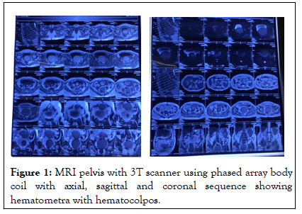
The patient is a 40 year old non pregnant para 3 live 2 female, who presented to the outpatient clinic on June 2019 with complaints of pelvic pain and fullness sensation for six days associated with four months amenorrhea. The patient reported that her last menstrual period was in January 2019. She gave a history of vaginoplasty, done at the age of 13 years, after which she got married and had three caesarean deliveries with the last child birth six years back. There was no history of any pelvic infection or any significant medical or surgical illness. Her previous cycles were normal with 28-30 days cycle duration, 4-5 day flow and soaking 2-3 pads/day with no dysmenorrhea. On per abdomen examination, a mass was palpable corresponding to 20 weeks gravid uterus. On per speculum examination external urethral meatus was dilated around 2 cm and dimple was seen at the vault. On per vaginal examination 3-4 cm vaginal length was felt with dimple felt at the vault, and the cervix was not felt. The uterus was soft and mobile. Bilateral fornices were free. Patient was severely anaemic on admission with hemoglobin of 6.8 gm%. Hemoglobin was built up and a Magnetic Resonance Imaging Scan was scheduled to locate the site of obstruction. The MRI scan revealed an anteverted uterus with gross dilatation of the endometrial canal (measuring 7.5 × 6.4 cm) along with stretching of the myometrium, endocervical canal as well as upper vagina (measuring 7.9 × 7.3 cm approximately) by fluid which appear hyperintense on T1W1 and heterogenous on T2W images along with rounding and ballooning of upper vaginal canal. Lower 15mm of vagina was not distended. Patient was prepared for hematometra drainage with vaginoplasty. During the surgery a uterine sound was introduced into the dimple seen and then dilated with serial dilators (upto a maximum of 11.5 mm). A constriction was felt at the junction of upper 1/3 and lower 2/3 of vagina. Ring cut was given anteriorly and laterally and dark colored blood was drained. Inner and outer vaginal mucosa was stitched using interrupted sutures (Figure 1). Finally patency was insured and a mould inserted. The postoperative course was uneventful and the patient achieved full recovery with resumption of menses in the month following the surgery.

Figure 1: MRI pelvis with 3T scanner using phased array body coil with axial, sagittal and coronal sequence showing hematometra with hematocolpos.
Hematometra is a medical condition in which there is retention of blood in the uterus and similarly, in hematocolpos there is retention of blood in the vagina. The obstruction causing retention can be primary (congenital), because of causes like imperforate hymen, transverse vaginal septum and partial vaginal agenesis or it may be acquired. This report describes an unusual case of a mid vaginal occlusion acquired in the Perimenopausal age, likely due to post inflammation fibrosis causing obstruction of vaginal outflow leading to formation of hematocolpos. Common symptoms of vaginal stenosis include chronic pelvic pain or pressure sensation, dysmennorhea, amenorrhea, infertility and endometriosis. Our patient developed vaginal stenosis due to gradual fibrosis post infection.
Ultrasound and MRI are two imaging modalities that can be used in the diagnosis of hematometra and hematocolpos [3-5]. On ultrasound, the uterus can be seen as an enlarged pelvic mass with an echo free lumen due to the presence of blood. Physical examination per abdomen reveals a palpable mass or an enlarged uterus. Additionally diagnosis of the level of obstruction and any congenital malformation is aided by MRI. Management of hematocolpos and hematometra usually involves serial dilatations with hegar’s dilator to drain accumulated blood [6]. The method of management chosen for our patient was serial dilatation with vaginoplasty. One of the greatest challenges associated with treatment is the high rate of recurrences. In an attempt to overcome this challenge, the use of catheters, pessaries and stents to maintain the patency of the cervix has been suggested by several authors. Yang, et al, published a case report of severe recurrent cervical stenosis and hematometra, refractory to both the surgical and conservative management [7]. There is a strong need for further organized study of suitable treatment modalities in order to prevent recurrent stenosis. In our patient as well, patency was ensured by placing a mould in the vagina for 4 weeks but during the follow visits, the patient started developing narrowing of the introitus and thus a decision for hysterectomy was taken as a definite treatment.
Vaginal stenosis is a rare complication post pelvic infection however with a history of vaginoplasty in our patient, the risk was high. Although other causes of vaginal stenosis should be considered, the history and evidence here pointed towards infective etiology. Post infection inflammatory fibrosis develops which can lead to vaginal stenosis. Ultrasonography is the best imaging modality for the diagnosis. The most appropriate initial approach is hematometra drainage with vaginoplasty. If recurrence is present, a definite treatment would be hysterectomy. Early recognition and treatment of this disorder may help to prevent severe complications like anemia, uterine rupture, endometriosis, etc.
[Cross Ref] [Google Scholar] [Pubmed]
[Cross Ref] [Google Scholar] [Pubmed]
[Cross Ref] [Google Scholar] [Pubmed]
[Cross Ref] [Google Scholar] [Pubmed]
[Cross Ref] [Google Scholar] [Pubmed]
[Google Scholar] [Pubmed]
Citation: Kashyap D, Yadav R, Kumar M (2022) Acquired Hematometra and Ematocolpos: A Rare Condition in a Perimenopausal Reproductive Female. Reprod Syst Sex Disord. 11:324.
Received: 26-May-2022, Manuscript No. RSSD-22-001-PreQc-22; Editor assigned: 30-May-2022, Pre QC No. RSSD-22-001-PreQc-22(PQ); Reviewed: 17-Jun-2022, QC No. RSSD-22-001-PreQc-22; Revised: 01-Jul-2022, Manuscript No. RSSD-22-001-PreQc-22(R); Published: 12-Jul-2022 , DOI: 10.35248/2161-038X.22.11.324
Copyright: © 2022 Kashyap D, et al. This is an open-access article distributed under the terms of the Creative Commons Attribution License, which permits unrestricted use, distribution, and reproduction in any medium, provided the original author and source are credited.