Journal of Leukemia
Open Access
ISSN: 2329-6917
+44 1300 500008
ISSN: 2329-6917
+44 1300 500008
Image Article - (2022)Volume 10, Issue 5
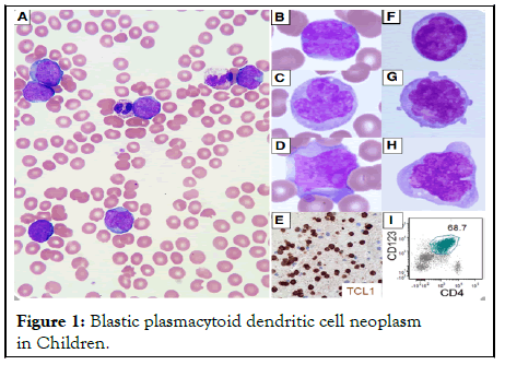
A previously healthy 2-year-old girl had a fever and cough for 2 days. With 76% blasts, peripheral blood showed marked leukocytosis (white blood cells, 75000/mL) (Figure 1).

Figure 1: Blastic plasmacytoid dendritic cell neoplasm in Children.
Blastic plasmacytoid dendritic cell neoplasm in children
Panels A-D: The blasts varied in size from medium to large, with agranular paleblue cytoplasm. The nuclei were irregular, folded or formed like flowers. A prominent nucleolus was seen in some of the specimens. Blasts were found in 87% of the cerebrospinal fluid [1].
Panel E: 47, X, der(X)t(X;8)(q24;q24.1), add(5)(p13), -6, del(6) (q13q27), +8, der(8), t(X;8), add(8) (q24.1) × 2, del(9)(q12q31), del(9)(q13q22), der(10;13)(q10;q10), add(12)(p13), -14, +3mar (20), del(9)(q12q31), del(9)(q13q22), der(10;13)(q10;q10), add(12)(p13),-14 MYC rearrangement t(X;8) (q24;q24.1) is confirmed by Fluorescence In Situ Hybridization (FISH).
FISH (Fluorescence In Situ Hybridization) showed a single deletion of the ETV6 gene but no MLL gene rearrangement, BCR/ABL1 translocation, or ETV6/RUNX1 translocation. The patient was diagnosed with blastic plasmacytoid dendritic cell neoplasm and was in complete remission 4 weeks after starting high-risk acute lymphoblastic leukaemia therapy, according to a bone marrow biopsy. The recognized distinguish this case: young age, negative CD56, acute leukaemia without skin lesions, and central nervous system involvement [2].
Panels F-H: The lactate dehydrogenase level in the patient's blood was 3759 IU/L. The patient's lymphadenopathy was significant, but she had no skin lesions or hepatosplenomegaly. The blasts were positive for CD4, CD7, CD36, CD38, dim CD45, CD123, and HLA-DR by flow cytometry, but negative for the tested lineage markers CD2, CD3 (surface and cytoplasmic), CD5, CD14, CD19, CD20, CD33, CD34, CD41, CD56, CD64, CD117, TdT, and myeloperoxidase by flow cytometry [3].
Panel I: The blasts tested positive for T-cell leukaemia 1 by immunohistochemistry.
[CrossRef] [Google Scholar] [PubMed]
[CrossRef] [Google Scholar] [PubMed]
[CrossRef] [Google Scholar] [PubMed]
Citation: Carugo S (2022) Acute Leukemic form of Blastic Plasmacytoid Dendritic Cell Neoplasm in Children. J Leuk. 10:304.
Received: 13-Jun-2022, Manuscript No. JLU-22-17918; Editor assigned: 17-Jun-2022, Pre QC No. JLU-22-17918(PQ); Reviewed: 07-Jul-2022, QC No. JLU-22-17918; Revised: 11-Jul-2022, Manuscript No. JLU-22-17918(R); Published: 18-Jul-2022 , DOI: 10.35248/2329-6917.22.10.304
Copyright: © 2022 Carugo S. This is an open-access article distributed under the terms of the Creative Commons Attribution License, which permits unrestricted use, distribution, and reproduction in any medium, provided the original author and source are credited.