Clinical & Experimental Cardiology
Open Access
ISSN: 2155-9880
ISSN: 2155-9880
Short Communication - (2021)Volume 12, Issue 5
Invasive physiological assessment of coronary lesions by hyperemic or resting indices was shown to improve the outcome of Ischemic Heart Disease (IHD) patients [1,2]. Treatment strategy was shown to change by more than 40% when using physiology assessment compared with angiography alone, thereby saving inappropriate stent implantations and bypass surgeries [3]. However coronary physiological assessment is performed in less than 10% of interventions [4]. This may be due to several reasons including setup time, cost, patients’ discomfort and operator preference. The invasiveness of the test was addressed by several angiography based Fractional Flow Reserve (FFR) software with good correlation to the pressure-wire based FFR [5-7]. These software however, require image capture at specific angle projections and manual annotation of the vessel of interest which may impact the result of the software.
The use of Artificial Intelligence (AI) in general, and especially in medicine is growing exponentially with several potential applications in various fields which cardiology is one of them [8]. The application of AI in medical imaging is growing and is considered a relatively classic niche for AI algorithms to provide important service for the physicians and patients. The use of AI in physiological studies, however, is in its launch.
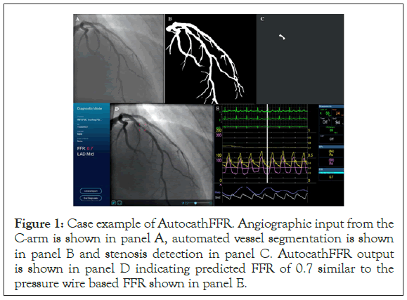
Recently, an automated algorithm (Autocath FFR by MedHub- AI, Tel-Aviv, Israel) to detect and examine the physiological significance of coronary lesions was tested in a pilot study in Israel. The algorithm was developed based supervised machine learning and computer vision algorithms according to vast angiographic and physiological data from multiple centers, using various angiographic machines and pressure wires. Autocath FFR analyzes online regular DICOM data from the catheterization laboratory and is able to automatically segment the coronary vessels and classify them according to each artery and part, it then detects narrowing and estimates their hemodynamic significance expressed as an estimated FFR value (Figure 1). The process doesn’t require specific projection angles or manual annotation and is performed fully automatically. However, the software does perform initially a self-testing procedure ensuring that the input image characteristics are within the range of the learned data. Low qualitative, damaged or incomplete images are detected and excluded. For optimal FFR prediction by the software, several views are needed. Another feature of the algorithm is that it can be used in an offline mode for cases that were performed during call hours for acute cases in which non-culprit lesions were left for later assessment and treatment or for further consultation. Lesion tracking can be used for assessment of the result following intervention in cases where another lesion was left untreated.

Figure 1: Case example of AutocathFFR. Angiographic input from the C-arm is shown in panel A, automated vessel segmentation is shown in panel B and stenosis detection in panel C. AutocathFFR output is shown in panel D indicating predicted FFR of 0.7 similar to the pressure wire based FFR shown in panel E.
The algorithm was tested in a pilot study including 31 patients that underwent coronary angiography with FFR assessment [9]. Most commonly the LAD was tested and mean FFR value was 0.82+0.08 with FFR ≤ 0.8, which is considered positive in 55%. In this study the discordance of Autocath FFR for lesion classification compared with pressure wire based FFR was noted in only 3 lesions. These results achieved a sensitivity of for identifying lesion with positive FFR of 0.88, specificity of 0.93 with positive predictive value of 0.93 and negative predictive value of 0.87, reaching an overall accuracy of 0. Mean difference between wire based FFR and Autocath FFR was 0.05+0.12 with a strong correlation coefficient of r=0.71. The results of this study were encouraging and provided the ground for CE approval and for a larger pivotal trial for an FDA approval.
Implementation of AI technology in the assessment of coronary lesions can potentially revolutionize the field by providing a highquality standard instead of the current practice of eye-ball assessment of lesions which result in a discrepancy rate of close to 50% [10]. While, coronary physiologic assessment is associated with less stenting procedures, it might be that we also miss significant lesions that otherwise would have been treated, however since the operator underestimates it, the lesion is left unexamined. It has been shown that even when patients are treated, the result is not sufficient and significant lesions are left untreated, and consequently patients left untreated and still suffering from angina [11]. AI can provide an anatomic and physiologic map of the coronary tree that can be both online and offline, which will result in disrupting the current practice and consequently will leap forward the diagnostic coronary angiography which is the basis for coronary artery disease therapeutic decisions and therefore outcome.
Despite the well-established evidence of FFR-guide revascularization therapy, its utilization is still low. There are many reasons for FFR underuse, including its cost, patient comfort and safety, and most of all, operator preference. However, a recent study showed a disturbing discrepancy rate between FFR result and operator decision reaching almost 50% of intermediate lesions [7,9]. Indeed, in the FAME trial, 35% of lesions with a stenosis grade of 50% to 70% were found significant by FFR while 20% of lesions graded 70% to 90% stenosis was found insignificant. Similar results were found among the current study population. This discrepancy was found also between AutoFFR and visual estimation in the lesions that were not assessed by pressure wire and may potentially indicate lesions that the operator missed or misinterpreted as hemodynamically insignificant. Various solutions have been developed including computerized tomography derived FFR [10,11] and angiography image-based FFR [5], relying on 3-dimensional reconstruction and implementation of flow dynamics algorithm, with various accuracy levels ranging from around 80% to above 90%. The main disadvantage of these solutions is the requirement for specific angiographic views and the need for vessel annotation for each artery, which requires some learning curve, interferes with the workflow of the catheterization laboratory and may result in significant prolongation of the procedure. The Autocath FFR is based on machine learning algorithm with automatic lesion detection and analysis, without the requirement of specific views or manual image selection and vessel annotation. These features enable a user-friendly device that can facilitate a straightforward appropriate decision process and treatment application with an easy incorporation in the catheterization laboratory workflow.
Our study main limitation is the small number of patients and lesions (especially those in arteries other than the LAD), as expected from a pilot feasibility study. Notwithstanding the need for further research in larger cohorts and various patient populations and settings, the results of this study point towards the potential of such systems to improve the decision process of the operator at the catheterization laboratory. In conclusion, AI-based angiography image derived FFR is a promising tool that can facilitate improved management of coronary artery disease patients. Further efforts to improve the algorithm and to increase its correlation with FFR are ongoing, along with planned larger scale studies of this technology for the evaluation of its clinical use.
Citation: Koifman E, Dogosh AA (2021) Artificial Intelligence in the Cathlab: Angiography Based Physiological Test. J ClinExp Cardiolog. 12:677.
Received: 01-Apr-2021 Accepted: 15-Apr-2021 Published: 22-Apr-2021 , DOI: 10.35248/2155-9880.21.12.677
Copyright: © 2021 Koifman E, et al. This is an open-access article distributed under the terms of the Creative Commons Attribution License, which permits unrestricted use, distribution, and reproduction in any medium, provided the original author and source are credited.