Journal of Clinical & Experimental Dermatology Research
Open Access
ISSN: 2155-9554
+44 1478 350008
ISSN: 2155-9554
+44 1478 350008
Case Report - (2020)Volume 11, Issue 5
Bier's spots are manifested by hypopigmented lesions of the upper limbs which appear in the declive position. Only a
few cases have been reported since its description by Bier in 1898.
We report a new case.
Bier’s spots; Limbs; Hypopigmented lesions
A 28 years old patient with no medical history, she presented with hypopigmented lesions of the upper limbs isolated and observed in the decline position, evolving for 2 years. The anamnesis did not find any notion of photosensitivity nor of Raynaud's syndrome nor of acrocyanosis [1].
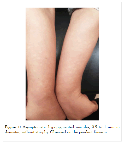
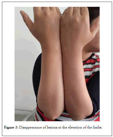
The clinical examination objectified hypopigmented macules of 5 to 10 mm in diameter irregular pale, scattered on the forearms in a sloping position of the limbs, Without any sign of atrophy (Figure 1) and quickly disappearing when the arms were raised (Figure 2), the remainder of the examination was normal.

Figure 1: Asymptomatic hypopigmented macules, 0.5 to 1 mm in diameter, without atrophy. Observed on the pendent forearm.

Figure 2: Disappearance of lesions at the elevation of the limbs.
The standard biological workup, cryoglobulinemia and immunoassay: Anti-neutrophilicpolynuclear cytoplasm antibody, Antinuclear antibodies, Soluble anti-nuclear antigen antibodies were normal.
Skin biopsy suggested vascular ectasia with extravasation of erythrocytes with minimal inflammatory lymphohistiocytic infiltrate consistent with Bier's stain.
The pathology was explained to the patient and it was decided to abstain from therapy.
Bier spots (vasospastic macules or physiological anemic macules) most often appear in young adults between the ages of 20 and 40. Women are most often affected, as is the case with our patient [2,3]. They are the consequence of an exaggerated physiological response of the small dermal vessels to venous hyperpressure. As a result, these white spots readily appear on the distal part of a sloping limb and disappear if the limb is raised and the venous pressure thus lowered. In the event of vascular stasis, such as during cryoglobulinemia, for example, Bier's spots may appear, hence the value of carrying out a workup in order to rule out an underlying etiology [4,5]. Note that an association with renal scleroderma crisis, lymphoma, mixed cryoglobulinemia have been reported in the literature. It is important to rule out differential diagnosis such as postinflammatory hypopigmentation, tinea versicolor, vitiligo, anemic nevus, pityiasisalba [6]. No treatment is necessary in the majority of cases, therapeutic abstention is always the rule in front of the asymptomatic and benign nature of this pathology [7].
Bier’ s spots correspond to a rare entity that reflects a physiological phenomenon, the diagnosis is clinical, an assessment is necessary in order to rule out an associated vascular pathology, no treatment is necessary in the majority of cases as in the case of our patient.
Citation: Palamino H (2020) Bier’s Spots: A New Case Report. J Clin Exp Dermatol Res. 11:532. DOI: 10.35248/2155-9554.20.11.532
Received: 11-Aug-2020 Accepted: 26-Aug-2020 Published: 31-Aug-2020 , DOI: 10.35248/2155-9554.20.11.532
Copyright: © 2020 Palamino H. This is an open-access article distributed under the terms of the Creative Commons Attribution License, which permits unrestricted use, distribution, and reproduction in any medium, provided the original author and source are credited.