Medical & Surgical Urology
Open Access
ISSN: 2168-9857
ISSN: 2168-9857
Case Report - (2020)Volume 9, Issue 5
Rectus sheath haematomas associated with anticoagulation are often self-limiting. When large, however, they can even extend into the pelvis and cause compression of adjacent organs. A 54-year-old female on treatment Coumadin; during her hospitalisation, she developed a spontaneous rectus abdominis haematoma and occurred bilateral obstructive uropathy. A large rectus sheath haematoma that extends into the bladder causing renal obstruction can be treated by minimale invasive surgery.
Rectus sheath hematoma; Abdominal wall mass; Obstructive anuria
Rectus Sheath Hematoma (RSH) is a relatively rare clinical condition, generally associated anticoagulant treatment [1]. RSH results from hemorrhage caused by rupture of one of the epigastric arteries or a tear of the muscle itself.
The patients may presents with local or diffuse abdominal wall mass, abdominal pain, tenderness, nausea, vomiting and fever [2]. Various complications have been reported depending on extent of the hematoma. We reported a case of large rectus sheath hematoma causing obstructive anuria and hydronephrosis.
A 54-year-old female patient, her medical history included hypertension and had cardiac valve replacement. She was using coumadin for ten years. The most common signs and symptoms of patient consist of abdominal pain, palpable abdominal wall mass, tenderness, nausea. Abdominal pain was acute onset and develops over hours.
Ultrasound (US) and Computed Tomography (CT) abdomen with contrast showed right rectus abdominis haematoma below the level of the umbilicus with active arterial bleeding in the extraperitoneal pelvis causing mass compression (Figure 1).

Figure 1: Computed Tomography (CT) scan shows blood in the left rectus sheath and bilateral hydronephrosis.
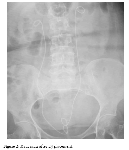
On the following day, she had moderate elevated serum creatinine (sCrea; 4.5 mg/dL) and the Hb level was down from 14.2 to 8.5 mg/dL. Three units of packed red blood cells were ordered. Systolic blood pressure diminished to around 80 mmHg, and urinary output decreased to 50 mL per 8 hours. She was admitted to the intensive care unit and found to be anuric and bilateral hydronephrosis were detected on US. Once her INR was adequately reduced to 1.5, a left and a right retrograde uretric double J stents were placed (Figure 2).

Figure 2: X-ray scan after DJ placement.
Decision was made to manage for the ruptured vessels conservatively. Normal urine output restored following the 6th hour. The patient was discharged sixth days following admission and urine output was started. Creatinine value decreased to the limit of normal levels after fourth day.
This case report presented of hemorrhagic shock and obstructive uropathy due to a large rectus sheath hematoma in a patient on anticoagulant therapy. Hemorrhage into the rectus muscle sheath is a relatively uncommon complication of anticoagulant therapy.
The most common signs and symptoms of RSH consist of abdominal pain, palpable abdominal wall mass, tenderness, nausea, vomiting, fever [2]. The pain is usually described as severe, sharp, persistent and non-radiating [2].
Ultrasound (US) is an in expensive, safe and easy to use diagnostic method that is used as a first-line test in acute abdominal pain and it has a sensitivity of 90% [3,4]. Typically, mass usually appears homogeneous and sonolucent, but in the presence of clot could appear.
CT imaging can also offer accurate anatomical delineation is reported to be a perfect predictor for acute RSH, with sensitivity and specificity reaching 100% [4-7].
RSH treatment could be conservative or invasive. Treatment choice is determined by the patient's condition. The vast majority of patients can be treated conservatively, because usually the hematomas are self-limiting, several recent studies have reported a satisfactory outcome of conservative treatments because of prognosis is favorable [8].
Management of this condition involves correcting any haemodynamic instability, followed by reversal of anticoagulation or treatment of bleeding diatheses. A conservative approach to management of RSH is highly effective in combination with double J stenting to relieve hydronephrosis.
Citation: Dede O (2020) Bilateral Obstructive Uropathy Due to Spontaneously a Large Rectus Sheath Hematoma in a Patient on Anticoagulant Treatment. Med Sur Urol 9:238. doi: 10.35248/2168-9857.19.9.238
Received: 10-Sep-2020 Accepted: 01-Nov-2020 Published: 07-Nov-2020 , DOI: 10.35248/2168-9857.19.8.214
Copyright: © 2020 Dede O. This is an open-access article distributed under the terms of the Creative Commons Attribution License, which permits unrestricted use, distribution, and reproduction in any medium, provided the original author and source are credited.