Medical & Surgical Urology
Open Access
ISSN: 2168-9857
ISSN: 2168-9857
Case Report - (2019)Volume 8, Issue 1
Bilateral duplicated system with ectopic ureter is a rare entity. Duplication and its associated anomalies are more common in females. Anomalies of the renal collecting system should be considered by clinicians and surgeons in patients presenting with urinary incontinence.
We present a 32-year-old female with a Bilateral Ureteral Duplication with Right Ectopic Ureter presenting with low volume Incontinence. We emphasized the importance of imaging in the diagnosis of anomalies of the renal collecting system presenting with incontinence.
In the absence of a dysplastic renal moiety, an ureteroneocystostomy is an ideal procedure of choice to correct incontinence from ectopic ureters in females.
Duplication; Ectopic; Incontinence; Ureters
Duplex renal system is described as the kidney has two pyelocaliceal systems with single, bifid (partial ureteral duplication) or double ureter (complete ureteral duplication) draining into the bladder [1]. The incidence of duplex renal system ranges from 0.5%-3% [1]. Bilateral complete renal duplication with ureteral ectopia is a rare entity, found more commonly in females [2]. The exact incidence is not known, as it is mostly an incidental finding [2]. Duplications are often completely asymptomatic and often come to light only in the course of investigations for other reasons [3]. Clinical problems are the result of obstruction, reflux, or ectopic openings, giving rise to hydronephrosis, infection and incontinence [3].
Ectopic ureter occurs in one in two thousand individuals, with the female to male ratio being 6:1 [2]. In females, 80% involve the duplex systems and most drain into the vagina or urethra and present as incontinence [2]. Most cases are diagnosed during childhood, as a result of continuous urinary dribbling or recurrent UTIs with a dysplastic upper pole renal moiety in duplex kidneys. Some patient may present during adulthood due to poor access to appropriate health care or due to milder symptoms over the years.
We therefore present a 32-year-old female with a bilateral ureteral duplication with right ectopic ureter presenting with low volume Incontinence.
A 32-year old female presented to our outpatient department with complaint of continuous low volume urinary incontinence for over 20 years requiring 1 to 2 pads daily. She could not admit whether the urine leakage was affected by positional changes or coughing. Physical examination smears of urine in the vagina with mild vagina inflammation. Intravenous Pyelogram revealed bilateral double renal collecting system without evidence of pelvicalyceal dilation with post micturition films showing contrast in the vagina.
We could not assess a dyplastic moiety in the absence of dimercapto-succinic acid and mercaptoacetyltriglycine (MAG-3) scans. However, the ultrasound and CT-scan showed an acceptable and contrast-secreting renal parenchyma on both sides. Further imaging with a contrast enhanced computed tomography along with a 3D reconstruction showed a complete duplicated ureter on the right with an ectopic ureter entering the vagina and a right lower pole moiety entering the bladder superolaterally.
On the left, showed a bifid ureter joining just above the pelvic brim. Therefore, the patient underwent a right ureteroneocystostomy over a double J stent. The right ectopic ureter was transected and ligated close to the ectopic insertion at the vagina. The non-refluxing Leadbetter-Politano technique was used for the repair.
The right ureter was tunneled under the mucosa superomedial into the bladder and anastomosed directly to the bladder mucosa with interrupted 3.0 PDS sutures. On postoperative day 7, a followup cystogram and post micturition films showed no contrast extravasation in the pelvis or vagina.
The drain and urinary catheter were subsequently removed on postop day 8. Her follow-up has been satisfactory (Figures 1-5).
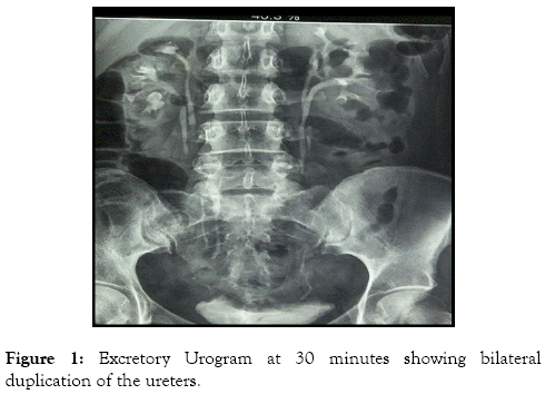
Figure 1: Excretory Urogram at 30 minutes showing bilateral duplication of the ureters.
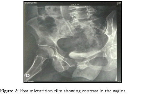
Figure 2: Post micturition film showing contrast in the vagina.
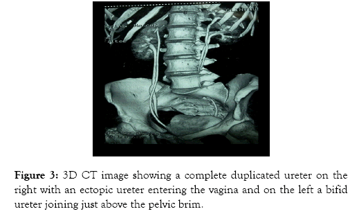
Figure 3: 3D CT image showing a complete duplicated ureter on the right with an ectopic ureter entering the vagina and on the left a bifid ureter joining just above the pelvic brim.
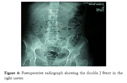
Figure 4: Postoperative radiograph showing the double J Stent in the right ureter.
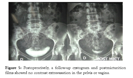
Figure 5: Postoperatively, a follow-up cystogram and postmicturition films showed no contrast extravasation in the pelvis or vagina.
Ureteral duplications are the result of premature splitting of the ureteric bud, a remnant of the Wolfian duct [3]. Partial duplication is observed in metanephric tissue that has not separated fully, but complete duplication may be result of two distinct ureteric buds.
The prevalence of complete or incomplete ureteral duplications in the urinary system has been stated 0.7% in autopsy series and between 2% and 4% in clinical series. It is two times more common in women than in men [4]. Incomplete duplication is seen three times more than complete duplication. Complete duplications are rare, occur in approximately less than 0.1% of the population, and are more common in females [2]. Only 25% of these are reported to be bilateral [2].
Ectopic ureter is defined as the abnormal opening of the ureter orifice outside of its normal site in the trigone. This case is more commonly seen in women and is 80% associated with duplex collecting system [4]. Weigert Mayer law is observed in complete ureteral duplication cases [5]. The ectopic ureter arrives from upper moiety is inferior and medial as compared to the lower pole moiety’s ureter [5]. Moreover, because the lower pole ureter makes a shorter tunnel into the bladder, it is more prone to reflux, while the anomalous insertion of the upper pole ureter makes this moiety more prone to obstruction [6]. The dilated upper pole infundibulum tends to push down on the functioning lower pole collecting system, creating the classically described “drooping lily” appearance [6].
The first presentation of congenital anomalies in later adulthood is more likely to be seen in rural settings or medically underserved areas [7].
The patient’s presentation symptoms depend on the insertion site of the ectopic ureter, and this differs between girls and boys [8]. Usually, males do not present urinary incontinence because of the ureter’s insertion above the external urinary sphincter. Like our patient, most girls present with urinary dribbling, as the insertion of the ectopic ureter will bypass the exterior urinary sphincter [9]. Usually, affected girls have normal voiding patterns with small volume leakage or spotting incontinence [9].
In females the most frequent sites of ectopic ureter insertion are bladder neck and upper urethra (33%), vaginal vestibule between urethra and vaginal opening (33%), vagina (25%), and less commonly the cervix or uterus (<5%) [8]. Only girls with a ureteral insertion at or above the bladder neck and upper urethra will be continent [10].
Renal ultrasound and excretory urography can almost never detect an ectopic insertion of the ureter and they do not provide enough data regarding the precise anatomy as well as in delineating the relationship between the ureter, bladder, and vagina [9]. Therefore, contrast-enhanced Computed Tomography (CT) or magnetic resonance imaging urography should be the method of choice for depicting or ruling out an ectopic ureter [9].
The best treatment in children and symptomatic patients is surgery, and it tries to resolve the incontinence, prevent further complications, preserve renal function and eliminate the UTIs [9]. Surgical treatment consists in upper pole hemi-nephrectomy in the non-functional duplex system or recently laparoscopic ureteral ligation (clipping) or ureteral re-implantation in case of a preserved renal function [11].
When there is a preserved kidney function or a functioning upper pole in duplex system kidney, the surgery technique consists on ureteroneocystostomy or distal and proximal ureteroureterostomy respectively [9]. In our patient, an ureteroneocystostomy was performed because a dysplastic moiety could not be proven; as the right upper moiety parenchyma (and also lower moiety) was acceptable with contrast-secretion. According to Duici et al. 5% of patients who underwent heminephrectomy, there was loss of renal function in the remaining ipsilateral moiety, as a result of vascular injury or torsion of the renal pedicle from extensive renal mobilization [9,12].
Bilateral duplicated system with ectopic ureter is a rare entity. Duplication and its associated anomalies are more common in females. Anomalies of the renal collecting system should be considered by clinicians and surgeons in patients presenting with urinary incontinence. In the absence of a dysplastic renal moiety, an ureteroneocystostomy is an ideal procedure of choice to correct incontinence from ectopic ureters in females.
Special thanks to the Department of Urology and Andrology of the Grand Yoff Hospital.
The authors declare no conflict of interest regarding this article.
Citation: Cassell AK, Traoré A, Jalloh M, Ndoye M, Diallo A, Labou I, et al. (2019) Bilateral Ureteral Duplication and Right Ectopic Ureter Presenting with Incontinence: A Case Report. Med Sur Urol 8:216. doi: 10.35248/2168-9857.19.8.216
Received: 09-Jan-2019 Accepted: 30-Jan-2019 Published: 06-Feb-2019 , DOI: 10.35248/2168-9857.19.8.216
Copyright: © 2019 Cassell AK, et al. This is an open-access article distributed under the terms of the Creative Commons Attribution License, which permits unrestricted use, distribution, and reproduction in any medium, provided the original author and source are credited.