Journal of Clinical and Experimental Ophthalmology
Open Access
ISSN: 2155-9570
ISSN: 2155-9570
Clinical image - (2024)Volume 15, Issue 1
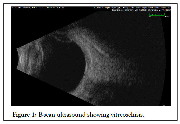
We report the case of a 65-year-old female patient, with a type 2 diabetes, who presented for a decreased visual acuity in both eyes. The examination of the right eye finds a Best Corrected Visual Acuity (BCVA) of hand motion, an intumescent white cataract, the fundoscopy is not possible. In the left eye, the examination finds a BCVA of 3/10 a nuclear and a severe nonproliferative diabetic retinopathy. B-scan of the right eye finds a vitreoschisis.
Vitreoschisis is a consequence of an abnormal separation in the thickness of the posterior vitreous cortex. During a normal Posterior Vitreous Detachment (PVD), two phenomena are associated such as a progressive liquefaction of the vitreous gel and a decrease in the strength of the adhesion of the cortex to the retina [1]. If liquefaction is abrupt with strong adhesion of the posterior cortex, the cortex may cleave leaving a posterior layer on the internal limiting membrane, forming the vitreoschisis.
On B-scan ultrasound, in vitreoschisis, two vitreous membranes are seen that appear to join in a single layer, forming the letter Y (Figure 1).

Figure 1: B-scan ultrasound showing vitreoschisis.
Recognition of vitreoschisis is important. Some echographic patterns of diabetic retinal detachment might be explained by this phenomenon [2]. If the inner wall of the vitreoschisis cavity were mistaken for the posterior hyaloid during pars plana vitrectomy, the outer wall of the cavity and intervening cortical vitreous cortex would remain intact. It could serve as a scaffold for continued neovascular proliferation and contribute to persistent tangential traction on the retina.
[Crossref] [Google Scholar] [PubMed]
[Crossref] [Google Scholar] [PubMed]
Citation: Elmansouri O, Bezza H, Munsif A, Kriet M, Elasri F (2024) B-Scan Ultrasound of a Vitreoschisis. J Clin Exp Ophthalmol. 15:967.
Received: 29-Dec-2023, Manuscript No. JCEO-23-28398; Editor assigned: 01-Jan-2024, Pre QC No. JCEO-23-28398 (PQ); Reviewed: 15-Jan-2024, QC No. JCEO-23-28398; Revised: 22-Jan-2024, Manuscript No. JCEO-23-28398 (R); Published: 29-Jan-2024 , DOI: 10.35248/2155-9570.24.15.967
Copyright: © 2024 Elmansouri O, et al. This is an open-access article distributed under the terms of the Creative Commons Attribution License, which permits unrestricted use, distribution, and reproduction in any medium, provided the original author and source are credited.