Journal of Clinical and Experimental Ophthalmology
Open Access
ISSN: 2155-9570
ISSN: 2155-9570
Clinical image - (2021)
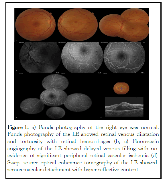
A 35-year-old female was referred to our department for sudden onset of blurred vision of the Left Eye (LE). Best corrected visual acuity of the LE was 20/1000. Anterior segment examination was normal. Fundus examination, fluorescein angiography and swept source optical coherence tomography findings concluded to the diagnosis of a non-ischemic CRVO of the LE. Right eye examination was unremarkable. Furthermore, the patient was positive to SARS COV 2 and symptomatic one week before the beginning of the symptomatology. An exhaustive workup including check for hypercoagulability, hematologic, immunologic and cardiac diseases came back normal. After two weeks of a regular follow up we noticed the disappearance of the CRVO signs and the macular edema with ad integrum restitution without any treatment. CRVO is one of the manifestations of COVID 19 infection. Cases reported varied in terms of presentation, severity, and management [1-5], in this case a spontaneous recovery was observed (Figure 1).

Figure 1: a) Funds photography of the right eye was normal. Funds photography of the LE showed retinal venous dilatation and tortuosity with retinal hemorrhages (b, c) Fluorescein angiography of the LE showed delayed venous filling with no evidence of significant peripheral retinal vascular ischemia (d) Swept source optical coherence tomography of the LE showed serous macular detachment with hyper reflective content.
Citation: Maamouri R, Nabi W, Somai M, Héla S, Monia C (2021) Central Retinal Vein Occlusion (CRVO) Associated to COVID-19 Infection. J Clin Exp Ophthalmol.12:898.
Received: 26-Nov-2021 Accepted: 10-Dec-2021 Published: 17-Dec-2021
Copyright: © 2021 Maamouri R, et al. This is an open-access article distributed under the terms of the Creative Commons Attribution License, which permits unrestricted use, distribution, and reproduction in any medium, provided the original author and source are credited.