International Journal of Physical Medicine & Rehabilitation
Open Access
ISSN: 2329-9096
ISSN: 2329-9096
Research Article - (2023)Volume 11, Issue 3
Objective: The purpose of present study was to investigate the effect of the ankle joint after the different taping (No Taping (NT), Lateral Taping (LT), Anterior-Posterior Taping (APT)) on the health adults balance ability and EMG activity around the ankle joint.
Materials and Methods: After the LT or APT of the ankle joint, the balance ability and EMG around the ankle joint of the healthy adults, which were single-leg standing with eyes open and closed were measured. Balance ability and EMG was compared before and after taped when the eyes were opened and closed.
Results: After LT and APT the body sway became less when eyes closed. In particular, LT methods, the trajectory length of X-direction shorter than NT (P<0.05); APT methods, the trajectory length of Y-direction shorter than NT (P<0.05). EMG activity of Tibialis Anterior and Peroneus Longus was decreased when single-leg standing with eyes closed (P<0.05).
Conclusion: The taping can enhance balance ability, and also can reduce the EMG activity of the muscles.
Exercise; Knee; Osteoarthritis; Bibliometric; Treatment
For athletes, taping is widely used in the sports arena, taping of the ankle is also used in injury treatment and prevention [1-7]. Ankle sprain could cause varus and valgus direction instability, taping could limit the instability of varus and valgus direction [8,9]. Taping can also improve lateral stability of the ankle joint. Ankle joint inversion sprain and Achilles tendonitis are representative motion ankle disorders. For the inversion sprain, the Lateral Taping (LT) technique can improve the lateral stability of the ankle joint; for the Achilles tendonitis, the Anterior-Posterior Taping (APT) technique can improve the stability of anterior-posterior of the ankle joint [10-13].
De Varis has shown that when healthy adults on single-leg were standing with eyes open, the total length of Center of Pressure (COP) with 20 seconds was 760 mm. After ankle sprain increased to about 800-860 mm [14]. In particular, after eyes closed, the total length of COP will greatly increase by 20 seconds, which was 1520 mm in healthy adults, after ankle sprain increased about 1660-1820 mm. Kobayashi′s [15] got similar results with De Varies. However, after taping with healthy adults, the change of total length of COP was unknown. Furthermore, there was no study on the affect concerning about the taping of the ankle joint of the body sway and EMG activity, and it is also unknown that LT and APT technique whether can improve the balance stability or change EMG activity. Therefore, it′s an interesting object and worthy to further study. The aim of this study was to examine the effect of ankle taping on balance ability and sEMG activity. We hypothesized that ankle taping technology also can improve the stability of ankle joint and body’s balance ability and EMG activity will reduced which was around the ankle joint. The results of the present study would be able to provide valuable theory and practice of rehabilitation technique for the athletes who have ankle injuries [16].
Study design
The balance ability and EMG activity of the left lower limb were measured under three conditions of bare foot, LT and APT under the condition of open and closed eyes. During the test, all subjects stood on single leg for 20 seconds. All the taping and measurements were performed by a senior physiotherapist, and subjects were blinded to our investigation regarding our purpose of identifying differences between the 2 taping.
Participants
Thirty healthy participants (16 male, 14 female) were recruited for this study, average age is 25.6 (male) and 26.8 (female) years old. Detailed information of subjects was shown in Table 1. People who had severe lower extremity injuries or cardiovascular diseases were excluded. All subjects in this study had no previous history of lower limb disorders, including ankle injury, Achilles tendonitis etc. and no pain or discomfort with lower limb when they were measured. This study was approved by the Ethics Committee of Beijing Rehabilitation Hospital, Capital Medical University (NO.2020bkky-037). And all participants provided written informed consent (Table 1).
| male(n=16) | female (n=14) | P | |
|---|---|---|---|
| Age (years old) | 25.6 ± 1.0 | 26.8 ± 1.6 | NS |
| Body height (cm) | 175.2 ± 0.1 | 161.3 ± 0.1 | NS |
| Body weight (kg) | 72.2 ± 4.9 | 52.4 ± 3.4 | NS |
| BMI (kg/m2) | 23.5 ± 2.7 | 20.1 ± 1.8 | NS |
Note: Values are presented as mean ± SD; NS: Not Significant.
Table 1: Basic information about study participants.
| Total length | X-direction | Y-direction | ||||
|---|---|---|---|---|---|---|
| EO | EC | EO | EC | EO | EC | |
| NT | 724.8 ± 112.8 | 1212.9 ± 215.9 | 523.4 ± 89.9 | 852.1 ± 170.0 | 402.4 ± 54.0 | 707.7 ± 107.9 |
| LT | 695.5 ± 106.1 | 1011.6 ± 162.8 | 468.6 ± 83.9 | 663.9 ± 153.1 | 417.2 ± 67.0 | 652.1 ± 85.8 |
Note: Values are presented as mean ± SD. EO, eyes open; EC, eyes closed; NT, no tape; LT, lateral taping. Compared eyes closed to eyes open, total length of COP with both NT and LT was significantly increased (*p<0.01); trajectory length of X-direction with both NT and LT was significantly increased (*p<0.01); trajectory length of Y-direction with both NT and LT was significantly increased (*p<0.01). Compared NT to LT when eyes closed, total length with LT was significantly reduced (*p<0.05); trajectory length of X-direction with LT was significantly reduced (*p<0.05).
Table 2: Trajectory length of COP, compared with NT and LT (mm).
| Total length | X-direction | Y-direction | ||||
|---|---|---|---|---|---|---|
| EO | EC | EO | EC | EO | EC | |
| NT | 724.8 ± 112.8 | 1212.9 ± 215.9 | 523.4 ± 89.9 | 852.1 ± 170.0 | 402.4 ± 54.0 | 707.7 ± 107.9 |
| APT | 649.5 ± 127.8 | 997.6 ± 180.7 | 457.3 ± 104.9 | 762.0 ± 154.0 | 373.4 ± 60.1 | 535.9 ± 127.0 |
Note: Values are presented as mean ± SD. EO, eyes open; EC, eyes closed; NT, no tape; APT, anterior-posterior taping. Compared eyes closed to eyes open, total length of COP with both NT and APT was significantly increased (*p<0.01); trajectory length of X-direction with both NT and APT was significantly increased (*p<0.01); trajectory length of Y-direction with both NT and APT was significantly increased (*p<0.01). Compared NT to APT when eyes closed, total length with APT was significantly reduced (*p<0.05); trajectory length of Y-direction with APT was significantly reduced (*p<0.05).
Table 3: Trajectory length of COP, compared with NT and APT (mm).
| TA | PL | MG | LG | TP | ||||||
|---|---|---|---|---|---|---|---|---|---|---|
| EO | EC | EO | EC | EO | EC | EO | EC | EO | EC | |
| NT | 16.1 ± 8.6 | 37.3 ± 12.3 | 49.8 ± 14.7 | 69.0 ± 17.2 | 55.1 ± 17.0 | 68.0 ± 15.2 | 21.1 ± 7.1 | 28.5 ± 7.6 | 35.1 ± 8.3 | 43.7 ± 5.7 |
| LT | 14.4 ± 9.8 | 31.0 ± 12.8 | 44.9 ± 11.2 | 52.9 ± 13.9 | 54.3 ± 14.3 | 67.6 ± 16.1 | 20.7 ± 5.9 | 27.2 ± 9.0 | 31.5 ± 7.1 | 42.8 ± 10.4 |
Note: Values are presented as mean ± SD. EO: Eyes open; EC: Eyes closed; NT: No tape; LT: Lateral taping. Compared eyes closed to eyes open, EMG activity of TA, PL, GSM, GSL and TP was significantly increased (*p<0.01). Compared LT to NT when eyes closed, EMG activity with both TA and PL was significantly induced (*p<0.05).
Table 4: EMG activity, compared with NT and LT (%MVC).
| TA | PL | MG | LG | TP | ||||||
|---|---|---|---|---|---|---|---|---|---|---|
| EO | EC | EO | EC | EO | EC | EO | EC | EO | EC | |
| NT | 16.1 ± 8.6 | 37.3 ± 12.3 | 49.8 ± 14.7 | 69.0 ± 17.2 | 55.1 ± 17.0 | 68.0 ± 15.2 | 21.1 ± 7.1 | 28.5 ± 7.6 | 35.1 ± 8.3 | 43.7 ± 5.7 |
| LT | 16.2 ±9.3 | 26.0 ± 10.8 | 43.6 ± 14.7 | 58.6 ± 13.1 | 53.4 ± 15.9 | 67.7 ± 16.2 | 20.1 ± 7.4 | 27.6 ± 6.8 | 34.3 ± 10.3 | 42.5 ± 6.6 |
Note: Values are presented as mean ± SD. EO: Eyes Open; EC: Eyes Closed; NT: No Tape; APT: Anterior-Posterior Taping. Compared eyes closed to eyes open, EMG activity of TA, PL, GSM, GSL and TP was significantly increased (*p<0.01). Compared APT to NT when eyes closed, EMG activity with both TA and PL was significantly induced (*p<0.05).
Table 5: EMG activity, compared with NT and APT (%MVC).
Taping methods
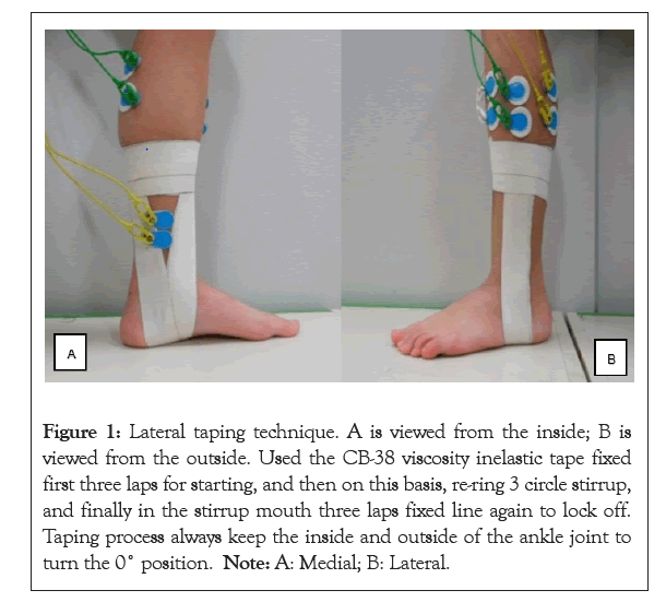
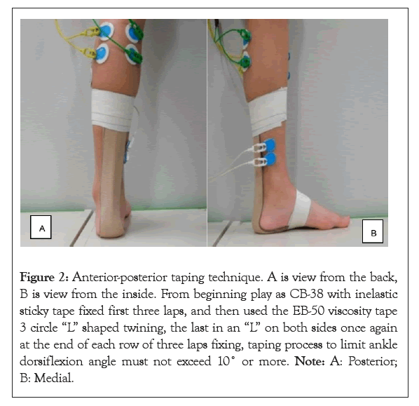
Sports Tape (NITREAT Company, Japan) was used in this study; the Sports Tape was sports special upper tape. In order to ensure the process of taping the same tension, we used unarmed gravity meter drawing each tap of the winding tension in the case of 40 N. Left foot of healthy body was measured. LT methods were shown in the Figure 1. In order to measure EMG activity, in the process of taping to avoid electrode paste location. Used the CB-38 viscosity inelastic tape fixed first three laps for starting, and then on this basis, re-ring 3 circle stirrups, and finally in the stirrup mouth three laps fixed line again to lock off. Taping process always keep the inside and outside of the ankle joint to turn the 0°position. APT methods were shown in Figure 2, like LP methods from beginning play as CB-38 with inelastic sticky tape fixed first three laps, and then used the EB-50 viscosity tape 3 circle “L” shaped twining, the last in an “L” on both sides once again at the end of each row of three laps fixing, taping process to limit ankle dorsiflexion angle must not exceed 10° or more (Figures 1 and 2).

Figure 1: Lateral taping technique. A is viewed from the inside; B is viewed from the outside. Used the CB-38 viscosity inelastic tape fixed first three laps for starting, and then on this basis, re-ring 3 circle stirrup, and finally in the stirrup mouth three laps fixed line again to lock off. Taping process always keep the inside and outside of the ankle joint to turn the 0° position. Note: A: Medial; B: Lateral.

Figure 2: Anterior-posterior taping technique. A is view from the back, B is view from the inside. From beginning play as CB-38 with inelastic sticky tape fixed first three laps, and then used the EB-50 viscosity tape 3 circle “L” shaped twining, the last in an “L” on both sides once again at the end of each row of three laps fixing, taping process to limit ankle dorsiflexion angle must not exceed 10˚ or more. Note: A: Posterior; B: Medial.

Figure 3: Electrode paste location. Note: TA: Tibialis Anterior; PL: Peroneus Longus; GSL: Lateral Head of Gastrocnemius; GSM: Medial Head of Gastrocnemius; TP: Tibialis Posterior; LG: Lateral head of Gastrocnemius; MG: Medial head of Gastrocnemius.
Measurement of balance ability
Body sway was measured using movable type balance measuring instrument UM-BAR (Younimeku,Tokyo,Japan). Subjects maintained 20 seconds of single-leg standing with eyes open and closed on the platform of movable type balance measuring instrument. To make sure the measurement not to fail, 20 seconds was chosen. Withdrawn from the center of gravity moving trajectory waveform the total length of COP, lateral (X-direction) trajectory length, anterior-posterior (Y-direction) trajectory length three indicators as criteria for evaluating the balance ability. In No-Tape (NT), LT methods and APT methods, three conditions measured the balance ability.
Record EMG activity
Electrical muscle activity was measured by Personal-EMG (Oi-zaka, Japan). The muscles for measurement included Tibialis Anterior (TA), Peroneus Longus (PL), Lateral head of Gastrocnemius (LG), Medial head of Gastrocnemius (MG), Tibialis Posterior (TP). The raw surface EMG waveform was converted into a Root Mean Square (RMS). For comparison of sEMG activity between subjects, sEMG activity in each muscle Maximal Voluntary Contraction (MVC) is assumed to be 100%, represented in %MVC16 [17]. The total sEMG activity with 20 seconds was calculated. Electrode paste location was shown in Figure 3. sEMG activity during MVC measurement position as follows: subjects sitting, the knee joint flexion 90°, natural ankle neutral position, and determination of subjects who do resistance movement (Figure 3).
Tibialis Anterior (TA): When ankle joint dorsiflexion TA strong contraction, lateral leg muscle which is most vulnerable touch at this time, then in the belly of the muscle electrode paste.
Peroneus Longus (PL): When ankle joint plantar flexion and valgus PL strong contraction.
Lateral head of Gastrocnemius (LG)/Mg: When ankle joint plantar flexion LG and MG strong contraction, MG begins on the medial epicondyle of femur, LG starting at the lateral epicondyle of femur, together with the Achilles ends heel.
Tibialis Posterior (TP): When ankle joint plantar flexion and varus TP strong contraction.
Synchronization
UM-BAR and Personal-EMG instruments can realize in-machine synchronization and simultaneously collect COP and sEMG.
Data analysis and statistics
Statistical test shows that all data conform to normal distribution, and the results are expressed as mean ± SD. The results of NT, LT and APY under open and closed eyes were analyzed by paired T-test; the results of pairwise comparison of NT, LT and APT under the condition of opened eyes were analyzed by one-way ANOVA. The results of pairwise comparison of NT, LT and APT under the condition of closed eyes were analyzed by one-way ANOVA. The level of significance was set at P<0.05. All statistical analyses were performed using SPSS Statistics version 22.0 (IBM Corp, Armonk, NY).
Balance ability
The results of total length of COP with NT as shown in Tables 2 and 3. When eyes open was 724.8 mm, eyes closed was 1212.9 mm, the later was increased by 67.3% compared to eyes open (P<0.01). Trajectory length of X-direction with NT, compared eyes closed to eyes open increased by 62.8%, Y-direction track length increased by 75.9% (P<0.01). Compared eyes closed to eyes open, total length of COP with LT methods increased 45.4% (P< 0.01), trajectory length of X-direction increased 41.7% (P<0.01), and trajectory length of Y-direction increased 56.3% (P<0.01). Total length of COP of the APT methods increased 53.6% (P<0.01) when comparison of eyes closed to eyes open, trajectory length of X-direction increased 66.6% (P<0.01), trajectory length of Y-direction increased 43.5% (P<0.01). Comparison of the APT and NT, LT and NT with eyes open (Tables 2 and 3).
The total length of COP of NT was 724.8 mm, the LT was its 96.0%, and APT was its 89.6%. The trajectory length of X-direction of NT was 523.3 mm, the LT was its 89.5%, and APT was its 87.4%. The trajectory length of Y-direction of NT was 402.4 mm, the LT was its 103.7%, and APT was its 92.8%. Comparison of the APT and NT, LT and NT with eyes closed. The total length of COP of NT was 1212.9 mm, the LT was its 83.4%, and APT was its 82.2% (P<0.05). The trajectory length of X-direction of NT was 852.1 mm, the LT was its 77.9% (P<0.05), and APT was its 89.4%. The trajectory length of Y-direction of NT was 707.7 mm, the APT was its 75.7% (P<0.05), and LT was its 92.1%. In summary, the total length of COP with LT, the trajectory length of X-direction decreased with eyes closed when compared to NT. The total length of COP of APT techniques, the trajectory length of Y-direction decreased with eyes closed when compared to NT. When eyes open no statistically significant changes with the reduction.
EMG activity
The results of sEMG activity were shown in Tables 4 and 5, whether NT, LT or APT, with eyes closed compared to eyes open showed a very large sEMG activity (P<0.01). When NT the sEMG activity of GSM and PL was relatively large, followed by TP slightly, TA and GSL was relatively small. TA and PL, with eyes open the sEMG activity of LT and APT slightly lower, the EMG activity of LT and APT was significantly lower when eyes closed (P<0.05). GSM and GSL, both eyes open and eyes closed, did not find any difference when compared LT and APT to NT. TP, when eyes open the sEMG activity of LT slightly lower, the sEMG activity of APT hardly changed; when eyes closed, there was no difference when compared LT and APT with NT (Tables 4 and 5).
Kobayashi suggested, when standing on single-leg with eyes open in 20 seconds, after ankle sprain the total length of COP was 773-878 mm which healthy adults’ measurement value lowers than this [15]. The total length of COP was 724.8 mm with NT when eyes open on present study. Therefore, we consider values are reasonable after comparing the results of present study with Kobayashi. In addition, this static body balance experiment was carried out under the premise of no statistically significant differences between male and female in the analysis and judgment. We summed up all the data before the implementation of resolution. X-direction movement was larger than Y-direction. This result prompted us to keep the body’s balance ability when standing with single-leg would control the instability of X-direction, when note the moving instability of Y-direction. However, the X-direction may be more important to keep balance ability [18,19].
We confirmed that when eyes closed the body sway was much larger than eyes open in this study. The trajectory length of COP increased when eyes closed compared with eyes open. There was no significant difference among NT, LT and APT when eyes open about the total length of COP. When eyes closed, total length of COP significantly reduced compared to NT. In particular, LT was used, the trajectory length of X-direction was significantly reduced; with APT techniques, the trajectory length of Y-direction was significantly reduced. The literature indicated that after ankle taping the sEMG activity of PL increased and the results of present study showed when eyes closed the sEMG activity of TA and PL significantly reduced [20,21]. When present study initially assumed after ankle taping around ankle muscles sEMG activity will reduce, exactly consistent with the experimental results. As agonist the PL strong contraction when ankle plantar flexion and valgus, after taping which limited ankle plantar flexion and valgus, it’s a strong possibility that the sEMG activity would reduce [22,23]. In addition, forming the total length of COP reduction to analysis, we also believe that after the lateral and anterior-posterior taping the sEMG activity will reduce. About TA, the same taping techniques, due to increased stability of the ankle joint, we consider the sEMG activity will reduced. Previous studies reported that simulate walking with ankle sprain, and then analyzed the sEMG activity of PL, may be necessary to consider analyze this dynamic and static analysis combined [24,25]. About sEMG activity of MG, there was almost no change at all after the taping; it’s possibly interrelated to keep the balance ability needs ankle lateral stability [22]. The sEMG activity of TP, although after the taping, either LT or APT techniques have been affected, when compared with NT did not appear much different. As the previous studies reported static dynamic combination of more rational analysis, this is the subject of present study in future.
In conclusion, when after taping with eyes closed, the trajectory of COP becomes smaller when compared with NT. Balance ability has improved. After the taping, when standing on single-leg with eyes closed, the EMG activity of TA and PL reduced. Therefore, we inferred the patients who were with ankle sprain after taping, in addition to reducing pain outside, which will not only improve the balance ability, but also can reduce the incidence of re-injury. The reasons of the limitation of the present study were presented as follows. Firstly, the subjects of this study were only form 20-30 years old, age range was limited. Thus follow 20 and over 30 years old, the changes of balance ability and EMG activities around the ankle joint after taping are not clear. Secondly, this experiment used taping patch are Japanese production of fine movement dedicated patch, different taping techniques and the problems of a different number of turns wound also need take into consideration.
This research was funded by the 2020-2022 Special Science and Technology Development Project of Beijing Rehabilitation Hospital, Capital Medical University (#2020-066).
All authors declare that there is no conflict of interest.
WG Gao and ZH Yu contributed equally to this work. W. G. Gao and Z. H. Yu wrote the manuscript; Z. J. Fan performed the systematic rehabilitation treatment; J. Zhang analyzed and interpreted the patient data; Y. B. Ma reviewed the manuscript and provided fund support; all authors read and approved the final manuscript.
[Crossref] [Google Scholar] [PubMed]
[Crossref] [Google Scholar] [PubMed]
[Crossref] [Google Scholar] [PubMed]
[Crossref] [Google Scholar ] [PubMed]
[Crossref] [Google Scholar] [PubMed]
[Crossref] [Google Scholar] [PubMed]
[Crossref] [Google Scholar] [PubMed]
[Crossref] [Google Scholar] [PubMed]
[Crossref] [Google Scholar] [PubMed]
[Crossref] [Google Scholar] [PubMed]
[Crossref] [Google Scholar] [PubMed]
[Crossref] [Google Scholar] [PubMed]
[Crossref] [Google Scholar] [PubMed]
[Crossref] [Google Scholar] [PubMed]
[Crossref] [Google Scholar] [PubMed]
[Crossref] [Google Scholar] [PubMed]
[Crossref] [Google Scholar] [PubMed]
[Crossref] [Google Scholar] [PubMed]
[Crossref] [Google Scholar] [PubMed]
[Crossref] [Google Scholar] [PubMed]
[Crossref] [Google Scholar] [PubMed]
[Crossref] [Google Scholar] [PubMed]
[Crossref] [Google Scholar] [PubMed]
[Crossref] [Google Scholar] [PubMed]
Citation: Gao WG, Yu ZH, Zhang J, Fan ZJ, Ma Y (2023) Changes in Balance Ability and Electromyography Activity after Ankle Taping in HealthyAdults. Int J Phys Med Rehabil.11:663.
Received: 26-Jan-2023, Manuscript No. JPMR-23-21678; Editor assigned: 30-Jan-2023, Pre QC No. JPMR-23-21678; Reviewed: 13-Feb-2023, QC No. JPMR-23-21678; Revised: 20-Feb-2023, Manuscript No. JPMR-23-21678; Accepted: 27-Feb-2023 Published: 27-Feb-2023 , DOI: 10.35248/2329-9096.23.11.664
Copyright: © 2023 Gao WG, et al. This is an open-access article distributed under the terms of the Creative Commons Attribution License, which permits unrestricted use, distribution, and reproduction in any medium, provided the original author and source are credited.