Journal of Clinical and Experimental Ophthalmology
Open Access
ISSN: 2155-9570
ISSN: 2155-9570
Case Report - (2022)Volume 13, Issue 6
Introduction: Neurofibromatosis type 1(NF1), is a neurodermal dysplasia. It is a multisystem hamartomatous disorder. The association of NF1 and uveal melanoma is also debatable, despite an embryological plausibility. We report a case of a choroidal melanoma in a patient with NF1.
Aim: Underline the importance of considering the diagnosis of choroidal melanoma when a choroidal mass is evident in patients with neurofibromatosis type1.
Observation: A 43-year-old woman with multiple cutaneous neurofibromas, cafe-au-lait spots, and a family history of NF who consults for rapidly progressive visual acuity on his right eye quantified at 1/10.
Results: The ophthalmologic examination revealed in the right eye, an elevated domed-shaped gray-yellowish colored lesion of the choroid with irregular margins not sharply demarcated, taking the macular region. Some orange pigmentation was noted.
Fluorescein angiography revealed multiple hyperfluorescent foci during the arteriovenous phase and progressive leaking, which resulted in marked staining of the mass late staining of the lesion and multiple pinpoint leaks at the level of the retinal pigment epithelium.
B-scan ultrasonography demonstrated an underlying choroidal mass, with an acoustically distinct inner boundary, and choroidal excavation. Optical coherence tomography demonstrated an underlying choroidal mass with serous retinal detachment and intra-retinal splitting. On the basis of the findings, a choroidal melanoma is retained. Assessment of tumor extension was negative for metastases. The patient was referred to a retinal oncologist.
Discussion: Hamartomas of the uveal tract may occur in patients with neurofibromatosis. These are mainly glial or melanocytic hamartomas. The possibility of an association between Neurofibromatosis type 1 (NF1) and uveal melanoma has been proposed on the basis of a common neural crest origin.
This case demonstrates the occurrence of malignant melanoma of the choroid in a patient with neurofibromatosis, and emphasizes the importance of considering this diagnosis when a choroidal mass is evident in such patients. Our case raises the question of the differential diagnosis of a fundus mass in a patient with neurofibromatosis. Entities to be considered include glial hamartoma of the retina, choroidal neurofibroma, choroidal nevus, and choroidal melanoma.
Conclusion: In all the major clinical forms of von Recklinghausen's disease, the association of NF1 and uveal melanoma is reported. Although choroidal melanoma in patient with NF1 is rare, ophthalmologists should think about it in front of any choroidal mass.
Von Recklinghausen’s Disease (VRD); Choroidal mass; Choroidal melanoma; Neurodermal dysplasia
The term Neurofibromatosis (NF) is referred to a group of genetic disorders that primarily affect the cell growth of neural tissues. There are two forms of NF: Type 1 (NF1) and type 2 (NF2). These two forms have few common features and are caused by mutations on different genes.
Neurofibromatosis type 1(NF1), also known as Von Recklinghausen’s disease, is a neurodermal dysplasia. This disease was first described by Friederich Daniel von Recklinghausen, the pathologist, in 1882 [1,2]. It is characterized by autosomal dominant inheritance with complete penetrance but variable expression [3]. NF1 is a multisystem hamartomatous disorder with protean expression of cutaneous, neurologic, skeletal, visceral, and ocular manifestations [4].
The ocular findings of NF1 include neurofibromas and neurilemmomas of the eyelids, conjunctiva, cornea and orbit, thickened corneal, conjunctival, or ciliary nerve, melanocytic and neuronal hamartomas of the trabecular meshwork, uvea, retina or optic nerve and glaucoma [5-9]. The association of NF1 and uveal melanoma is also debatable, despite an embryological plausibility [10,11]. We report a case of a choroidal melanoma in a patient with NF1 and discuss the possible causal association.
A 43-year-old woman with multiple cutaneous neurofibromas, cafe-au-lait spots and a family history of NF who consults for rapidly progressive visual acuity on her right eye quantified at 1/10 with flashes (Figure 1).
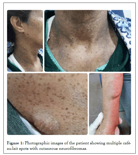
Figure 1: Photographic images of the patient showing multiple café- au-lait spots with cutaneous neurofibromas.
The ophthalmologic examination was normal in the left eye. It revealed, in the right eye, an elevated dome-shaped gray-yellowish colored lesion of the choroid with irregular margins not sharply demarcated, taking the macular region to the optic disc which overflows it above and below. Some orange pigmentation was noted (Figure 2).
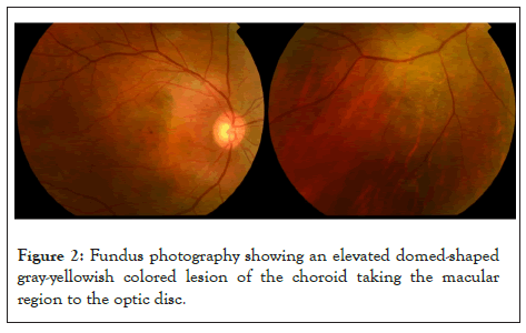
Figure 2: Fundus photography showing an elevated domed-shaped gray-yellowish colored lesion of the choroid taking the macular region to the optic disc.
Fluorescein angiography revealed multiple hyper fluorescent foci during the arteriovenous phase and progressive leaking, which resulted in marked staining of the mass late staining of the lesion and multiple pinpoint leaks (hot spots) at the level of the retinal pigment epithelium [12,13] with the overlying serous fluid in the later angiograms (Figure 3).
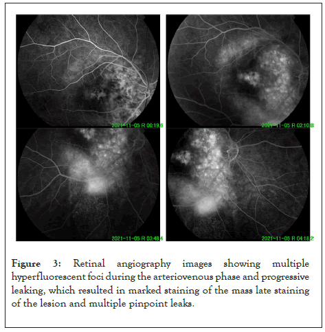
Figure 3: Retinal angiography images showing multiple hyperfluorescent foci during the arteriovenous phase and progressive leaking, which resulted in marked staining of the mass late staining of the lesion and multiple pinpoint leaks.
B-scan ultrasonography demonstrated an underlying choroidal mass, with an acoustically distinct inner boundary, and choroidal excavation (Figure 4).
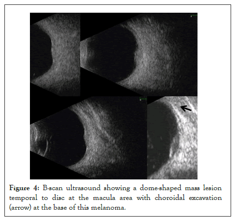
Figure 4: B-scan ultrasound showing a dome-shaped mass lesion temporal to disc at the macula area with choroidal excavation (arrow) at the base of this melanoma.
Optical coherence tomography demonstrates an underlying choroidal mass, serous retinal detachment around and overlying the tumor with intra-retinal splitting (Figure 5).
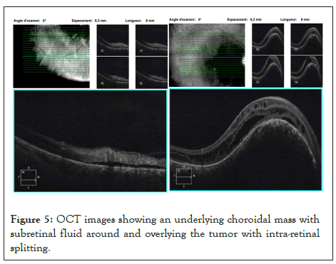
Figure 5: OCT images showing an underlying choroidal mass with subretinal fluid around and overlying the tumor with intra-retinal splitting.
On the basis of the findings (elevated choroidal mass, orange pigment, subretinal fluid, decreased vision with flashes, and margin of the tumor within 3 mm of the optic disc, ultrasonographic hollowness, without drusen or surrounding halo) choroidal melanoma is retained. Assessment of tumor extension was negative for metastases. The patient was referred to a retinal oncologist.
Hamartomas of the uveal tract may occur in patients with neurofibromatosis. These are mainly glial or melanocytic hamartomas. Several clinical and pathological studies are consistent with the hypothesis that Von Recklinghausen’s disease represents a dysontogenetic state of cells of the neural crest origin derived from the neural crest [14]. Many various tumors may be associated with NF1 such as neurofibroma, schwannoma, pheochromocytoma, glioma, meningioma and possibly melanoma. The possibility of an association between Neurofibromatosis type 1 (NF1) and uveal melanoma has been proposed on the basis of a common neural crest origin [15-19]. A predisposition to uveal malignant melanoma may also exist because of the increased incidence of choroidal nevi in Von Recklinghausen’s disease and the supposition that malignant melanomas arise from these nevi may also explain the possible association. Despite the theoretical possibility of an association, the frequency of patients with co-existing NF1 and uveal melanoma is rare [20-32]. To our knowledge, only 15 cases of choroidal melanoma in association with NF1 have been documented in the literature. Our case is the 16th (Table 1). This case demonstrates the occurrence of malignant melanoma of the choroid in a patient with neurofibromatosis and emphasizes the importance of considering this diagnosis when a choroidal mass is evident in such patients. Our case raises the question of the differential diagnosis of a fundus mass in a patient with neurofibromatosis. Entities to be considered include glial hamartoma of the retina [32]; choroidal neurofibroma [33]; choroidal nevus [34] and choroidal melanoma.
| Author (year) | Age (years)/Sex |
|---|---|
| Bair (1937) [21] | 50/M |
| Gartner (1940) [22] | 54/F |
| Strachov (1941) [23] | 35/F |
| Nordmann (1961) [24] | 39/F |
| Nordmann (1963) | 39/F |
| Yanoff (1967) | 31/F |
| Wiznia (1978) [25] | 60/F |
| Croxatto (1981) [26] | 68/F |
| Arnesen (1985) [27] | 52/M |
| Arnesen (1985) [27] | 73/F |
| Antle (1990) [28] | 53/M |
| Bacin (1993) [29] | 16/F |
| Warwar (1998) [30] | 64/F |
| Friedman (1998) [31] | 69/F |
| Current case | 43/F |
| Note: M: Male; F: Female | |
Table 1: Reported cases of choroidal melanoma associated with Neurofibromatosis type 1 (NF1).
The glial hamartoma (astrocytoma) should present no diagnostic problem, for it is retinal rather than choroidal in location, white rather than pigmented, and identical to lesions noted in tuberous sclerosis. A choroidal neurofibroma or a large nevus may be impossible to distinguish from a malignant melanoma, both clinically and histologically. In such cases, therefore, a period of observation for growth seems justified.
Choroidal melanoma is the most common primary intra-ocular malignant tumour. His Current diagnosis is based on both the clinical experience of the specialist and modern diagnostic techniques such as indirect ophthalmoscopy, ultrasonography scans, and fundus fluorescein angiography. In all the major clinical forms of von Recklinghausen's disease, the association of NF1 and uveal melanoma is reported. Although choroidal melanoma in patient with NF1 is rare, ophthalmologists should think about it in front of any choroidal mass.
[Crossref] [Google Scholar] [PubMed]
[Crossref] [Google Scholar] [PubMed]
[Crossref] [Google Scholar] [PubMed]
[Crossref] [Google Scholar] [PubMed]
[Crossref] [Google Scholar] [PubMed]
[Crossref] [Google Scholar] [PubMed]
[Crossref] [Google Scholar] [PubMed]
[Crossref] [Google Scholar] [PubMed]
[Crossref] [Google Scholar] [PubMed]
[Crossref] [Google Scholar] [PubMed]
[Crossref] [Google Scholar] [PubMed]
[Crossref] [Google Scholar] [PubMed]
[Crossref] [Google Scholar] [PubMed]
[Crossref] [Google Scholar] [PubMed]
[Crossref] [Google Scholar] [PubMed]
[Crossref] [Google Scholar] [PubMed]
[Google Scholar] [PubMed]
[Crossref] [Google Scholar] [PubMed]
[Crossref] [Google Scholar] [PubMed]
[Crossref] [Google Scholar] [PubMed]
[Crossref] [Google Scholar] [PubMed]
Citation: Benelkadri F, Filali ME, Ouidani B, Kriet M (2022) Choroidal Melanoma in a Patient with Neurofibromatosis: A Case Report. J Clin Exp Ophthalmol. 13:952.
Received: 31-Oct-2022, Manuscript No. JCEO-22-19019; Editor assigned: 02-Nov-2022, Pre QC No. JCEO-22-19019 (PQ); Reviewed: 16-Nov-2022, QC No. JCEO-22-19019; Revised: 23-Nov-2022, Manuscript No. JCEO-22-19019 (R); Published: 02-Dec-2022 , DOI: 10.35248/2155-9570.22.13.930
Copyright: © 2022 Benelkadri F, et al. This is an open-access article distributed under the terms of the Creative Commons Attribution License, which permits unrestricted use, distribution, and reproduction in any medium, provided the original author and source are credited.