Journal of Bone Research
Open Access
ISSN: 2572-4916
ISSN: 2572-4916
Research - (2023)Volume 11, Issue 6
Study design: Skeletal relapse following mandibular advancement using the Sagittal Split Osteotomy (SSO) is associated with the degree of advancement and method of fixation. Objective: The study purpose is to compare the biophysical properties of two fixation techniques for the SSO advancement.
Methods: This cadaveric in vitro study was conducted at a single tertiary care centre. Paired hemimandibles underwent SSO with 10 mm of advancement then fixated using two techniques. Group 1 used a single 3D ladder miniplate and group 2 used a miniplate and two bicortical screws (hybrid technique). Hemimandibles were loaded in the first molar region using an Instron mechanical testing unit. The primary predictor variable is the fixation method. The outcome variables were peak load in Newtons (N) defined as the load at which permanent deformation started, displacement value at peak load, and load (N) necessary for a specific amount of displacement. Donor demographic and anatomic variables were age, gender, dental status, mandibular body height, ramus width, and thickness of mandibular angle. Descriptive statistics and paired two-sided t-tests were performed with P-value ≤ 0.05.
Results: The fifteen human mandibles were 53% male and 47% female with mean age of 82.2 years (range 78-92 years). Mean ± Standard Deviation (SD) peak load was 75.3 ± 43.3 N for 3D ladder plate technique and 116.2 ± 57.9 N for the hybrid technique, the difference of means (group 1–group 2) was -40.9 ± 73.3 N (T-value=2.15, P-value=0.048).
Conclusion: The hybrid technique showed a significantly greater peak load compared to the 3D plate for the SSO advancement, suggesting the former may provide superior resistance to relapse in the clinical setting. This cadaveric model can be utilized for additional clinical questions and be a bridge between synthetic in vitro models and clinical studies.
Sagittal Split Osteotomy (SSO); Cadaveric study; Bicortical screw; Instron
The Sagittal Split Osteotomy (SSO) is the most commonly used technique in orthognathic surgery [1,2]. The SSO allows for three dimensional manipulation of the mandible to correct craniofacial abnormalities as well as treat Obstructive Sleep Apnea (OSA) via Maxillomandibular Advancement (MMA) [3-6]. Large advancements and the counterclockwise rotation of the mandible are often required when the SSO is utilized MMA. However, these movements are more highly associated with relapse thus risking unstable post-operative outcomes in patients [7-10]. The consequences of relapse including malocclusion and suboptimal esthetic and functional outcomes have led to efforts to optimize post-surgical stability of the SSO [11].
Skeletal relapse is influenced by surgical and biomechanical factors. It is the role of the surgeon to identify modifiable factors that can be clinically optimized such as fixation technique. The most common fixation techniques for the SSO utilize bicortical screws alone, miniplates alone, or a hybrid model utilizing bicortical screws in addition to a miniplate. Literature supports the mechanical strength of 3 bicortical screws in the inverted-L arrangement, however, the hybrid technique offers similar strength while providing additional benefits particularly in SSO advancements [12-14]. Three-dimentional (3D) miniplates are affective in the fixation of mandibular fractures [15-17], and would be expected to offer similar or improved mechanical properties to two traditional miniplates for SSO fixation. Current in vitro biomechanical studies have utilized synthetic mandibles [12,13,18- 22], and less commonly sheep mandibles [23,24], or finite element analysis [14,25], allowing for reproducibility and application to a myriad of surgical questions. While useful, they are unable to mimic the unique anatomic properties of the human mandible.
The purpose of the present study is to utilize a human cadaveric model [26], to compare SSO fixation techniques and thus provide valuable biomechanical rational for surgical decisions. As large advancements show the strongest correlation with relapse, the model will perform the SSO with a 10 mm advancement. The aim of the present study is to compare the biomechanical stability of the SSO advancement between the single plate with two bicortical screws and three-dimensional ladder plate osteosynthesis fixation techniques.
Study design
The study followed an in vitro cadaveric study design [26], utilizing human mandibles obtained from the Anatomical Donor Program at the University of Alabama at Birmingham, School of Medicine (Birmingham, AL). Due to the study design, institutional review board approval was not required. The ethics of the Declaration of Helsinki were followed. The inclusion criteria were intact edentulous, partially edentulous, or dentate mandibles. Exclusion criteria were any mandibles where the donor had a history of osteoporosis, metastatic bone cancer, metallic bone disorders, mandibular tumors or the presence of mandibular third molars.
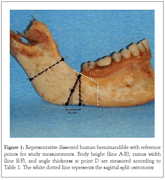
All cadaveric skulls and mandibles were previously hemisected in the midsagittal plane, generating hemimandibles that were paired for each study arm. These mandibles were dissected by oral and maxillofacial surgery residents and a single attending surgeon as seen in Figure 1. Mandibles were cleaned and mounted in acrylic bone cement in 9.5 cm × 3 cm × 3 cm block molds made of silicone. The acrylic was allowed to set in a pneumatic curing unit set to 20 psi for 5 minutes. Additional acrylic cement was placed from the sigmoid notch to the angle encompassing the condyle and posterior border of the ramus. Blocks with mounted hemimandibles were removed from their mold and labeled with the donor number. Record of anatomic variables described below was acquired prior to completion of SSO (Table 1).
| Measurement | Description |
|---|---|
| Mandibular Height | Straight distance from area of confluence of the alveolar crest and external oblique ridge (Point A) to the inferior border of the mandibular body (Point B) |
| Ramus Width | Straight distance from coronoid notch of the anterior ramus (Point E) to the posterior border of the mandible (Point F) |
| Angle Thickness | Straight distance between the lateral and medial aspect of mandibular angle at Point D. Point D is the midpoint of the line A-C whereby Point C is 10 mm posterior to Point B along the inferior border of the mandible. |
Note: SSO: Sagittal Split Osteotomy
Table 1: Anatomical measurements (mm) of hemimandibles prior to SSO (Figure 1).

Figure 1: Representative dissected human hemimandible with reference points for study measurements. Body height (line A-B), ramus width (line E-F), and angle thickness at point D are measured according to Table 1. The white dotted line represents the sagittal split osteotomy.
SSO was performed via standard technique on each hemimandible by the same surgical residents. All hemimandibles underwent 10 mm advancement then paired hemimandibles would go to each of the study arms for alternative fixation techniques as seen in Figure 2. In the group 1, fixation was performed with a 3D curved ladder titanium 2.0 mm, 12-hole non-compression plate and 7.0 mm in length, 8 monocortical titanium fixation screws (KLS Martin, Freiburg, Germany) along the lateral aspect of the mandible. In the group 2, fixation was performed with 2 bicortical positional screws and 1 horizontal lateral titanium plate 2.0 mm, 4-holes non- compression plate, 10.0 mm bicortical and 7 mm monocortical titanium fixation screws (KLS Martin, Freiburg, Germany) along the lateral aspect of the mandible.
Figure 2: Hemimandibles from single donor split at the mandibular symphysis that underwent two methods of fixation after sagittal split osteotomy and mounted into acrylic cement blocks. A) Anatomical right mandible fixated with single plate with two bicortical screws then B) fixated in acrylic cement block. C) Anatomical right mandible fixated with 3D ladder plate then D) fixated in acrylic cement block.
Biomechanical evaluation was performed on each advanced and fixated hemimandible using an Instron 5565 mechanical testing unit (Instron Corp, Norwood, MA) as seen in Figure 3. Force was applied to the first molar region and performed in compression mode at a cross-head speed of 1 mm per minute. Loading was continued at the aforementioned rate until hardware distortion occurred. The maximum load in Newtons (N) and displacement of the hemimandible were recorded. The force at increasing displacement after peak load was also recorded.
Figure 3: Representative example of Instron applying force to premolar region of hemimandible with fixation after sagittal split osteotomy.
Variables
Heterogenous donor variables were age, gender, dental status, mandibular body height, ramus width, and thickness of mandibular angle. The primary predictor variable was the fixation method as described above. The outcome variables were peak load in Newtons (N) defined as the load at which permanent deformation started, displacement value at peak load, and load (N) necessary for a specific amount of displacement at 1 mm, 3 mm, 5 mm, 6 mm.
Data collection methods
Deidentified donor information was collected and paired with appropriate hemimandible specimen. Outcome variables were measured by Instron 5565 mechanical testing and recorded. Data were acquired and stored using Bluehill 2 software (Instron Corp).
Data analysis
Descriptive statistics including mean, standard deviation and range were obtained for demographic data and outcome variables. Paired two-sided t-tests were performed to compare biophysical properties. A significance level of 0.05 was used. Statistical analyses were performed using SAS, version 9.4 (SAS Institute, Cary, NC).
Fifteen human mandibles were included in the study. Donors were comprised of 53% male and 47% female with mean age of 82.2 years (range 78-92 years). Dental status of mandibles was 20% edentulous, 53.3% partially edentulous, and 26.7% fully dentate. Descriptive statistics for anatomic and biophysical measurements are shown in Tables 2 and 3, respectively. Comparison of biophysical measurements between fixation techniques is shown in Table 4.
| Measurements | 3D ladder plate | Plate and bicortical screws |
|---|---|---|
| Mean ± SD | Mean ± SD | |
| Body height (mm) | 23.4 ± 3.2 | 22.9 ± 2.9 |
| Ramus width (mm) | 31.0 ± 2.8 | 31.0 ± 3.0 |
| Angle thickness (mm) | 7.7 ± 1.5 | 7.2 ± 1.4 |
Note: SD: Standard Deviation; SSO: Sagittal Split Osteotomy.
Table 2: Anatomical measurements of hemimandibles prior to SSO.
| Measurements | 3D ladder plate | Plate and bicortical screws |
|---|---|---|
| Mean ± SD | Mean ± SD | |
| Peak load (N) | 75.3 ± 43.3 | 116.2 ± 57.9 |
| Displacementa (mm) | 7.7 ± 3.9 | 8.1 ± 4.3 |
| Force at 1mm (N) | 19.7 ± 14.3 | 27.4 ± 16.4 |
| Force at 3 mm (N) | 41.7 ± 21.2 | 50.7 ± 30.3 |
| Force at 5 mm (N) | 53.7 ± 30.5 | 74.3 ± 41.2 |
Note: SSO: Sagittal Split Osteotomy; SD: Standard Deviation aAt peak load
Table 3: Biophysical properties of hemimandibles that underwent alternative fixation techniques following SSO with 10 mm advancement.
| Measurements | Difference of meansa Mean ± SD |
T-Valueb | P-Value |
|---|---|---|---|
| Peak Load (N) | -40.9 ± 73.3 | 2.15 | 0.048* |
| Displacementc (mm) | -0.4 ± 5.8 | 0.28 | 0.781 |
| Force at 1 mm (N) | -7.7 ± 16.0 | 1.87 | 0.082 |
| Force at 3 mm (N) | -9.0 ± 33.1 | -1.05 | 0.310 |
| Force at 5 mm (N) | -20.6 ± 44.5 | 1.79 | 0.094 |
Note: SSO: Sagittal Split Osteotomy; SD: Standard Deviation; N: Newtons aDifference: 3D ladder plate-Plate and Bicortical Screws bPaired 2-tailed t-test, *denotes statistical significance cAt peak load
Table 4: Comparison of biophysical properties for hemimandibles alternatively fixated after SSO and 10 mm advancement.
Relapse from the SSO in orthognathic surgery is driven by multifactorial etiologies therefore it is necessary for surgeons to identify which factors can be optimized. The most important contributor to risk of relapse is the amount of advancement but this factor is dictated by functional and esthetic goals [7-10]. Fixation method is second in its influence on relapse, making it the appropriate modifiable factor that should be investigated. The author’s previously described cadaveric model used for fixation of mandibular angle fractures was applied for the present study [26].
The present in vitro cadaveric study sought to evaluate the comparative biomechanical strength of two fixation techniques for SSO advancement. The authors chose to compare fixation with a miniplate and two bicortical screws with the use of a single 3D ladder plate, given the underrepresentation of 3D ladder plates for orthognathic surgery in current literature. The results showed asignificantly higher peak load in the single plate with 2 bicortical screws compared to the 3D ladder plate (Table 3), suggesting the former would be more resistant to relapse in clinical use. The displacement at peak load and force applied at each amount of displacement were not significantly different between the two methods, suggesting that resistance to displacing forces is equivocal after initial deformation. Sato et al. [12], performed linear loading and photoelastic analysis to evaluate both mechanical resistance and stress distribution of synthetic hemimandibles that underwent SSO advancement with subsequent fixation of either 3 bicortical screws, a miniplate, or the hybrid technique. They found that using a bicortical screw in addition to fixation with a miniplate improved the mechanical resistance as well as offset some of the force generated in the screws closest to the osteotomy compared to a miniplate alone. The use of 3 bicortical screws had the greatest mechanical resistance of the three fixation techniques and produced similar levels of stress to the hybrid technique. Other studies similarly support the use of 3 bicortical screws, especially when compared to the use of single plates using only monicortical screws [13,14]. Peterson et al. [13], demonstrated this same relationship for both incisal and molar loading models. Kuik et al. [19], supported the use of 3 bicortical screws in the inverted L pattern for advancements of 10 mm, but the use of 2 miniplates in advancements greater than 10 mm.
Clinical studies have demonstrated that the amount of advancement is positively correlated to relapse, but have not shown strong relationships between fixation method and relapse [27,28]. Rocha et al. [29], compared the use of the hybrid technique using a miniplate with 2-3 bicortical screws and the use of 2 miniplates and found there to be no significant difference in postoperative stability. Ultimately, some factors disallow for the use of certain fixation techniques regardless of their mechanical properties. Factors that may exclude the use of 3 bicortical screws are large advancements or unfavorable splits, both of which can limit the amount of bone overlap required for the use of bicortical screws. The use of miniplates also carries advantages including the avoidance of stab incisions and ease of use if adjustments are required [30]. Additionally, the increased risk of nerve injury with the use of bicrotical screws may favor the use of miniplates and 3D plates [30,31]; however, bicortical screws are not shown to carry increased risk in some clinical studies [32].
The reasons for undergoing the SSO for mandibular advancement vary from correcting retrognathia or hemifacial microsomia to increasing airway volume in patients with OSA, therefore this patient cohort encapsulates a wide range of anatomical variance. The polyurethane hemimandible model is most commonly used as it allows for reproducibility and lower costs while demonstrating comparable strength to bone [33,34]. However, this model’s homogeneity is a detriment as it does not account for the variable anatomies of the human mandible. The cadaver model in the present study aids in this shortcoming and can best be utilized to strengthen associations found with synthetic models.
The present study is subject to weaknesses that hold true for any in vitro study. First, this model is unable to mimic the intricate and physiologically dynamic nature of human mandibular function. Second, the use of hemimandibles undermines the strength and structural integrity as compared to the intact mandible. These first two factors are comparable to the vast majority of present literature, and thus are important to maintain in order to strengthen associations made between synthetic and cadaver models. Anotherweakness is that the small sample size limits the power to further investigate how specific anatomical differences may relate to the efficacy of fixation techniques.
Future iterations of the general study design could be used to evaluate many biomechanical factors such as a greater degree of advancement, rotational movements and varied fixation techniques. It would also be useful to elucidate how specific anatomical factors influence the relationship between fixation technique and stability. The present study demonstrated higher peak load in the single plate with bicortical screws compared to the 3D ladder plate in cadaver hemimandibles that underwent SSO with 10 mm of advancement. These findings offer evidence in support of this hybrid fixation technique that may guide the clinical practice of orthognathic surgery.
No funding was obtained for this study.
The authors have no conflict of interest to disclose.
IRB Statement: The principles of the Declaration of Helsinki were followed for cadaveric specimens.
[Crossref] [Google Scholar] [PubMed]
[Crossref] [Google Scholar] [PubMed]
[Crossref] [Google Scholar] [PubMed]
[Crossref] [Google Scholar] [PubMed]
[Crossref] [Google Scholar] [PubMed]
[Crossref] [Google Scholar] [PubMed]
[Crossref] [Google Scholar] [PubMed]
[Crossref] [Google Scholar] [PubMed]
[Crossref] [Google Scholar] [PubMed]
[Crossref] [Google Scholar] [PubMed]
[Crossref] [Google Scholar] [PubMed]
[Crossref] [Google Scholar] [PubMed]
[Crossref] [Google Scholar] [PubMed]
[Crossref] [Google Scholar] [PubMed]
[Crossref] [Google Scholar] [PubMed]
[Google Scholar] [PubMed]
[Crossref] [Google Scholar] [PubMed]
[Crossref] [Google Scholar] [PubMed]
[Crossref] [Google Scholar] [PubMed]
[Crossref] [Google Scholar] [PubMed]
[Crossref] [Google Scholar] [PubMed]
[Crossref] [Google Scholar] [PubMed]
[Crossref] [Google Scholar] [PubMed]
[Crossref] [Google Scholar] [PubMed]
[Crossref] [Google Scholar] [PubMed]
[Crossref] [Google Scholar] [PubMed]
[Crossref] [Google Scholar] [PubMed]
[Crossref] [Google Scholar] [PubMed]
[Crossref] [Google Scholar] [PubMed]
[Crossref] [Google Scholar] [PubMed]
[Crossref] [Google Scholar] [PubMed]
[Crossref] [Google Scholar] [PubMed]
[Crossref] [Google Scholar] [PubMed]
[Crossref] [Google Scholar] [PubMed]
Citation: Zhang J (2023) Comparing Peak Load Capacity between Bicortical Screws and 3D Plates in Human Cadaveric Sagittal Split Ramus Osteotomy. J Bone Res. 11:245.
, Manuscript No. BMRJ-23-27317; , Pre QC No. BMRJ-23-27317 (PQ); , QC No. BMRJ-23-27317; , Manuscript No. BMRJ-23-27317 (R); , DOI: 10.35248/2572-4916.23.11.245
Copyright: © 2023 Zhang J. This is an open-access article distributed under the terms of the Creative Commons Attribution License, which permits unrestricted use, distribution, and reproduction in any medium, provided the original author and source are credited.