Journal of Clinical Trials
Open Access
ISSN: 2167-0870

ISSN: 2167-0870
Case Report - (2022)
Background: Congenital type R-I Left Coronary Artery (LCA) absence, combined with Myocardial Bridge (MB) over the right coronary artery, is a rare congenital coronary artery malformation, and may have fatal consequences.
Patient: A 52-year-old man presented with chest pain for 3 days and 2 hours. The patient was advised for admission. He underwent Computed Tomography Angiography (CTA) examination of the coronary artery to screen for Coronary Artery Disease (CAD).
Results: The coronary artery CTA showed the absence of LCA and the MB was confined to the second right ventricular branch. Coronary Angiography (CAG) examination confirmed congenital absence of LCA combined with MB over the right coronary artery and 20% stenosis in the right coronary artery. Treatment was given according to the secondary prevention of Coronary Heart Disease (CHD). After 3 days of treatment, the symptoms of chest pain did not recur, and the Electrocardiogram (ECG) was normal.
Conclusion: If the patient has ischemia changes in ECG but no detectable changes in myocardial enzymes, myoglobin and serum troponin T, a coronary artery Computed Tomography Angiography (CTA) examination can be utilized to screen for single coronary malformations and other varieties of cardiovascular malformation. CAG can be performed to confirm the diagnosis.
Coronary artery malformatione; Coronary artery disease; Single coronary artery; Myocardial bridge
Single Coronary Artery (SCA) malformation is rare, and if combined with other coronary artery malformations, is even rarer. This is the first case report of the world's first patient with left coronary artery absence combined with Myocardial Bridge (MB) over the right coronary artery. Patients with such coronary artery malformations are often misdiagnosed before examination of coronary Computed Tomography Angiography (CTA) or coronary angiography (CAG). At present, no systematic studies have shown whether the secondary prevention of Coronary Heart Disease (CHD) will improve the long-term prognosis of the patients with complex coronary artery malformations if they are complicated with coronary atherosclerosis.
A 52-year-old man presented with chest pain for 3 days and 2 hours on July 30, 2021. Three days prior, the patient suffered from chest pain, which was a paroxysmal dull pain that was located in the lower section of the sternum and lasted more than 10 seconds to 2 minutes with no connection to physical activity. The symptom was not accompanied by palpitations, shortness of breath, dizziness, or hemoptysis. The outpatient treatment following the "coronary heart disease" treatment had been poor. Symptoms occurred for another 2 hours, with the same nature as before. The patient was then transferred from the emergency department to our department for systematic diagnosis and treatment. The patient had hypertension for 6 months and took amlodipine tablets (5 mg) once daily. His maximum blood pressure was to 170/102 mmHg and was not monitored. He denied use of any tobacco or alcohol and did not have other known chronic diseases. There were no similar diseases or any other genetic history of disease in his family.
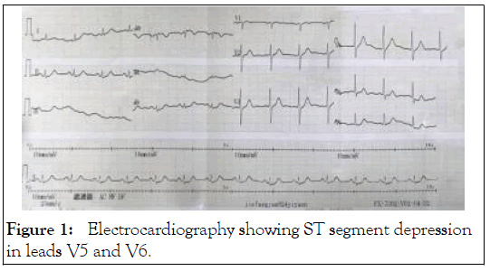
The patient’s vital signs were normal upon admission. On physical examination: the patient’s body temperature was 36.5° C, pulse rate: 77/min, respiratory rate: 18/min, blood pressure: 137/91 mmHg, weight: 62 kg, and height: 1.68 meters. The patient had a clear mind, upright posture, no cyanosis of lips, no dilation of jugular veins, and there was no negative sign of hepatic jugular venous reflux. The respiratory sound of both lungs was clear; there were no dry or wet rales. The cardiac boundary was normal with a heart rate of 77/min. The heart rhythm was uniform, and there was no murmur in the auscultation area of each valve. The abdomen was flat and soft, without tenderness and rebound pain, and the bowel sound was normal. The radial arteries of both upper limbs had consistent pulsation. The pulse of dorsalis pedis artery of both lower limbs was consistent. There was no edema in either of the lower limbs. Auxiliary examination results from the emergency department showed no detectable changes in myocardial enzymes, myoglobin, serum troponin T, blood routine, coagulation function, liver and kidney function, serum glucose, and or electrolytes. Electrocardiographic recordings during sinus rhythm showed an ST change in Figure 1.

Figure 1: Electrocardiography showing ST segment depression in leads V5 and V6.
Admission diagnosis
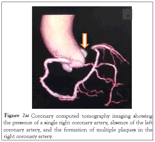
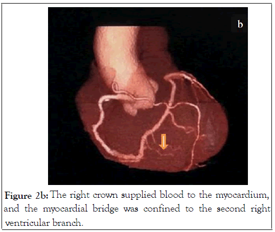
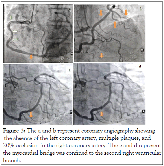
Causes of chest pain to investigate: CHD, Acute Coronary Syndrome (ACS), and hypertension grade 2, at high risk. On the sixth hour post-admission, re-examination of myocardial enzyme and serum troponin T were normal. The Electrocardiogram (ECG) showed no change from before. No abnormalities were found in thyroid function or glycosylated hemoglobin. Echocardiography results showed: 1) No significant abnormalities in the size of each chamber in the heart; 2) Low calcification in the right and non-coronary aortic valves; 3) Tricuspid valve micro regurgitation; 4) No obvious abnormalities in the left ventricular overall contractile function. On the second and third day in the hospital, the patient underwent CTA examination of the coronary artery and CAG, respectively. Coronary artery Computed Tomography Angiography (CTA) (Figures 2a and 2b) and Coronary Angiography (CAG) (Figure 3a-3d) showed the absence of the left coronary artery and 20% stenosis in the right coronary artery. The distal blood vessel supplied blood to the left heart. The MB was confined to the second right ventricular branch (Figure 2b and Figure 3c and 3d). CAG showed a TIMI glow of grade 3.

Figure 2a: Coronary computed tomography imaging showing the presence of a single right coronary artery, absence of the left coronary artery, and the formation of multiple plaques in the right coronary artery.

Figure 2b: The right crown supplied blood to the myocardium, and the myocardial bridge was confined to the second right ventricular branch.

Figure 3: The a and b represent coronary angiography showing the absence of the left coronary artery, multiple plaques, and 20% occlusion in the right coronary artery. The c and d represent the myocardial bridge was confined to the second right ventricular branch.
Definite diagnosis: 1) Absence of left coronary artery (type R-I) 2) MB (the second right ventricular branch) 3) Coronary atherosclerosis 4) Hypertension grade 2, at high risk. During his hospitalization, the patient was treated with dirthiazem (90 mg, qd), fosinopril (10 mg, qd), atorvastatin (20 mg, qn), and aspirin (100 mg, qd). Amlodipine tablets were stopped, which is a secondary prevention of CHD. After 3 days, the patient’s chest pain disappeared. Re-examination of the ECG indicated that it was normal. The patient improved and was discharged on August 9, 2021.
With the development of medical technology, the detection of coronary artery malformations has increased, both in quantity and in quality. Coronary artery malformations include abnormal initiation, distribution, and termination. An abnormal origin of a coronary artery indicates that the coronary artery originates from the contralateral sinus, posterior sinus, sinus bottom, above the sinus ridge, or pulmonary artery, often accompanied by abnormal proximal deformation. If it is between the aorta and pulmonary artery, it is prone to be compressed, resulting in myocardial ischemia or even sudden death. The opening of coronary artery with abnormal origin is generally small, and it is at an acute angle or tangential position in the beginning, which also makes it easy to cause myocardial ischemia and this kind of malformation, is easily missed and frequently misdiagnosed. The arteriole directly opening in the right coronary sinus is called the accessory coronary artery, which is also attributed to an abnormal coronary origin, and generally does not cause hemodynamic changes.
Coronary artery malformations include Single Coronary Artery (SCA), coronary artery fistula, and MB. The incidence of coronary malformations is 0.6%-1.6%, and single coronary artery malformations account for approximately 3.3% of all coronary artery malformations [1]. SCA is a rare congenital coronary artery abnormality. The prevalence reported in the general population is 0.0240%-0.66%, diagnosed by invasive coronary angiography [2]. Single coronary malformations are classified into types I, II, and III according to the classification of Lipton et al. [3] Type I is classified into type L-1 and R-I. This case was categorized as type R-I. At present, right coronary artery absence is relatively common in single coronary artery (SCA) malformations, while left coronary artery absence is rare incidence. Dong Shen et al. counted the results of CAG in 48,158 cases globally [4]. The results showed that the incidence of single coronary artery malformation was 0.032%, including 0.022% of type L and 0.01% of type R. SCA often causes syncope, arrhythmias, angina pectoris, myocardial infarction, congestive heart failure, and even sudden death. SCA’s clinical and ECG manifestations lack specificity, so it is prone to be misdiagnosed [5]. Yan et al. presumed that during an early age, the absence of risk factors such as atherosclerosis, hypertension, and diabetes may be what caused the lack of clinical symptoms. Patients are symptomatic only when CAD begins to occur [6]. If patients with SCA malformation have ischemic symptoms, they should receive the secondary prevention of CHD, whether it is complicated with coronary atherosclerosis or not. Gaoshang Wang et al. believe that the supply area of SCA branches is generally wide, and the probability of adverse cardiovascular events in cases of acute myocardial infarction is higher than in normal people [7]. For the treatment of patients with SCA malformation combined with occlusive vessels, the indications for Percutaneous Coronary Angioplasty (PCI) should be expanded so that the patients benefit more, and this may provide additional benefits.
MB is a congenital anomaly in which a segment of a major epicardial coronary artery course through the myocardial wall [8]. The detection rate of MB in CAG is 0.52%-2.5%. MB mostly occurs in the Left Anterior Descending (LAD), in 98.08% of cases. MB over the Left Circumflex (LCX) artery accounts for the remaining 1.92% of cases. Hence, MB over the right coronary artery is particularly rare. In this case, type R-I left coronary artery absence combined with MB over the right coronary artery is a rare congenital coronary malformation not previously reported in the literature.
To analyze the relationship between PTEN mutation and EC patient prognosis, Kaplan-Meier survival analysis was performed in PTEN-mutant patients and PTEN-wild patients using R ‘survival’ package. Then R ‘survminer’ package was used to draw the Kaplan-Meier Overall Survival (OS) curve. In addition, we compared the prognosis difference of different mutation types of PTEN in PTEN-mutant patients using the same method. Furthermore, correlation between clinical stage and PTEN mutation was analyzed.
Clinically, if the patient has ischemia changes in ECG, we need to consider whether it is SCA, MB, or other coronary malformations in addition to acute coronary syndrome of CHD, so as to avoid misdiagnosis.
Follow-up and outcomes
At the outpatient follow-up of about 7 months, the patient stated that he was taking his medicine regularly, with no attack of chest pain and stable blood pressure control.
The most important limitation of this study is its lack of longterm follow-up, and subsequent inability to assess patient prognosis.
Funding information is not applicable.
The authors declare that they do not have any conflicts of interest.
Written informed consent was obtained from the patient.
The work is approved by all the authors for participation.
The work is approved by all the authors for publication.
Some or all data and material generated or used during the study are available in a repository or online (Provide full citations that include URLs or DOI).
Some or all code generated or used during the study are available in a repository or online (Provide full citations that include URLs or DOI).
Conceptualization: Mei-lian Cai, Data curation: Mei-lian Cai, Investigation: Mei-lian Cai, Resources: Mei-lian Cai, Supervision: Wei Zhang, Writing-original draft: Mei-lian Cai, Writing-review & editing: Mei-lian Cai
We thank LetPub (www.letpub.com) for its linguistic assistance during the preparation of this manuscript.
[Google Scholar] [Pubmed]
[Crossref] [Google Scholar] [Pubmed]
[Crossref] [Google Scholar] [Pubmed]
Citation: Cai M, Zhang W (2022) Complex Congenital Coronary Artery Malformations: A Case Report. J Clin Trials. S16:005.
Received: 07-Mar-2022, Manuscript No. JCTR-22-16147; Editor assigned: 09-Mar-2022, Pre QC No. JCTR-22-16147 (PQ); Reviewed: 22-Mar-2022, QC No. JCTR-22-16147; Revised: 28-Mar-2022, Manuscript No. JCTR-22-16147 (R); Published: 04-Apr-2022 , DOI: 10.35248/2167-0870.22.S16.004
Copyright: © 2022 Cai M, et al. This is an open access article distributed under the terms of the Creative Commons Attribution License, which permits unrestricted use, distribution, and reproduction in any medium, provided the original author and source are credited.