Clinical Pediatrics: Open Access
Open Access
ISSN: 2572-0775
ISSN: 2572-0775
Case Report - (2024)Volume 9, Issue 5
Odontogenic oral lesions in pediatric patients, it is essential to understand the pathogenesis of odontogenic tumors. The clinical presentation, microscopic features and prognosis are addressed for odontogenic lesions in the newborn, but familiarity with these entities is essential due to the different therapeutic implications of these diagnoses.
The case we present is a five-day-old newborn in the Neonatology service of the Dos de Mayo National Hospital, Peru. The reason for consultation was "he was born with a lump in his mouth." On clinical inspection, a palatine lobe was found. The palatine lobe was presented for inspection. Conventional outpatient surgery is performed to remove the foreign body, completely removing the tumor. The surgical specimen was sent to pathology, which confirmed the definitive diagnosis; congenital epulis of the newborn.
Newborn; Gingival neoplasms; Gum diseases; Epulis
Congenital epulis of the newborn is a benign tumor of unknown etiology present at the time of birth. It is also called congenital tumor of granulosa cells, granulosa cell gingival tumor, Newman tumor or simply congenital epulis of the newborn [1,2]. On clinical examination, it is a mass pedunculated pink, inserted into the crest of the alveolar ridge or process. It can be uni-or multilobar. The prevalence is higher, in proportion (2:1), in the upper jaw and in women (8:1) [3]. Histologically, the tumor is characterized by being encapsulated, with a proliferation of cells with polygonal morphology, oval nucleus and granular cytoplasm covered by a thin stratified epithelium and without projections on the underlying epithelium [4,5]. Congenital epulis usually presents as a solitary lesion. Also there have been reported few cases of spontaneous regression [6]. Complications caused by epulis congenital prevents feeding and breathing and therefore, treatment recommended is surgical removal under local or general anesthesia, to reduce the risk of damage to underlying alveolar bone and developing tooth buds. The treatment will be performed with minimally invasive surgery in the area of the tumor; the identification of the lesion must be managed with an analysis anatomopathological. Therefore, it is important for parents to educate them about oral health during pregnancy and the treatments that can be performed on newborn, also in the causes of these pathologies with their development at their feeding and breathing [7-10].
Clinical case report
Male patients five days old, who attend the pediatric dentistry service for performing the evaluation. The mother's reason for consultation is that “her baby has a lump in the mouth on the palate". The mother says that he was born with the lump on the palate and she realized at two days after he was born that he had lump in his mouth. The mother informed us about the history, normal delivery at term with a weight of 3455 g and a height of 50 cm, family history of the parents does not refer systemic diseases, other congenital anomalies or developmental disorders (Figure 1).
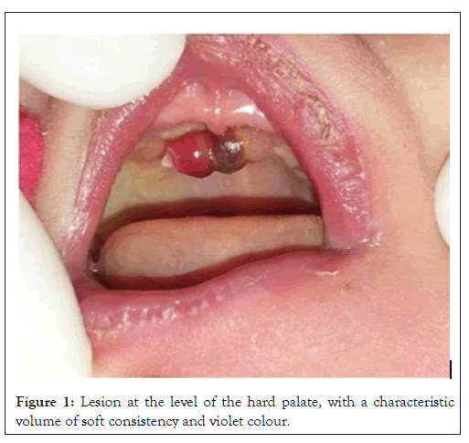
Figure 1: Lesion at the level of the hard palate, with a characteristic volume of soft consistency and violet colour.
In the intraoral clinical examination, a lobulated tumor was found in the palate area. The size of the pre maxilla 0.7 × 0.3 x 0.2 cm of a light brown color with brown areas dark. No other alteration was perceived. A presumptive diagnosis of congenital epulis of the newborn was reached. The parents are informed of the procedure and laboratory tests are performed presenting Hb of 12 g/dl, Hct of 35.01%, platelets of 382,500 mm3. Mother of Family signs the informed consent explaining the advantages and disadvantages of the clear and precise procedure (Figure 2). Scheduling the surgery will allow for a biopsy, which will confirm the diagnosis, definitive with the laboratory study (Figure 3). In the inspection after surgery, it is noted that the lobe that was present in the palate was removed (Figure 4).
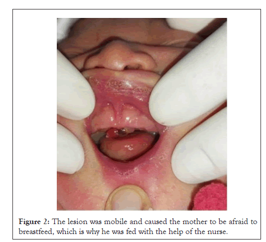
Figure 2: The lesion was mobile and caused the mother to be afraid to breastfeed, which is why he was fed with the help of the nurse.
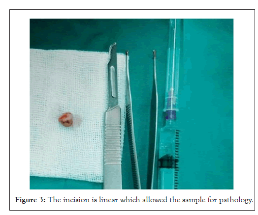
Figure 3: The incision is linear which allowed the sample for pathology.
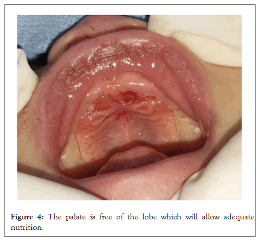
Figure 4: The palate is free of the lobe which will allow adequate nutrition.
It is evident after the incision, the palatine rugae is not affected. Careful instructions are given to the mother in writing and verbally, advice is given on how to clean the baby after 24 h and about breastfeeding due to the properties, it presents, including immunity and anti-inflammatory protection for the baby. The baby is evaluated after 24 h and after a week an excellent recovery is seen. Parental care is evaluated and it is explained that the baby must be brought for a follow-up check every month to continue with his evaluation (Figure 5).
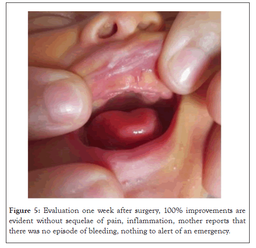
Figure 5: Evaluation one week after surgery, 100% improvements are evident without sequelae of pain, inflammation, mother reports that there was no episode of bleeding, nothing to alert of an emergency.
The subsequent evolution was satisfactory. Intact surface epithelium was observed in the histological sections of the material obtained, with features of parakeratosis, areas of acanthosis marked with elongation of nails epithelial cells anastomosed to each other and areas of pseudoepitheliomatous hyperplasia. The tissue Sub epithelial connective tissue presents abundant polygonal cells with cytoplasm intensely granular, oval and hyperchromatic nuclei with a moderate amount of vascular channels. In the deepest area, bands of fibroconnective tissue arranged in bundles with spindle-shaped fibroblasts and some chronic inflammatory cells. In conclusion, it is a congenital granular cell tumor (congenital epulis), thus confirming the clinical diagnosis (Figure 6).
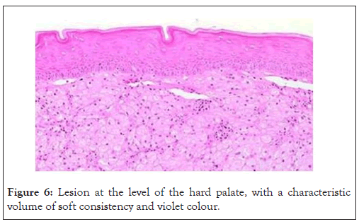
Figure 6: Lesion at the level of the hard palate, with a characteristic volume of soft consistency and violet colour.
Histological characteristics of the overlying epithelium may present atrophy with focal ulceration. They are pedunculated mucosal nodules, largely formed by polygonal cell sheets with a granular and eosinophilic cytoplasm, cell borders distinct, centrally located nuclei and discrete nucleoli. Remains can be seen odontogenic.
In most cases, recognition of the injury occurs after birth, in other situations the lesion is of considerable size and it is possible to visualize it in uterus by ultrasound. Some authors state that when it is small, it can return without sequelae. Congenital epulis can be a cause of extreme anxiety in parents and health professionals, since difficulties in feeding, Sucking and breathing can cause systemic compromise in newborns who present it. Therefore, it is necessary that specialists in fetal anomalies, such as gynecologists and perinatologists, as well as specialists in charge of the first door of newborn care, such as paediatricians, neonatologists and pediatric dentists, have the necessary information to identify, diagnose and promptly treat this rare congenital disorder with this type of approach, without affecting the dental or systemic development of the child [5,6,11].
In the literature, we find both cases that have been treated surgically and those who have preferred only observation. This case meets the criteria commonly found in medical literature in shape, color and number of the tumor; even in the possibility of resolution without surgery if it had not been for the feeding problem [12,13].
Congenital epulis in newborns is a rare but benign tumor that can often be managed through careful observation. However, in some cases, surgical intervention may be necessary. This can typically be performed using local anesthesia and without the need for hospitalization. The importance of the diagnosis lies in how serious the problem is for the family. In this case the clinical examination and the pathological examination with the report pathology was helpful in achieving a diagnosis, due to the experience of the observers. However, it is necessary to always have the means that allow, with a histological examination.
[Google Scholar] [PubMed]
[Crossref] [Google Scholar] [PubMed]
[Crossref] [Google Scholar] [PubMed]
[Crossref] [Google Scholar] [PubMed]
[Crossref] [Google Scholar] [PubMed]
[Crossref] [Google Scholar] [PubMed]
[Crossref] [Google Scholar] [PubMed]
[Crossref] [Google Scholar] [PubMed]
[Crossref] [Google Scholar] [PubMed]
Citation: Vera JF (2024). Congenital Epulis in the Newborn: Report of a Clinical Case. Clin Pediatr. 09:279.
Received: 08-Aug-2024, Manuscript No. CPOA-24-33442; Editor assigned: 12-Aug-2024, Pre QC No. CPOA-24-33442 (PQ); Reviewed: 26-Aug-2024, QC No. CPOA-24-33442; Revised: 02-Sep-2024, Manuscript No. CPOA-24-33442 (R); Published: 09-Sep-2024 , DOI: 10.35248/2572-0775.24.09.279
Copyright: © 2024 Vera JF. This is an open-access article distributed under the terms of the Creative Commons Attribution License, which permits unrestricted use, distribution and reproduction in any medium, provided the original author and source are credited.