Immunome Research
Open Access
ISSN: 1745-7580
ISSN: 1745-7580
Research Article - (2023)Volume 19, Issue 2
Objective: To investigate the correlation between autoimmune antibodies, ultrasonic elasticity score, and elasticity coefficient in patients with Hashimoto’s Thyroiditis (HT).
Methods: The thyroid function, the titer of the Thyroid globulin Antibody (TgAb) and Thyroid Peroxides Antibody (TPOAb) were detected by chemiluminescence method from the fasting serum of 258 patients suffering with HT; the ultrasonic elasticity score and elasticity coefficient were evaluated after detecting the patients’ thyroid with GELOGIQ3 type color doppler ultrasound diagnostic system and getting a satisfactory elastosonography. Then the correlation between autoimmune antibodies, ultrasonic elasticity score and elasticity coefficient were analyzed respectively.
Results: Serum Thyroid Stimulating Hormone (TSH), TGA and TPO in 258 patients with HT increased significantly, the titer of TgAb was positively correlated with elastic coefficient, r=0.62, P<0.01, and also positively correlated with the elastic score, r=0.65, P<0.01; TPOAb was positively correlated with elastic coefficient, r=0.63, P<0.01, and positively correlated with elastic score as well, r=0.62, P<0.01.
Conclusions: It’s helpful to increase the accuracy rate of HT diagnosis with the detection of thyroid autoimmune function combined with real-time ultrasound elastosonography score.
Hashimoto’s Thyroiditis (HT); Autoimmune; Elastosonography; Elastic score; Elastic coefficient
Hashimoto’s Thyroiditis (HT), also known as chronic lymphocyte thyroiditis, is a common type of Autoimmune Thyroid Disease (AITD). The notable characteristic feature of HT is presence of extensive lymphocyte and plasma cell infiltration in thyroid tissue, as well as thyroid specific autoantibodies could be found in the serum [1].
At present, it is considered as a chronic autoimmune disease with auto thyroid tissue as antigen with the main cause of high iodine in heredity and diet. Cellular and humoral immunity play roles during this process, which could change the structure and function of the thyroid gland. Goiter and thyroid nodule are the typical clinical manifestations, as well as the main causes of hypothyroidism. At the same time, it is easy to be accompanied with other autoimmune diseases and damages of organs and systems besides thyroid gland [2]. It is a disease with a long course, high incidence and poor treatment, because the clinical manifestation in the thyroid inflammation disease is intricate. In fact, with the help of thyroid function and ultrasound examination, especially real-time ultrasound elastography, also known as “electronic palpation”, the diagnosis could provide information about the internal elastic characteristics of tissues and could reflect the soft and hard tissues objectively. The purpose of this study is to examine the ultrasound elastography of 258 patients with HT, by analyzing the elastic score and elastic strain coefficient, and the changes of serum thyroid autoantibodies, and to investigate the correlation between ultrasound elastography and thyroid autoimmunity [3].
General Information
A total of 258 HT patients with detailed physical examination data from January 2019 to June 2020 were selected and confirmed according to fisher's clinical diagnostic criteria by the serum autoantibodies level, fine needle biopsy and/or pathological examination [4]. Among them, 182 female, 76 male, age span 19-72, average age (40.29 ± 11.32). HT is divided into 4 phases according to the results of thyroid function test: Hypothyroidism is characterized by reduced TSH with elevated serum Free Thyroxine (FT4) or Triiodothyronine (FT3) level; Normal thyroid function is characterized by the normal level of TSH with normal FT3 or FT4 level; subclinical hypothyroidism is characterized by elevated TSH with normal FT3 and FT4 levels; Clinical hypothyroidism is characterized by elevated TSH with reduced FT3 or FT4 level [5].
The data regarding 258 patients were analyzed. The patients were divided in four different groups: HT hyperthyroidism group (n=52); HT normal group (n=86); HT subclinical group (n=64); HT hypothyroidism group (n=56) [6].
Inclusion criteria: Compliance with fisher diagnostic criteria, the age was between 19 and 72 years old, gender was not limited; other therapies were discontinued during the study period; the subjects had detailed information about their condition, and the project was examined and approved by the medical ethics committee of the hospital [7].
Exclusion criteria: Those who do not conform to the above criteria; those who coexist with other diseases during the period of observation; those who have received other related treatment that may affect the results of this study; those who are pregnant and lactating women [8].
Instruments and methods
The detector is color doppler ultrasonic diagnostic instrument (GE LOGIQ3 type) with linear array probe and the frequency of the probe is about 7.5 MHz-13 MHz. Firstly, subjects took supine posture, padded the shoulder to make the neck fully exposed. Secondly, subjects were performed routine examination of the two dimensional ultrasonography, as well as observed thyroid contour, size and thickness of the thyroid gland, the echo of thyroid parenchyma, and the internal blood flow signals of the gland tissue [9]. Then the subjects were told to hold their breath. At the same time, the two dimensional and elastic images of the thyroid were displayed in real time. The Region of Interest (ROI) was selected and the probe was manually pressured, the frequency was 2 times/s. The composite index of the pressure vibration frequency near the elastic image was maintained to be between 3-4 levels, lasted for 3 seconds, and then the relatively stable values and images were obtained. When the pressure was appropriate, the connective tissue around the thyroid capsule was displayed as a continuous banded red, and the muscles around the thyroid gland were uniformly yellowish green, and the images were relatively clear and stable. On the basis of satisfied elasticity image, all elasticity images were scored according to the color that the ROI displayed. To classify elasticity images, we evaluated the color pattern both in the lesion and in the surrounding thyroid tissue. On the basis of the overall pattern, we assigned each image an elasticity score on a five point scale. A score of 1 indicated even strain for the entire lesion (i.e., the entire lesion was evenly shaded in green). A score of 2 indicated strain in most of the lesion, with some areas of no strain (i.e., the lesion had a mosaic pattern of green and blue, and was given priority to with green). A score of 3 indicated strain at the periphery of the lesion, with sparing of the center of the lesion (i.e., the periphera part of lesion was green, and the central part was blue, and the proportion of green and blue part was similar). A score of 4 indicated no strain in the entire lesion (i.e., the entire lesion was blue, but its surrounding area was not included). A score of 5 indicated no strain in the entire lesion or in the surrounding area (i.e., both the entire lesion and its surrounding area were blue with/without a little green in the internal) [10].
The dual real time display function of color Doppler ultrasound was enabled again. Then the images were replayed, and the elastic images were observed and analyzed. A frame of relatively stable images was selected, and the regions of interest were drawn on the two-dimensional images. HT thyroid parenchyma lesion area was set as (A) with the ipsilateral sternocleidomastoid muscle (B) was used as the reference area [11]. The average elastic strain data of the ROI region are calculated by the instrument system. Taking the ratio of the two parts, that is the elastic strain rate of the lesion region (SR), or the elastic coefficient. The image score and SR were measured by double blind method by two ultrasound diagnostics. If any dispute should arise over this process, we may submit it for comprehensive evaluation by the third ultrasonic doctor with diagnostic qualification [12].
Thyroid function test: 3 ml of peripheral venous blood was collected from every patient on empty stomach at early morning in vacuum non-anticoagulant test tube. And then, serum was separated by centrifuging for 10 min at 1500 r/min speed. The serum sample test must be accomplished on the same day [13].
The thyroid function, the titer of TgAb and TPOAb were detected by chemiluminescence method from the fasting serum of 216 patients suffering with HT. The reference range drawn up by the laboratory is as follows: FT3 1.45 pg/mL-3.48 pg/mL, FT4 0.71 ng/mL-1.85 ng/mL, TSH 0.35 μIU/mL-4.94 μIU/mL, TPOAb 0.00 IU/mL-35.00 IU/mL, TgAb 0.00 IU/ mL-40.00 IU/mL [14].
Statistical analysis
Statistical analysis was performed with SPSS 25 statistical analysis software. Results were expressed as means ± SEM. Student’s t test for two groups and a one way Analysis of Variance (ANOVA) for more than two groups were used to determine statistically significant differences [15]. The correlation between elastic image score, elastic strain coefficient and thyroid function was analyzed by Spearman correlation analysis. Differences achieving values of P<0.05 were considered statistically significant [16].
Thyroid function and autoimmune antibodies in different HT groups
The serum levels of TgAb, TPOAb and TSH were increased in 258 patients with HT. The level TSH was normal in HT hyperthyroidism group and HT normal group. But there were significant differences among the groups (P<0.01). In addition, as the level of FT3 and FT4 decreased, the level of TSH, TgAb and TPOAb increased gradually (P<0.05 or 0.01). The results were shown in Table 1 [17].
| Groups | n | FT3 (pg/mL) | FT4 (ng/mL) | TSH ((µIU/mL)) | TgAb (IU/mL) | TPOAb (IU/mL) |
|---|---|---|---|---|---|---|
| Hyperthyroidism groups | 52 | 4.32 ± 0.36 | 2.19 ± 0.27 | 0.19 ± 0.13 | 98.62 ± 21.36 | 119.59 ± 98.36 |
| Normal groups | 86 | 2.74 ± 0.26 | 1.58 ± 0.34 | 2.75 ± 0.62 | 128.36 ± 31.12 | 187.18 ± 79.46 |
| Sub clinical groups | 64 | 2.36 ± 0.31 | 1.04 ± 0.25 | 5.68 ± 0.56 | 169.67 ± 36.54 | 256.68 ± 102. 38 |
| Hypothyroidism groups | 56 | 1.26 ± 0.32 | 0.64 ± 0.27 | 10.78 ± 1.02 | 261.12 ± 42.26 | 329.48 ± 158.12 |
| HT groups | 258 | 3.16 ± 2.14 | 0.98 ± 0.56 | 8.52 ± 3.14 | 181.66 ± 124.39 | 254.88 ± 186.05 |
Table 1: Comparison of thyroid function and autoimmune antibodies indifferent HT groups.
Ultrasonic elastic imaging score and elastic coefficient in different HT groups
In addition to the HT hyperthyroidism group and HT normal group, there were significant differences in SR among the other groups (P<0.01 or P<0.05) [18]. In other words, with the decline of thyroid function, the SR of thyroid tissue increased. The elastic score of thyroid tissue was significant differences between the other groups (P<0.01 or P<0.05), except the HT hyperthyroidism group and the HT normal group. That is to say, with the decline of thyroid function, the elastic score of thyroid tissue increased, which indicated that the hardness of thyroid increased. The results were shown in Table 2 and Figures 1-4 [19].
| Groups | n | Strain Ratio (SR) | Elasticity score |
|---|---|---|---|
| Hyperthyroidism groups | 52 | 1.53 ± 1.12 | 1.73 ± 1.36 |
| Normal groups | 86 | 1.86 ± 1.26 | 1.92 ± 1.38 |
| Sub clinical groups | 64 | 2.28 ± 1.42 | 2.39 ± 1.27 |
| Hypothyroidism groups | 56 | 2.69 ± 1.25 | 2.92 ± 1.29 |
| HT groups | 258 | 2.32 ± 1.35 | 2.51 ± 1.37 |
Table 2: Comparison of ultrasonic elasticity imaging score and elastic coefficient in different HT groups.
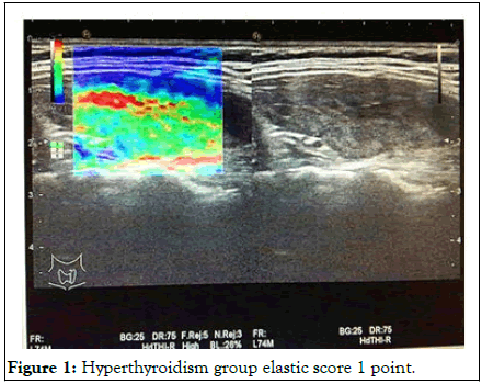
Figure 1: Hyperthyroidism group elastic score 1 point.
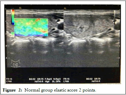
Figure 2: Normal group elastic score 2 points.
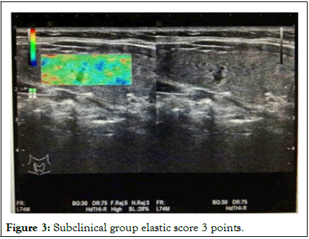
Figure 3: Subclinical group elastic score 3 points.
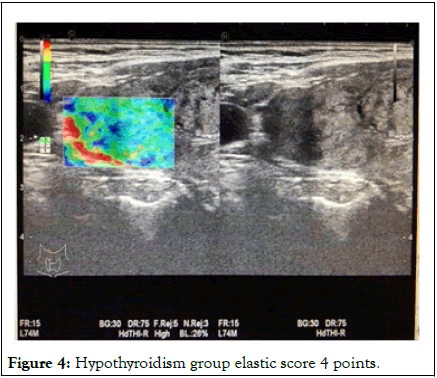
Figure 4: Hypothyroidism group elastic score 4 points.
Correlation between thyroid ultrasound elastic score, elastic coefficient and autoantibodies in HT patients
In 258 patients, TgAb was positively correlated with SR (r=0.62, P<0.01), as well as elasticity score (r=0.65, P<0.01); TPOAb was positively correlated with SR (r=0.63, P<0.01), as well as elastic score (r=0.62, P<0.01). The results were shown in Figures 5-8 [20].
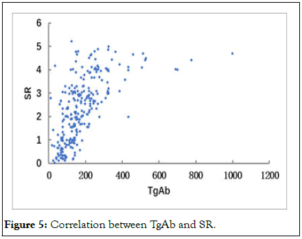
Figure 5: Correlation between TgAb and SR.

Figure 6: Correlation between TgAb and elastic coefficient.
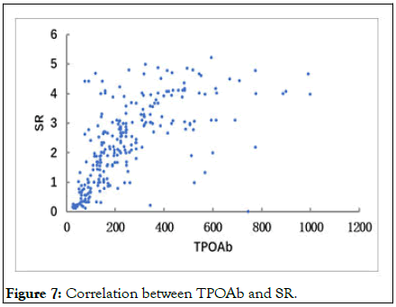
Figure 7: Correlation between TPOAb and SR.
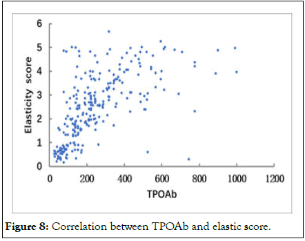
Figure 8: Correlation between TPOAb and elastic score.
HT is a kind of common clinical autoimmune thyroid disease with unclear etiology and various clinical manifestations. In addition, iodine excess intake is an important environmental factor in the development of the disease. In recent years, with the popularization of iodized salt, iodine intake gradually increased, which were accompanied by the increased incidence of HT. The typical pathological features are the infiltration of a large number of lymphocytes and the proliferation of connective tissue in different degrees. There are high autoantibodies titer of thyroid resistant components could be detected in the patients’ serum, such as TPOAb and TgAb. Because the disease is concealed, the patients don’t usually come to see a doctor until hyperthyroidism or hypothyroidism symptoms exists. There are some patients with HT are found via the routine examination. And there are some are confirmed by histological diagnosis during the operation of thyroid nodule of tumor [21].
HT may not have any clinical symptoms in the early stage, and may present only as elevated TPOAb and TgAb, or may present only as some manifestations of hyperthyroidism, and then powerless, hypothyroidism appears in the late stage. HT related thyroid specific antibodies include TPOAb, TgAb, TmAb. Among them, TPOAb is the main antibody that mediates the cytotoxic effect of cell mediated antibody. On the basis of T cell mediated cell damage, thyroid follicular epithelial cells are further damaged, resulting in hypothyroidism. TPO is an indispensable enzyme in the synthesis of thyroid hormones, and does not overflow into the blood from the thyroid gland normally. When the structure of thyroid follicular cells is destroyed, TPO leaks from the top of the thyroid cell, namely the edge of the cavity of the thyroid follicular cell out of the peripheral blood. As an important autoantigen of autoimmune thyroid diseases, TPO stimulates the body’s immune system, producing thyroid tissue component antibody TPOAb and activating complement and antibody dependent cell mediated cytotoxicity, resulting in thyroid immune damage. With the discovery of new effector T cell subsets and the further understanding of T cell subsets, it has been found that thyroid organ specific autoimmune diseases are mainly related to Th1 cells. Th17 cells, as independent CD4 T cell subsets, play a pathogenic role by releasing IL-17, IL-17F and IL-22. It could induce local cells to synthesize chemical factors and proinflammatory factors, and affect the production of autoimmune antibodies. The abnormal secretion of IL-17 may lead to the disorder of thyroid autoimmune function and then induce the occurrence and development of HT. In addition, it has been found that the immune imbalance of peripheral blood Treg/Th17 cell axis ran through the different stages of HT, and could shown abnormal changes in the normal thyroid function stage, as well as a qualitative change in subclinical hypothyroidism. The ratio of Th17/Treg cells are positively correlated with the thyroid autoantibody TPOAb-TgAb, which means the imbalance of Th17/Treg cell axis might play an important role in the production of thyroid autoantibodies and the abnormal immune response of T cells mediated by thyroid autoantibodies, and participate in the autoimmune injury of thyroid tissues. Furthermore, up-regulation of IL-35/IL-35 expression mediated by inducible T cells to induce Treg immunosuppression might play an important role in the autoimmune model. In addition, memory T cells and autoreactive T cells are interdependent and can be transformed into each other, and participate in the development of various autoimmune diseases, including Multiple Sclerosis (MS) and Systemic Lupus Erythematosus (SLE). What’s more, the proportion of CD4+CD45RO+ memory T cells, IFN-γ, IL-17, TPOAB and TgAb in peripheral blood of HT patients are significantly higher than those in normal subjects. So, it is suggested that CD4+CD45RO+ memory T cells may be involved in the pathogenesis of HT.
Normal thyroid tissue is composed of follicular structure and connective tissue between follicles. The follicular cavity contains colloid and with a soft texture. However, because of the infiltration of numerous lymphocytes and plasma cells in the stroma of HT gland, the normal follicles are destroyed and even the structure of follicles is atrophied, tissue fibrosis, and thyroid stiffness gradually increases. Currently, the preferred imaging modality for thyroid disease is ultrasound, which has a wide range of clinical applications. Real time ultrasound elastography is a new ultrasonic diagnostic technique, which can objectively provide the characteristic information of hardness in tissues, coined by Ophir, et al. The basic principle is that according to the different elastic coefficients (stress/strain) of various tissues, the tissues undergo different degrees of deformation after being compressed by local external forces, and then, the changes in the movement amplitude of the echo signals before and after the tissue compression are converted into real time color images, which can reflect the softness and hardness of the tissue. The real time ultrasound elastography of the thyroid gland can indirectly reflect the hardness of the thyroid parenchyma by measuring SR, providing a new auxiliary information for the ultrasound diagnosis of HT, which could be used as a supplement to conventional ultrasound examinations and could better assess the functional status and pathological progress of HT. Because the disease evolution process of HT is more complicated and changeable, and individual differences are large, it is often divided into normal, hyperthyroid, subclinical hypothyroidism and hypothyroidism in clinical practice. Different clinical processes have different thyroid morphologies and with different imaging patterns. The ultrasound images of HT usually show diffuse and uniform hypoechoic, which may appear as reticular fiber intervals, or focal hypoechoic with ill-defined boundaries, or diffuse and uneven hypoechoic with nodules and other complex ultrasound manifestations. During the progression of HT, there is often proliferation of fibrous tissue and separation of thyroid tissue, resulting in a large number of nodules. The nodules are squeezed and fused with each other, which often results in atypical ultrasound images. Some studies have shown that the echo levels, echo, texture, calcification, halo sign and boundary regularity of thyroid nodule in the HT and non-HT context are similar. Benign thyroid nodules in the HT context are often hypoechoic, while hypoechoic is often one of the sonographic manifestations of thyroid cancer, and the sensitivity of the diagnosis of thyroid cancer is high. So, it is necessary to pay enough attention to the ultrasonic examination.
Ultrasound elastography is based on the different elastic coefficients between normal tissue and diseased tissue to be a diagnostic method. It relies on external force or alternating vibration method and could avoid the influence of subjective factors on the diagnosis. This technology uses the tissue’s own elastic characteristics and the difference in tissue hardness to image, and evaluates and analyzes the different colors of the lesions, which has a high diagnostic accuracy rate. Ultrasonic elastic strain rate ratio measurement mainly evaluates the relative hardness of the tissue in the form of numerical values, so as to avoid the influence of artificial selection of the size and range of the sampling frame on the detection of lesion hardness. Ultrasound elastography can reflect the hardness of thyroid nodules, in which the ratio of elasticity score and elastic strain rate has high diagnostic value.
In the study of ultrasonic elastic strain ratio, the hyperthyroidism group and the normal thyroid function group were mainly composed of localized hypochondria and diffuse hypochondria without cords. The subclinical hypothyroidism group and hypothyroidism group were mainly characterized by diffuse hypochondria with cord and nodule formation, and with different ultrasonic elastic strain ratio of different echo types (E2/E1). The difference was statistically significant. The results has shown that E2/E1 could reflect the hardness of the thyroid parenchyma to a certain extent, evaluate the patient’s thyroid function status and the progress of the disease, and could prevent the occurrence of hypothyroidism in the early stage. According to the study, E2/E1 in hyperthyroidism group<normal thyroid function group<subclinical hypothyroidism group<hypothyroidism group; E2/E1 in limited echo reduction type<diffuse echo reduction without cord strip type<diffuse echo reduction with cord strip type<nodule formation; and E2/E1 of different thyroid function groups is positively correlated with serum TSH, indicating that the larger the E2/E1, the higher the TSH, and the course of the disease gradually develops in the direction of hypothyroidism.
TGAB and TPOAb are the marker antibodies of thyroiditis Hashimoto. The detection of TGAB and TPOAB is convenient, accurate and sensitive, which is of great significance for diagnosis, prognosis and follow up after treatment. The results of this study has showed that the increase of TGA and TPO in serum of HT patients, and the increase of SR and elasticity score of thyroid tissues, suggesting that due to the continued existence of TGA and TPO, the hardness of thyroid tissue increased. Therefore, TgAb, TPOAb and thyroid ultrasonography should be performed once the thyroid tissue is hard by palpation. In conclusion, the diagnosis of HT can be confirmed by ultrasonography combined with the determination of anti-thyroid autoantibodies and thyroxine levels in serum. However, there is no objective quantitative index reflecting the progress of the disease in the conventional two-dimensional ultrasound examination, and real time ultrasound elastography technology combined with thyroid autoimmune function detection may be helpful in improving the diagnosis of Hashimoto’s thyroiditis.
The present study was supported by the national natural science foundation of China (grant no. 81473687; 82274538), the academic promotion program of Shandong first medical university (grant no. 2019 QL017), the natural science foundation of Shandong province (grant no. ZR2020MH357, ZR2020 MH312), Tai’an science and technology innovation development project (grant no.2020NS092).
The authors declare that they have no known competing financial interests or personal relationships that could have appeared to influence the work reported in this paper.
The present study was approved by the research ethics committee of the second affiliated hospital of Shandong first medical university (Tai’an, China). The procedures performed were in accordance with the ethical standards of the institutional and/or national research committee and with the 1964 Helsinki declaration and its later amendments or comparable ethical standards.
Informed consent was obtained from all individual participants included in the study for patients’ study participation and publication of identifying information and images.
The authors thank the national natural science foundation of China (grant no. 81473687; 82274538), the academic promotion program of Shandong first medical university (grant no. 2019QL017), the natural science foundation of Shandong provi nce (grant no. ZR2020MH357, ZR2020MH312), Tai’an science and technology innovation development project (grant no. 2020NS092) for funding the project.
[Crossref] [Google Scholar] [PubMed]
[Crossref] [Google Scholar] [PubMed]
[Crossref] [Google Scholar] [PubMed]
[Crossref] [Google Scholar] [PubMed]
[Crossref] [Google Scholar] [PubMed]
[Crossref] [Google Scholar] [PubMed]
[Crossref] [Google Scholar] [PubMed]
[Crossref] [Google Scholar] [PubMed]
[Crossref] [Google Scholar] [PubMed]
[Crossref] [Google Scholar] [PubMed]
[Crossref] [Google Scholar] [PubMed]
[Crossref] [Google Scholar] [PubMed]
[Crossref] [Google Scholar] [PubMed]
[Crossref] [Google Scholar] [PubMed]
[Crossref] [Google Scholar] [PubMed]
Citation: Li X, Ran Z, Sun X, Wang Y, Wei C (2023) Correlation between Autoimmune Antibodies and Elastosonography Score in Patients with Hashimoto’s Thyroiditis. Immunome Res. 19:231.
Received: 14-Feb-2023, Manuscript No. IMR-23-21807; Editor assigned: 17-Feb-2023, Pre QC No. IMR-23-21807 (PQ); Reviewed: 03-Mar-2023, QC No. IMR-23-21807; Revised: 19-Apr-2023, Manuscript No. IMR-23-21807 (R); Published: 26-Apr-2023 , DOI: 10.35248/1745-7580.23.19.231
Copyright: © 2023 Li X, et al. This is an open-access article distributed under the terms of the Creative Commons Attribution License, which permits unrestricted use, distribution, and reproduction in any medium, provided the original author and source are credited.