Journal of Clinical Trials
Open Access
ISSN: 2167-0870

ISSN: 2167-0870
Research Article - (2024)
Introduction: Patients with Hepatitis C Virus (HCV) develop different types of renal injuries. Membranoproliferative Glomerulonephritis (MPGN) is the most common type occurs in HCV patients who are complicated with small- vessel vasculitis cryoglobulinaemia. The aim of this work is to study the detection of HCV-RNA in the renal tissue using both Polymerase Chain Reaction and In Situ Hybridization (PCR and ISH) Techniques. In addition, we investigate the different patterns of renal affection in association with HCV infection.
Methods: The material of this work consisted of 50 native renal biopsies taken from HCV seropositive patients with renal lesions. All cases were collected as paraffin-embedded blocks. All cases were examined using a light microscope and evaluated according to the type of renal tissue as well as assessment of the glomeruli, tubules, interstitium and Vessels. In Situ Hybridization (ISH) and Reverse Transcription Polymerase Chain Reaction (RT-PCR) were used for the detection of HCV RNA in renal biopsies.
Results: Our study sample showed male predominance which represented 86% of cases. The age range was 19-67 years with a mean age of 47.7 ± 8.8 years. Most cases showed glomerular diseases (46 cases representing 92%) and the most predominant glomerular disease was Membranoproliferative Glomerulonephritis (MPGN) (58%). There was a significant relation between the degree of tubular atrophy and interstitial fibrosis (p-value=0.036). Regarding vascular changes, most cases showed no vascular changes (62%). However, the predominant vascular lesion was vascular thickening (34%) with seven cases above 50 years old. Ten cases were found to have cryoglobulinaemia and the main associated clinical presentations were impaired renal functions (40%) and nephrotic syndrome. In this study detection of HCV-RNA in the included 50 renal biopsies was tested by using 2 techniques ISH and RT-PCR. Regarding the ISH technique, positive hybridization signals were detected in the tubular epithelium of 28 cases (56%). RT-PCR detected HCV-RNA in 40 cases (80%).
Conclusion: Renal affection represents a major entity of extra-hepatic affection in patients with HCV. Most of renal affection is in the form of MPGN, but other forms of affection may occur and progress to end-stage renal disease. In addition, renal affection is due to viral invasion of renal tissue as proved by the detection of HCV-RNA within the tissues. Moreover, RT-PCR was found to be more sensitive than ISH.
Hepatitis C virus; Glomerulonephritis; RT-PCR; In situ hybridization; Infected patients
The incidence of Hepatitis C virus (HCV) has shown a rising trend over the years, where in 2020 it was estimated that over 71 million individuals across the globe are at risk of acquiring chronic HCV infection [1]. HCV can cause disease either by direct effect on infected cells, or through the host’s immune response. Both mechanisms are often integrated in the pathogenesis of disease, as a complex interaction [2]. HCV virus ultimately causes liver cirrhosis in most of the infected patients. Pathogenesis of cirrhosis is due to the viral replication within hepatocytes, which stimulates an immunogenic reaction that leads to inflammatory, necrotic and fibrotic changes in the hepatocytes [3].
In addition to hepatic affection, the pathological consequences of HCV virus may extend to other organs. Acute Kidney Injury (AKI) is an important consequence of HCV. Its main pathogenesis is attributed to the excess synthesis of cryoglobulins following HCV infection. Consequently, this can induce immune complex- mediated vascular inflammation, arterial thrombosis and cryoglobulin accumulation [4].
More commonly encountered, patients with HCV develop Membranoproliferative Glomerulonephritis (MPGN). MPGN usually occurs in HCV patients who are complicated with small-vessel vasculitis cryoglobulinaemia [5-7]. Like AKI, the pathogenesis of MPGN depends on the raised level of circulating antigens within the blood that became eventually accumulated within renal glomeruli [8,9]. The incidence of death in HCV patients significantly rise among patients who develop cryoglobulinaemia. Recent research findings indicate a 74% overall survival rate after 10 years, with a substantial difference between type III cryoglobulinaemia (84%) and type II cryoglobulinaemia (71%) [10].
Other kidney diseases due to HCV also include MPGN without cryoglobulinaemia, membranous nephropathy, focal segmental glomerulosclerosis, IgA nephropathy, thrombotic micro-angiopathy, interstitial nephritis and polyarteritis nodosa [11-13].
The aim of this work is to study the different patterns of renal affection in association with HCV infection and the availability of detection of HCV-RNA in the renal tissue using both PCR and ISH techniques.
Study population
The material of this work consisted of 50 native renal biopsies taken from HCV seropositive patients with renal lesions. These cases were selected from renal biopsies in (University hospital, Faculty of medicine, Fayoum University). All cases were collected as paraffin embedded blocks. In addition, 2 control cases were selected for the molecular biology methods (1st one was renal biopsy from 1.2 years old male child diagnosed as no change under light microscopy and the 2nd one was liver biopsy from 58 years old male with chronic active hepatitis C, HAI was 9/18 and stage was 3/6).
Cases with history of other systemic diseases which may contribute in renal affection such as SLE, Diabetes mellitus, Bilharziasis, HBV infection and hypertension were excluded.
All cases were examined using a light microscope and evaluated according to type of renal tissue as well as assessment of the glomeruli, tubules, interstitium and vessels.
Molecular biology methods
Tissue were then prepared for RT-PCR as follows:
RNA extraction from paraffin sections: 1 ml Xylene was added to 30 µm of the paraffin sections in autoclaved Eppendorf tubes and the tubes incubated for 10 min at room temperature. The fluid was removed following centrifugation for 2 min and then 1 ml of 95% ethanol added to the tubes which were incubated for 15 min at room temperature. Total RNA was extracted from kidney biopsies using total RNA extraction kit provided by Qiagene Corporation, Madison, WI, USA. The amount of RNA was quantitated by reading the Optical Density (OD) at wavelength of 260 nm by using a spectrophotometer.
cDNA synthesis and PCR of HCV: The RT-PCR method used was the one step method. The kit was provided by Qiagen. The RT-PCR amplification protocol was formed of Qiagen one step RT- PCR buffer (5x), dNTPs (10 µM), primer (1CH, 10 µM), primer (2CH, 10 µM), Qiagen one RT-PCR enzyme mix and template RNA in a total volume of 50 µL. The PCR cycling condition was as follows: One cycle of 50°C for 30 min, one cycle of 95°C for 15 min followed by 40 cycles of 95°C for 1 min, 45°C for 1 min and 72°C for 1 min. Then extension for 10 min at 72°C.To increase the sensitivity of the RT-PCR assay of HCV, 3 µL of the first amplified product was subjected to nested PCR.
Agarose gel electrophoresis: 10 µL of the PCR product was separated electrophoretically in a 1.5% agarose gel stained with ethidium bromide and observed under UV light.
In Situ Hybridization (ISH)
The biotinylated RNA probe was allowed to come to room temperature. It was thoroughly mixed before use. One drop (approximately 40 µl) of probe was added to the appropriate tissue section and a glass cover slip was placed over the section and the probe. Care must be taken to avoid bubble formation. Large sections required 2 drops of the probe and larger cover slips to adequately cover the section. Slides were placed flat on the (pre- warmed) heating block and were incubated at 100°C ± 2°C for 5 min. The slides were taken off the heating block and placed flat in a humidified incubation chamber. The slides were incubated at 37°C for 24 h. 800 ml of buffer 2 were pre-warmed to 37°C ± 2°C. The slides were removed from the incubation chamber. The slides were individually rinsed with gentle agitation in the first container of buffer 2 in order to remove the coverslips and rinse off excess probe. The coverslips floated off the slides and sank to the bottom of the staining dish. The slides were placed in the staining rack and washed in three changes of buffer 2 or 3 min each at 37°C ± 2°C.
Statistical analysis
Data were handled using the statistical package SPSS© (Statistical Package for Social Sciences) version 16. Data were summarized using mean and the standard deviation for quantitative variables while the frequency and percentages were used for the qualitative ones.
Comparisons between groups were done using Chi-square and Fisher’s exact test for small numbers for qualitative (nominal) variables. While student’s t test and one-way Analysis of Variance (ANOVA) were used to compare two groups and more than two groups respectively for the quantitative variables.
The significance level of the statistics was assessed by determining the p-value (Probability Value) where p ≤ 0.05 denotes Significant data.
Baseline characteristics
Our study sample showed male predominance as the male patients represented 68% of cases with a male-to-female ratio of 2.1:1 (Table 1). The age range was 19-67 years with a mean age of 47.7 ± 8.8 years. The most frequent age group was in the fifth decade (54%). The most prevalent clinical presentation was the impairment of renal function (50%). Most cases showed glomerular diseases (46 cases representing 92%) and the most predominant glomerular disease was MPGN (58%).
| Parameters | Cases (n=50)* | |
|---|---|---|
| Age | <10-50 | 34 (68%) |
| <50-70 | 16 (32%) | |
| Sex | Males | 34 (68%) |
| Females | 16 (32%) | |
| Clinical presentation | Nephrotic syndrome | 20 (40%) |
| Impaired renal function | 25 (50%) | |
| Edema | 9 (18%) | |
| Microscopic hematuria | 7 (14%) | |
| Proteinuria | 10 (20%) | |
| Hepatosplenomegaly | 1 (2%) | |
| Oliguria | 1 (2%) | |
| Histopathological diagnosis | MPGN | 11 (22%) |
| MPGN with crescent formation | 4 (8%) | |
| MPGN with intraluminal hyaline thrombi | 7 (14%) | |
| MPGN with accentuated lobular architecture | 9 (18%) | |
| FSGS | 5 (10%) | |
| MN | 4 (8%) | |
| Focal proliferative GN | 3 (6%) | |
| Mesangial proliferative GN | 3 (6%) | |
| No change under L.M | 2 (4%) | |
| Acute interstitial nephritis | 1 (2%) | |
| Chronic interstitial nephritis | 1 (2%) | |
| Amyloidosis | 2 (2%) | |
Note: *Indicates number and frequency.
Table 1: Baseline characteristics, clinical presentation and histological diagnosis of the included sample.
Results of HCV detection in the renal tissue
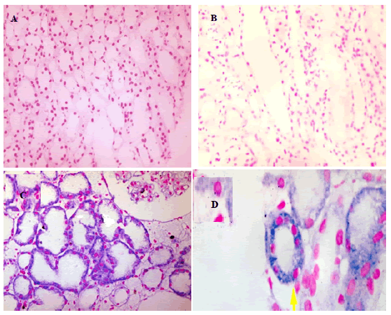
Only 28 cases (56%) showed positive hybridization signals for HCV-RNA detected in the tubular epithelial cells (Table 2). Whereas RT-PCR detected viral RNA in 40 cases (80%). This highlights the higher sensitivity of RT-PCR in the detection of viral RNA within renal tissue.
| Positive | Negative | |
|---|---|---|
| ISH | 28 | 22 |
| RT-PCR | 40 | 10 |
Table 2: Detection of HCV-RNA virus within renal tissue.
Characteristics of histopathological examination
Tubular atrophy was detected in 29 cases where the majority of them were of moderate degree (Table 3). It was found that 34 cases of glomerular diseases (73.9%) were associated with interstitial inflammation. Moreover, twenty-eight cases showed a certain degree of interstitial fibrosis, mainly mild or moderate.
| Mild | Moderate | Marked | Total | |
|---|---|---|---|---|
| Tubular atrophy | 9 (31.03%) | 13 (44.8) | 7 (21.14) | 29 |
| Interstitial fibrosis | 12 (42.86%) | 10 (35.71%) | 6 (21.43%) | 28 |
| Interstitial inflammation | 16 (47.06%) | 16 (47.06%) | 2 (5.88%) | 34 |
Table 3: Histopathological examination data.
It was found that 85.7% of cases with marked atrophy were presented with impaired renal function (Table 4). In addition, the serum creatinine level increased with increasing degree of tubular atrophy, however, this relation didn't reach a significant value (p=0.295).
| Tubular atrophy (n=29) | |||
|---|---|---|---|
| Mild | Moderate | Marked | |
| Nephrotic syndrome | 2 | 4 | 0 |
| Impaired renal function | 6 | 6 | 6 |
| Edema | 0 | 3 | 0 |
| Microscopic hematuria | 0 | 0 | 1 |
| Proteinuria | 2 | 0 | 0 |
| Serum creatinine level* | 4.00 ± 2.71 | 4.56 ± 3.13 | 8.10 ± 0.14 |
| Mild interstitial fibrosis | 3 | 4 | 0 |
| Moderate interstitial fibrosis | 2 | 3 | 2 |
| Marked interstitial fibrosis | 0 | 1 | 5 |
Note: *Serum creatinine level was reported in only 16 patients with tubular atrophy.
Table 4: Relationship between tubular atrophy, clinical presentation, serum creatinine level and interstitial fibrosis.
Table 5, showed that there was a significant relation between the degree of tubular atrophy and interstitial fibrosis (p-value=0.036). As shown in the table, 60% of the cases of mild tubular atrophy showed mild interstitial fibrosis, while 71.4% of the cases of marked tubular atrophy showed marked interstitial fibrosis.
| Interstitial Fibrosis | Mild fibrosis | Moderate fibrosis | Marked fibrosis | Total | ||||
|---|---|---|---|---|---|---|---|---|
| Tubular atrophy | ||||||||
| No. | % | No. | % | No. | % | No. | % | |
| Mild atrophy | 3 | 60% | 2 | 40% | 0 | 0% | 5 | 100% |
| Moderate atrophy | 4 | 50% | 3 | 37.50% | 1 | 12.50% | 8 | 100% |
| Marked atrophy | 0 | 0% | 2 | 28.60% | 5 | 71.40% | 7 | 100% |
Table 5: Relationship between the degree of tubular atrophy and degree of interstitial fibrosis.
It was found that serum creatinine level increased with an increasing degree of interstitial fibrosis, however, this relation didn't reach a significant value (p=0.952) (Table 6). Regarding vascular changes, most cases showed no vascular changes (62%). However, the predominant vascular lesion was vascular thickening (34%) with seven cases above 50 years old. Two cases showed vascular amyloid deposition.
| Degree of interstitial fibrosis | No. of cases | % | Serum creatinine level |
|---|---|---|---|
| Mild fibrosis | 3 | 27.20% | 4.15 ± 3.40 |
| Moderate fibrosis | 4 | 36.40% | 4.57 ± 3.16 |
| Marked fibrosis | 4 | 36.40% | 5.00 ± 4.24 |
Table 6: Relationship between the degree of interstitial fibrosis and serum creatinine.
Table 7, showed that the most common clinical presentation in cases of MPGN was impaired renal function but with no significant value (p=0.160).
| Clinical presentation | Histopathological diagnosis | Total | ||||||||
|---|---|---|---|---|---|---|---|---|---|---|
| MPGN | Mesangial Proliferative GN | Focal Proliferative GN | No change under light microscopy | FSGN | MN | Amyloidosis | Acute interstitial nephritis | Chronic interstitial nephritis | ||
| Nephrotic syndrome | 10 | 1 | 1 | 1 | 2 | 3 | 2 | 0 | 0 | 20 |
| Impaired renal function |
17 | 2 | 2 | 0 | 2 | 1 | 0 | 0 | 1 | 25 |
| Edema | 5 | 0 | 1 | 1 | 0 | 2 | 0 | 0 | 0 | 9 |
| Microscopic hematuria | 2 | 2 | 0 | 0 | 1 | 0 | 0 | 1 | 1 | 7 |
| Proteinuria | 6 | 0 | 0 | 0 | 1 | 2 | 1 | 0 | 0 | 10 |
| Total | 40 | 5 | 4 | 2 | 6 | 8 | 3 | 1 | 2 | 50 |
Table 7: Relation between clinical presentations and histopathological diagnoses.
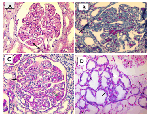
Ten cases were found to have cryoglobulinaemia and the main associated clinical presentations were impaired renal functions (40%) and nephrotic syndrome (40%) (Table 8). The most predominant glomerular disease in cases of cryoglobulinaemia was MPGN (80%), two of which showed intraluminal hyaline thrombi and single case showed crescent formation (Figures 1 and 2).
| Cryoglobulinaemia cases (n=10) | ||
|---|---|---|
| Clinical presentation | Nephrotic syndrome | 4 (40%) |
| Impaired renal function | 4 (40%) | |
| Edema | 1 (10%) | |
| Proteinuria | 1 (10%) | |
| Glomerular disease | MPGN | 8 (80%) |
| MN | 1 (10%) | |
| FSGS | 1 (10%) |
Table 8: The prevalence of Cryoglobulinaemia and its association to clinical presentations and glomerular disease.

Figure 1: A case of Cryoglobulinaemic MPGN showing, Note: A) Mesangial proliferation and matrix expansion with capillary thrombi (black arrow) (H&E x400); B) By Masson trichrome stain the same case shows double contour appearance (black arrow) with intracapillary Cryoglobulinaemic thrombi (yellow arrow) (x400); C) By PAS stain there are endocapillary cellular proliferation, basement membrane thickening with double contour appearance (thick arrows) and intracapillary Cryoglobulinaemic thrombi (thin arrows); D) The same case shows positive hybridization signals for HCV RNA particles (perinuclear purplish blue precipitate) ISH x400.

Figure 2: Regarding ISH interpretation. Note: A) Negative control case (renal biopsy from 1-year-old infant showing no hybridization signals in the perinuclear space) (nuclear fast red x400); B) A negative case showing no hybridization signals, (ISH x400); C) Positive hybridization signals for HCV RNA particles (perinuclear purplish blue precipitate) ISH x400; D) Positive hybridization signals for HCV RNA particles (perinuclear purplish blue precipitate) ISH x1000.
The findings of this work adds insights into different patterns of renal affection associated with HCV infection and the availability of detection of HCV-RNA in the renal tissue using both PCR and ISH techniques.
In this study detection of HCV-RNA in renal tissue was tested by using 2 techniques ISH and RT-PCR. Regarding the ISH technique, positive hybridization signals were detected in the tubular epithelium of 28 cases (56%). Detection of HCV-RNA in renal tissue by using ISH was also reported by previous reports where they found that HCV-RNA was detected by ISH in all involved cases [14,15]. On the other hand, Davda et al., (1993) were unable to detect HCV RNA by ISH in renal biopsies of two cases of HCV associated with membranous nephropathy, probably due to low viral load within the tissue [16]. Thus, the level of viremia and sensitivity of the techniques should be taken into consideration when comparing the results of the present work and previous work in literature. The second technique used in this study was nested RT-PCR technique which detected HCV-RNA in 40 cases (80%). These results confirm the presence of HCV RNA in the kidney tissue of the HCV-infected patients and were in agreement the previously published articles [17-20]. These results help to prove that HCV detection in tissue by using the PCR technique is more accurate and reliable than ISH technique. The difference in the sensitivity of the techniques of ISH may affect the results.
According to clinical and laboratory presentations, the most common clinical presentation in this study was impaired renal function (50%). This came in agreement with the results of Mesquita et al., which reported that a larger proportion of patients with HCV presented with renal failure (58.1%) [21]. In addition, this high prevalence of impaired renal function among the patients was similar to the results noticed by Dalrymple et al., (50%) [22]. The observed association of HCV infection with renal insufficiency might be explained by two potential mechanisms. The first one is direct HCV-related renal injury due to the deposition of immune complexes within renal glomeruli. Another possible mechanism might be HCV-related cirrhosis with subsequent renal impairment [23,24]. In addition, in Egypt, the prevalence of impaired renal function may be due to delay in seeking medical advice.
Histopathological examination of the 50 cases under light microscope revealed variable glomerular, tubular and interstitial affection. The most common glomerular disease was MPGN (58%). These results regarding the predominance of MPGN in cases of HCV-nephropathy were in accordance with previously published reports where all of them reported a prevalence over 50% [21,25-28]. Furthermore, MPGN represented the vast majority of renal affection among diabetic patients with HCV [29]. In contrast, the study of Coccoli et al., the most frequent renal lesion concomitant with HCV was diabetic nephropathy [30]. The second most frequent lesion was MPGN, with or without cryoglobulinaemia [30]. In the present study out of the 29 cases of MPGN, 7 cases showed intraluminal hyaline thrombi representing (24.1%). Nine cases (31%) showed accentuation of lobular architecture and 4 cases (13.8%) showed crescent formation. Likewise, a previous study found that 6 cases showed MPGN with intraluminal hyaline thrombi (22.2%) and 5 cases showed MPGN with accentuation of lobular architecture (18.5%) [25].
Regarding tubulointerstitial affection, 46 cases (92%) showed variable degrees of tubulointerstitial damage. Only 2 cases (4%) were diagnosed as interstitial nephritis. One of them was acute interstitial nephritis and the other was chronic. The remaining 44 cases (88%) showed secondary interstitial affection in the form of chronic interstitial inflammation and interstitial fibrosis, both of which were graded as mild, moderate and marked. Four cases (8%) showed no interstitial involvement. Tubular lesions detected were in the form of tubular atrophy of variable degrees in 58% of cases. These results confirm two facts, the first one is that there is a significant relationship between HCV infection and tubulointerstitial damage. The second one is that this relation may be primary (without glomerular affection) or secondary (with glomerular affection). Kasuno et al., examined renal biopsies from 13 HCV seropositive cases, out of which 10 cases were having tubulointerstitial affection (77%) [14]. Recently, it was discovered that the use of anti-retroviral drugs could significantly reduce the rate of tubulointerstitial inflammation and fibrosis [31].
In the present work serum creatinine level was found to increase in association with the degree of tubular atrophy and interstitial fibrosis. Despite that these relations didn't reach significant values, it’s important the degree of renal affection before starting anti- retroviral therapy significantly affects the response to the drug [32].
In the present work ten cases were found to have cryoglobulinaemia (20%) and the main associated clinical presentations were impaired renal functions (40%) and nephrotic syndrome (40%). The most predominant glomerular disease was MPGN (80%), two cases showed intraluminal hyaline thrombi and one cases showed crescent formation. The presence of cryoglobulinaemia in association with HCV infection is a fact that was established by several studies [33-35]. The predominance of renal affection in the form of MPGN in the present work was also reported by Garini et al. [27]. However, in their work there was a higher incidence (100% of cases), but this can be explained by limited number of cases included in their work (only 4 cases) [27]. Also, vascular affection was reported in 17 cases (34%) in the form of thickening of blood vessels (7 cases <50 years old) and 2 cases (4%) showed vascular amyloidosis. Amyloidosis may be due to HCV stimulation of monoclonal B-cell proliferation through chromosomal rearrangement, which leads to the production of amyloid proteins [28].
One important strength of this study is that it enhances the understanding of the pathology of renal affection in HCV patients. However, we are limited by the small number of the sample included. Thus, further studies are needed with a larger sample size. In addition, we recommend studying the reason for HCV presence in the kidney cells and detecting whether it is due to viral replication or only homing of circulating virus in the renal tissue.
In conclusion, renal affection represents a major entity of extra- hepatic affection in patients with HCV. Most of renal affection is in the form of MPGN, but other forms of affection may occur and progress to end-stage renal disease. In addition, renal affection is due to viral invasion of renal tissue as proved by the detection of HCV-RNA within the tissues. Moreover, RT-PCR was found to be more sensitive than ISH.
[Crossref] [Google Scholar] [PubMed]
[Crossref] [Google Scholar] [PubMed]
[Crossref] [Google Scholar] [PubMed]
[Crossref] [Google Scholar] [PubMed]
[Crossref] [Google Scholar] [PubMed]
[Crossref] [Google Scholar] [PubMed]
[Crossref] [Google Scholar] [PubMed]
[Crossref] [Google Scholar] [PubMed]
[Crossref] [Google Scholar] [PubMed]
[Crossref] [Google Scholar] [PubMed]
[Crossref] [Google Scholar] [PubMed]
[Crossref] [Google Scholar] [PubMed]
[Crossref] [Google Scholar] [PubMed]
[Crossref] [Google Scholar] [PubMed]
[Crossref] [Google Scholar] [PubMed]
[Crossref] [Google Scholar] [PubMed]
[Crossref] [Google Scholar] [PubMed]
[Crossref] [Google Scholar] [PubMed]
[Crossref] [Google Scholar] [PubMed]
[Crossref] [Google Scholar] [PubMed]
[Crossref] [Google Scholar] [PubMed]
[Crossref] [Google Scholar] [PubMed]
[Crossref] [Google Scholar] [PubMed]
[Crossref] [Google Scholar] [PubMed]
[Crossref] [Google Scholar] [PubMed]
[Crossref] [Google Scholar] [PubMed]
[Crossref] [Google Scholar] [PubMed]
[Crossref] [Google Scholar] [PubMed]
[Crossref] [Google Scholar] [PubMed]
[Crossref] [Google Scholar] [PubMed]
[Crossref] [Google Scholar] [PubMed]
Citation: Elmahdi MH, Rashed AM, Elhamid ASA (2024). Detection of Hepatitis C Viral Particles in Renal Tissue in Patients with HCV Associated Nephropathy: A Clinic Pathological Study. J Clin Trials. S30:001.
Received: 26-Oct-2024, Manuscript No. JCTR-24-34825; Editor assigned: 28-Oct-2024, Pre QC No. JCTR-24-34825 (PQ); Reviewed: 12-Nov-2024, QC No. JCTR-24-34825; Revised: 20-Nov-2024, Manuscript No. JCTR-24-34825 (R); Published: 28-Nov-2024 , DOI: 10.35248/2167-0870.24.S30.001
Copyright: © 2024 Elmahdi MH, et al. This is an open-access article distributed under the terms of the Creative Commons Attribution License, which permits unrestricted use, distribution, and reproduction in any medium, provided the original author and source are credited.