Journal of Agricultural Science and Food Research
Open Access
ISSN: 2593-9173
ISSN: 2593-9173
Research Article - (2018) Volume 9, Issue 4
BBTV was used as an antigen to produce single chain fragment variable (scFv) antibody using phage display technology. Two clones showing highest reading in monoclonal ELISA were selected viz., pBSNMAB5 and pBSNMAB40. Single chain antibody fragments of pBSNMAB5 and pBSNMAB40 were subcloned into pQUANTa body expression vector which enabled to transcriptionally fuse with scFv monoclonal antibody fragment and the gene coding for alkaline phosphatase (PhoA) enzyme. The clones were sequenced with LMB3 forward and pHEN reverse primer and characterized. The scFv genes of both clones were 795 bp long and they found to share similar homology sequence. BLASTn and BLASTx analysis results indicated 85 percent homology with Synthetic construct anti-TNF alpha single chain Fv antibody gene, partial cds and further the BLASTx analysis showed that the 86 per cent homology with circulating B cell antibody heavy chain variable region (Homo sapiens). Upon expression of clone, it has produced fusion protein of ALP along with pBSNMAB5 and pBSNMAB40 monoclonal antibody. The scFv-ALP conjugate has been produced against BBTV protein. Using this antibody conjugate, a detection kit was developed. This Rapidot Immunodiagnostic kit detects the BBTV as low as 0.9 g/ml concentration in both NC membrane and membrane cassette and it was also cross checked with other proteins to evaluate the specificity of mono clones raised against BBTV.
Keywords: Rapidot; Antibodies; Phagemid; Antibody library; Immunoassay and detection
Banana (Musa spp. ) belongs to the family Musaceae and globally important fruit crops. India is the largest producer of banana with total annual production of 28.45 MT. Due to intensification of banana cultivation in the recent years the viral diseases have been causing considerable damage to banana production. Banana productivity is generally reduced by viral diseases; the most deleterious disease which lemmatizes banana production is banana bunchy top nanovirus (BBTV), the causal agent of Banana Bunchy Top Disease (BBTD) [1].
Prokaryotic (E. coli ) expression of plant viral antigen provides several advantages in antigen preparation over the traditional methods and the recombinant antigen has utilized to develop immunodiagnostics of several plant viruses [2]. The resistance to the BBTV can be produced by scFv genes which produce antibodies against the crude protein of antigen through phage display techniques several scFv antibody clones were produced against BBTV. The recombinant single-chain variable fragment (scFv) antibodies offer several advantages over mouse monoclonal antibodies (mAbs) for use in biotechnological applications due to their availability for genetic manipulation, which allows fusion of effectors protein such as fluorescent proteins or toxins. Antibody libraries, with their wide range of antigen-binding specificities, have increasingly been a source of specific recombinant antibody fragments via phage display technology. The monoclonal EL Lowry ISA reveals that the two clone’s pBSNMAB5 and pBSNMAB40 had shown highest affinity for BBTV-CP [3].
The ELISA is a highly specific and sensitive serological techniques introduced for identification of plant viruses [4]. The ELISA technique is based on principle in which the viral antigens are recognized by their specific antibodies (IgG) in association with colorimetric properties. Thus the present investigation has been initiated to detect BBTV by utilizing scFv antibodies in RapiDot Immuno Assays.
Antigen used
Protein isolated from E. coli M15 clone carrying bbtv gene using IPTG induction method.
Antibody used
PBSNMAB5 and pBSNMAB40 scFv antibody fragment from pIT2 phagemid Vector. The plasmid was isolated using the alkaline lysis protocol from the phagemid vector and used for further restriction digestion to obtain scFv gene. In the Tomlinson scFv library, scFv genes were cloned into the phagemid vector between NotI and SfiI restriction sites [5]. Therefore scFv gene can be eluted by digesting the phagemid DNA with NotI and SfiI restriction enzymes and cloned into pQUANTabody expression vector as follows.
Forty four μl of pBSNMAB5, pBSNMAB40 and pQUANTabody vector DNA added into 3 separate tubes. Five μl of 10x Not-I restriction enzyme buffer, 0.5 μl of purified BSA, 1 μl of NotI enzyme were added into each tube separately and mixed. These tubes were incubated at 37°C for 2 h in water bath. Five μl of 3M Sodium acetate and 130 μl of 70% ethanol were added to precipitate the DNA. The tubes were incubated on ice for 1 h and spun at 5,000 rpm for 30 min. The pellet was dissolved in 25 μl sterile T10E0.1. Twenty five μl of Not- I restriction digested pQUANTabody plasmid DNA, monoclones pBSNMAB5 and pBSNMAB40 DNA were taken in three tubes. Three μl of 10x SfiI restriction digestion buffer, 1 μl of SfiI restriction enzyme and sterile distilled water was added to final volume of 30 μl. The tubes were incubated in water bath at 50°C for 2 h. The digested product was then ligated into pQUANTabody vector using 2 μl of 10x T4-DNA ligase buffer and 1 μl of T4-DNA ligase (5 U/μl) enzyme and the ligated mixture was transferred into E. coli DH5α.
Screening of the transformants
Transformed anti-BBTV-ALP clones were designated as pBSNMALP5 and pBSNMALP40 and was confirmed by PCR amplification after 30 cycles at 94.0°C 5.0 min, 94.0°C 1.0 min, 56.0°C 1.0 min, 72.0°C 2.0 min, 72.0°C 10 min using following ALP primers.
PhoA forward GCACTGGCACTCTTACCGTTAC
PhoA reverse CAGTCTGATCACCCGTTAAAC
Protein expression analysis
The transformants were further tested for the presence of scFv gene by expressing the protein in E. coli and using that protein for detecting BBTV coated on the ELISA plate. In order to determine the clone that has full length functional antibody gene, Blue clones were picked and tested for their ability to bind BBTV in an ELISA experiment.
Standardization of immunoassay for BBTV-CP
The extracted protein (BBTV) concentration was estimated [6]. Serial dilution (10-8) of protein was made and 2 μl volumes from each dilution was doted onto PVDF strip. A negative control without antibody was used for comparison. The PVDF strip was then reacted with scFv antibodies which later lead to development of visible color dots. Intensity of color development against different concentration of antibody protein was then recorded.
Cloning of scFv from pBSNMAB5 and pBSNMAB40 into pQUANTabody vector raised against BBTV
The pBSNMAB5 and pBSNMAB40 phagmids were subjected to restriction digestion. In both the cases fragment of approximately 795 bp was released (plate 1). These restricted products along with pQUANTabody vector were ligated, transformed and screened via blue white selection method on specific medium. The initial screening of pBSNMALP40 and pBSNMALP5 clones were done by PCR using phoA forward and phoA reverse primers. The clones were confirmed by restriction digestion with SfiI and NotI enzymes. A fragment of 795 bp was released (Plate 2). scFv gene in pIT2 vector is present in fused form with gIII gene. The antibody protein produced consists of both scFv and gIII. So the scFv fragment when restricted from pIT2 vector and cloned into expression vector like pQUANTabody will produce highly expressed and specific antibody protein. The pQUANTabody vector contains alkaline phosphatase gene which acts as secondary antibody in determining its specificity to antigen in less time (Figure 1 and 2).
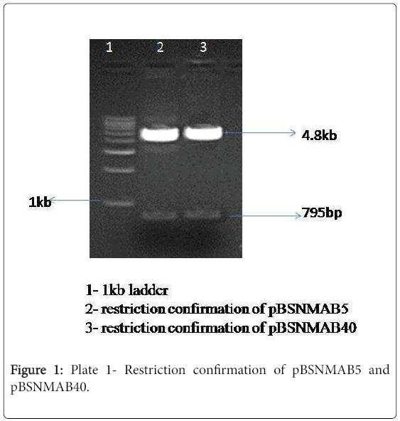
Figure 1: Plate 1- Restriction confirmation of pBSNMAB5 and pBSNMAB40.
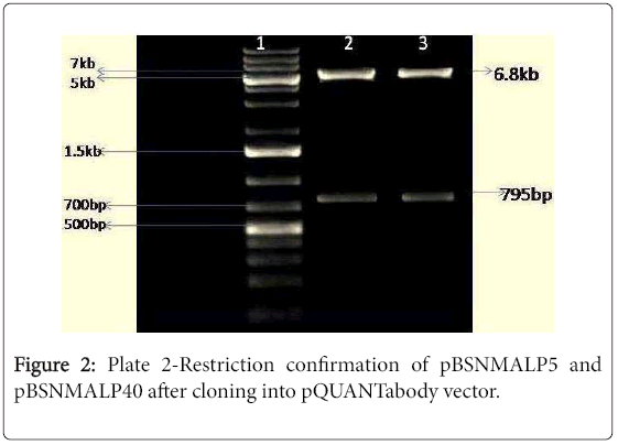
Figure 2: Plate 2-Restriction confirmation of pBSNMALP5 and pBSNMALP40 after cloning into pQUANTabody vector.
In PCR, an amplicon of 990 bp was observed. This confirms the transformants using PCR.
scFv Sequence result
The clones were sequenced and characterized, the clone pBSNMAB5 was of 795 bp long and clone pBSNMAB40 had same length base pairs. The pBSNMAB5 and pBSNMAB40 sequence results of both primers were assembled in Bio Edit software. It was found that the sequences of both clones are hundred percent similar (Figure 3). The assembled sequence was found to have SfiI and NotI restriction sites; the sequence between these two restriction sites is responsible for production of scFv for BBTV coat protein. The homology search through BLASTn programme for similar nucleotide and BLASTx programme for similar amino acid was carried out. Results are depicted in Table 1.
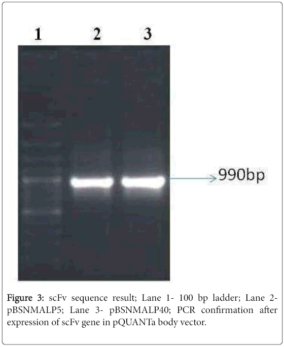
Figure 3: scFv sequence result; Lane 1- 100 bp ladder; Lane 2- pBSNMALP5; Lane 3- pBSNMALP40; PCR confirmation after expression of scFv gene in pQUANTa body vector.
| BLAST Type | Accession No. | E value | Match | % Identity |
|---|---|---|---|---|
| BLASTn | JN887438.1 | 0.00 | Synthetic construct anti-TNF alpha single chain Fv antibody gene, partial cds | 85 |
| BLASTn | DQ375454.1 | 0.00 | Synthetic construct clone D1 anti-TREM-like transcript-1 antibody gene, complete cds | 85 |
| BLASTn | DQ375453.1 | 0.00 | Synthetic construct clone C10 anti-TREM-like transcript-1 antibody gene, complete cds | 85 |
| BLASTp | BAJ52218.1 | 5e -20 | Immunoglobulin gamma heavy chain (Homo sapiens) | 86 |
| BLASTp | ABF83223.1 | 1e -19 | Circulating B cell antibody heavy chain variable region (Homo sapiens) | 86 |
Table 1: Blast and results.
One step detection of BBTV using fusion antibodies
The Direct ELISA was carried out for the above antibody conjugated clones such as pBSNMALP5 and pBSNMALP40. In this experiment, it was noticed that all the crude conjugate protein detected BBTV antigen efficiently (1.35 by pBSNMALP5 and 1.76 by pBSNMALP40). Almost 22 fold increase in absorbance value when compared to control value 0.061 (Figure 4). The antibody conjugated with ALP reduced the cost of the diagnosis and time (Table 2).
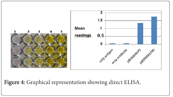
Figure 4: Graphical representation showing direct ELISA.
| ELISA plate wells | Mean readings |
|---|---|
| Only antigen | 0.061 |
| Only antibody | 0.065 |
| pBSNMALP | 51.35 |
| pBSNMALP40 | 1.76 |
Table 2: Direct ELISA readings of scFv-ALP conjugated antibodies.
Standardization and development of immunoassays for Banana Bunchy Top Virus
The low volume of the sample required for Dot Immuno-Binding Assays is an important advantage over ELISA in analyzing samples without dilution. One advantage of the immune-dot blot technique is that the membranes used to bind the proteins also allow their retention of antigenicity and accessibility to antibody [7].
The antigen coated on NC membrane at different concentration was allowed to react with antibody-ALP conjugates. When antigen was spotted on NC membrane it is revealed that at concentration of 0.9 μg/ml, the antibody conjugate was able to detect. In case of ELISA the antibody conjugate was able to detect antigen at the concentration of 3 μg/ml. At this concentration, there was significant color development, the ELISA reading was 0.080 and control value was 0.052 (Table 3).
| ELISA plate well | Mean readings |
|---|---|
| A1B1C1 | 0.052 |
| A2B2C2 | 0.048 |
| A3B3C3- 1 μg | 0.071 |
| A4B4C4- 2 μg | 0.078 |
| A5B5C5- 3 μg | 0.08 |
| A6B6C6- 4 μg | 0.089 |
| A7B7C7- 5 μg | 0.092 |
| A8B8C8- 6 μg | 0.098 |
| A9B9C9- 7 μg | 0.101 |
| A10B10C10- 8 μg | 0.118 |
| A11B11C11- 9 μg | 0.123 |
| A12B12C12- 10 μg | 0.135 |
Table 3: Standardization of antigen concentration with ELISA.
Standardization of antigen concentration using Dot Blotting Immuno Assay
The RapiDot immunoassay have developed using monoclonal antibody (pBSNMAB5 and pBSNMAB40 as capture and pBSNMALP5 and pBSNMALP40 conjugates as detection) proved to be efficient and reliable for BBTV detection. The antibody conjugates able to detect the antigen BBTV in the span of 20-25 minutes. Similar results were obtained against Phytophthora nicotianae spores and against Cry2B [8,9].
The assay does not require any sophisticated equipment nor can highly trained technical staff for carrying out this assay. It was used to validate different infected field samples to detect the presence of BBTV infection (Figure 5).
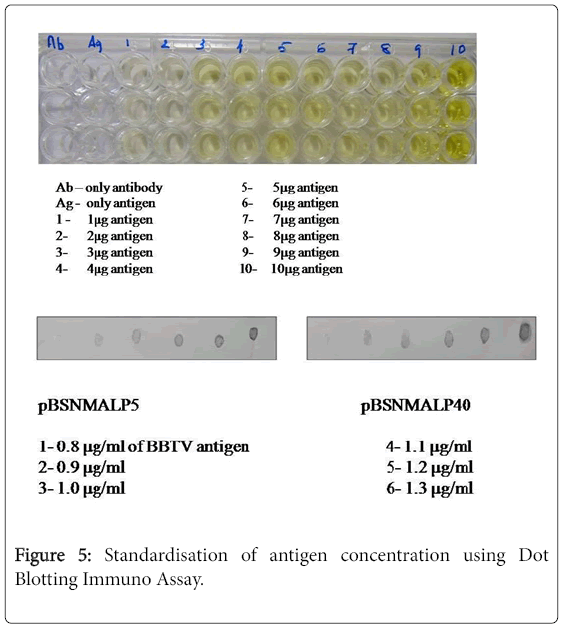
Figure 5: Standardisation of antigen concentration using Dot Blotting Immuno Assay.