Virology & Mycology
Open Access
ISSN: 2161-0517
ISSN: 2161-0517
Research Article - (2022)Volume 11, Issue 6
Human samples of human Coxsakievirus, Adenovirus and COVID-19 virus were collected during October, 1st, 2021 to 8 of December 2021. Human Coxsakievirus, Adenovirus and COVID-19 virus was including (1 month, 1-15 years). The RT-PCR method was detected of all samples, the results showed sixty eight (68%) while thirty two (32%) negative cases, sixty four (64%) while thirty six (36%) negative cases and twenty five (25%) while seventy five (75%) negative cases. The Population groups studied samples subject groups were distribution into (6) groups including (1 month, 1-15, 16-31 and 32-47 and 48-63 and 64-79 and 80-95) year, changed age too gender. Our study also showed that the fourth groups (48-63) years is the largest percentage (41.18) of coxsakievirus, adenovirus (43.75) and COVID-19 virus (44) in compare of aged groups then [1 month, 1-15(4.41%) coxsakievirus, (4.68%) Adenovirus, (0) COVID-19; 16-31 (7.35%) Coxsakievirus, (9.37%) Adenovirus, (12%) COVID-19 virus; 32-47 (22.05%) Coxsakievirus, (17.18%) Adenovirus, (20%) COVID-19 virus; 64-79 (25%) Coxsakievirus, (25%) Adenovirus, (20%) COVID-19 virus; 80-95 (0) for Coxsakievirus and Adenovirus, (4%) COVID-19 virus years. The samples were isolated from the hospitals including (Al-Sadr, AL Hakeem, AL Sajjad), the first study in Iraq to diagnose of human Coxsakievirus, Adenovirus, COVID-19 virus with myocarditis.
Coxsakievirus; Adenovirus; COVID-19 virus; Myocarditis; Real time PCR
Coxsackievirus group B (CVB) belongs to the genus Enterovirus of the Picornaviridae family, which is one of the main pathogens of viral myocarditis and dilated cardiomyopathy. There are six serotypes of CVBs (1–6), which are reported to have a role in the development of chronic diseases such as chronic myocarditis and Type 1 Diabetes Mellitus (T1DM). CVB are the most common cause of infectious myocarditis that can lead to Dilated Cardio Myopathy (DCM) and cardiac failure [1]. The family Picornaviridae comprises a variety of RNA viruses, many of which are important pathogens of human and livestock, affecting the CNS, liver, heart, and the respiratory and gastrointestinal tracts. All members of the family Picornaviridae are nonenveloped, single, positive-stranded RNA viruses with genomes ranging from 7 kb to 10 kb, which consists (from 5’ to 3’) of a 5’ UnTranslated Region (UTR), a single Open-Reading Frame (ORF), a 3’UTR, as well as a poly (A) tail. The single long ORF encodes a polyprotein, which is processed by viral proteases into structural proteins (VP1, VP2, VP3, and VP4) and nonstructural proteins (2A, 2B, 2C, 3A, 3B, 3Cpro, and 3Dpol, and in some genera, also containing Lpro). Structural proteins play a central role in viral capsids assembly, whereas nonstructural proteins are involved in cleavage of viral polyprotein, viral replication, translation, hijacking host-cell machinery, and multiple processes [2]. These viruses are transmitted by the fecal-oral and respiratory routes [3]. Human Aadeno Viruses (HAdVs) are members of the Adenoviridae family. The name derives from the initial isolation of the virus from human adenoids in 1953. Adenoviruses are medium-sized (70e100 nm), nonenveloped viruses with an icosahedral nucleocapsid containing a double-stranded linear DNA genome 34e36 kbp length. The icosahedral shell is composed primarily of 240 capsomeres of hexon trimers, 12 pentameric penton capsomeres at each vertex of the icosahedron, and 12 fibers extending from the pentons. The hexon has been established to carry the antigen specificity markers a and ε with group, subgroup and type-specific immunogenicity and neutralization. The penton base carries b epitope and reacts as a minor group-specific antigen. It has been associated with cellular toxicity and interacts with the inner surface of endosomes during disruption of internalized vesicles. The fiber contains a major antigen, g, and is responsible for type specificity, cell attachment, and hemagglutination. Because of their important roles in cell entry and establishment of host infection, these structural proteins are crucial in the pathogenesis of HAdV infections [4]. Transmission occurs from an infected person to other individuals via respiratory routes, fecal-oral contamination, and/or direct contact. Respiratory transmission via a cough or a sneeze is the most common mode of transmission. Fecal-oral transmission occurs through contaminated food or water, and transmission via water can occur in public swimming pools due to ineffective chlorine treatment [5]. The family Picornaviridae comprises a variety of RNA viruses, many of which are important pathogens of human and livestock, affecting the CNS, liver, heart, and the respiratory and gastrointestinal tracts. All members of the family Picornaviridae are nonenveloped, single, positive-stranded RNA viruses with genomes ranging from 7 kb to 10 kb, which consists (from 5’ to 3’) of a 5’ UnTranslated Region (UTR), a single Open-Reading Frame (ORF), a 3’UTR, as well as a poly (A) tail. The single long ORF encodes a polyprotein, which is processed by viral proteases into structural proteins (VP1, VP2, VP3, and VP4) and nonstructural proteins (2A, 2B, 2C, 3A, 3B, 3Cpro, and 3Dpol, and in some genera, also containing Lpro). Structural proteins play a central role in viral capsids assembly, whereas nonstructural proteins are involved in cleavage of viral polyprotein, viral replication, translation, hijacking host-cell machinery, and multiple processes [6]. The SARS-CoV-2 Genome Coronaviruses are named for the crown-shaped spikes on their outer surface. The novel coronavirus (SARS-CoV-2) is an enveloped, 29.9 kb-long, positive-sense, single-stranded RNA virus belonging to the β-coronavirus genus. The Open-Reading Frames (ORFs) 1a and 1b represent ∼70% of the complete viral genome and possess several conserved nonstructural protein sequences. A frameshift between these ORFs encodes two polypeptides (1a and 1ab) that are processed by viral proteases to produce non-structural proteins, which are involved in viral replication and suppression of host innate immune defenses [7]. The coronaviruses constitute a group of viruses belonging to the Coronaviridae family. SARS-CoV-2, which belongs to the β-coronavirus genus with MERS-CoV and SARS-CoV, has a round or elliptic and often pleomorphic form, enveloped, and measuring between 60-140 nm in diameter. CoVs are positive-stranded RNA viruses; they contain four major structural proteins on their surface: the membrane protein M, the envelope protein E, and the spicule S. In fact, the projection of the spike on the virus surface that causes the corona (Latin: crown) appearance. Inside the virion, a vacuum separating the inner core from the envelope was observed using cryoelectron microscopy. These cores are formed by the genomic RNA associated with the nucleoprotein N [8]. The new coronavirus is transmitted from person to person through aerosols dispersed in the air and through fomites. Once in contact with the respiratory epithelium, the virus invades the cell and begins its viral replication process. The degree of clinical repercussions varies not only by the direct cytopathic lesion caused by the virus, but also by the systemic inflammatory reaction triggered by the host’s immune system [9-11]. The symptoms for COVID-19 include fever, fatigue and respiratory symptoms like cough, sore throat, dyspnea, anosmia, pneumonia for mild to severe cases. However, the patients develop Acute Respiratory Distress Syndrome (ARDS) which eventually leads to multiple organ failure [12]. Myocarditis is characterized by inflammation of the heart muscle tissue. Clinical manifestations range from minor (isolated chest pain) to life-threatening conditions (acute heart failure, cardiogenic shock, ventricular arrhythmia) [13]. Myocarditis is an inflammatory heart disease associated with an increased risk of dilated cardiomyopathy and heart failure. Typically, myocardial injury is caused by either infectious (e.g., coxsackieviruses and adenoviruses) or non-infectious agents (e.g., autoimmune activations and toxins) [14]. Myocarditis is an inflammatory cardiac disorder induced predominantly by viruses but also by other infectious agents including bacteria (such as Borrelia spp.), protozoa (such as Trypanosoma cruzi) and fungi. Myocarditis can also be induced by a wide variety of toxic substances and drugs (such as immune checkpoint inhibitors) and systemic immune-mediated diseases. Importantly, the aetiopathogenesis, induction and course of myocarditis related to different infectious agents vary considerably. The most common viruses associated with inflammatory cardiomyopathy include: primary cardiotropic viruses that can be cleared from the heart, including adenoviruses and enteroviruses (such as coxsackie A viruses or coxsackie B viruses, and echoviruses); vasculotropic viruses that are likely to have lifelong persistence, including parvovirus B19 (B19V; from the erythrovirus family); lymphotropic viruses with lifelong persistence that belong to the Herpesviridae family (such as Human Herpes Virus 6 (HHV6), Epstein–Barr virus and human cytomegalovirus); viruses that indirectly trigger myocarditis by activating the immune system including Human Immunodeficiency Virus (HIV), Hepatitis C Virus (HCV), influenza A virus and influenza B virus; and viruses from the Coronaviridae family, including Middle East Respiratory Syndrome Coronavirus (MERS-CoV), Severe Acute Respiratory Syndrome Coronavirus (SARS-CoV) and SARS-CoV-2, which have Angiotensin Converting Enzyme 2 (ACE2) tropism and can potentially mediate direct cardiac injury. These coronaviruses are also suggested to indirectly trigger myocarditis, in a similar manner to influenza A and B viruses, via cytokine-mediated cardiotoxicity or by triggering an autoimmune response against components of the heart. The exact pathological mechanisms underlying SARS-CoV-2 associated heart disease are so far unknown and require in-depth investigation of EMB and autopsy samples from affected patients [15]. The disease, which can affect individuals of all ages, although it is more frequent in young people, has several clinical manifestations:
• Asymptomatic forms.
• Acute forms, which resolve in about 50% of cases within 2-4 weeks: patient develops dyspnea or orthopnea, palpitations, efort intolerance/malaise, heart failure, chest pain with or without cardiac troponin I or T release and has unobstructed coronary arteries at coronary angiography. A pleuritic chest pain may be present in case of concomitant pericarditis. Palpitation, syncope or aborted sudden death due to unexplained new-onset atrial or ventricular tachy or bradyarrhythmias can be observed. In the case of viral agents, a respiratory or gastrointestinal syndrome, with or without increased systemic inflammatory markers and fever, may precede (days or weeks) the clinical onset of cardiac signs and symptoms.
• Fulminant forms, presenting with unexplained acute heart failure.
• Chronic forms (about 25% of myocarditis) manifest with persistent cardiac dysfunction and in 12–25% may progress to end-stage inflammatory DCM. In these cases, patients present with symptoms of chronic or acute heart failure; more severe forms meet the indications for heart transplantation [16].
Collect affected specimens of human parvovirus B19
Samples were collected of Coxsakievirus, Adenovirus and COVID-19 virus through a start interval 1 October 2021 up to 8 December 2021. Sixty eight for Coxsakievirus, sixty four for Adenovirus, twenty five for COVID-19 virus positive cases including with infected human patients of age ranged one month up to ninty five years of specimens.
Real time PCR technique
This method was used to diagnose human Coxsakievirus, Adenovirus, COVID-19 virus via (this primer was designed based on the NCBI which is about mixed gen in the same primer). Viral DNA was extracted by using Viral Nucleic Acid Extraction Kit (gSYNC TM DNA extraction kit) (Geneaid, Lot No.FA30411-GS, USA) and for RNA (Cat.Lot.0234845744001, promega, USA, Addbio, Lot 2001A, Korea). This technique was performed in Alamin center for advanced research and biotechnology by using (Analytik Jena\Qtower3G) device.
Genetic analytic method for diagnosis of parvovirus B 19 through RT- PCR
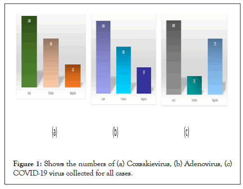
Only (68) cases were positive while (32) negative as in Coxsakievirus, while in Adenovirus only (63) cases were positive while (37) negative, and in COVID-19 virus only (25) cases were positive while (75) negative in Figures 1a-1c. The Population groups studied samples subject groups were distribution into (6) groups including (1 month, 1-15, 16-31, 32-47, 48-63, 64-79 and 80-95) year (Table 1). The age group (48-63 years) compared to other totals in terms of age in Figures 2a-2c.

Figure 1: Shows the numbers of (a) Coxsakievirus, (b) Adenovirus, (c) COVID-19 virus collected for all cases.
| Oligo\Name | Sequence | Bases | PCR product size |
|---|---|---|---|
| Coxsakievirus-F sequence | 5-TAATGCAGTGGCTGCTTGTC -3 | 20 | 234 |
| Coxsakievirus-R sequence | 5-TCGCACCTGAAGGCTTAACT-3 | 20 | - |
| Adenovirus-F sequence | 5-TGCAACATCAGGTAGGGTCA-3 | 20 | 215 |
| Adenovirus-R sequence | 5-TCGCACCTGAAGGCTTAACT-3 | 20 | - |
| Covid19- F sequence | 5-GAGGCGGAGGTACAAATTGA-3 | 20 | 138 |
| Covid19-R sequence | 5-GTGGTAGCCCTTTCCACAAA-3 | 20 | - |
Table 1: Gens of human Coxsakievirus, Adenovirus, COVID-19 virus depended to NCBI.

Figure 2: The figure shows the method for diagnosing of human (a) Coxsakievirus, (b) Adenovirus, (c) COVID-19 virus through real time-PCR.
Diagnosis of human myocarditis the study is considered in Najaf Governorate and at the level of Iraq as well by designing primers depending on the Location "NCBI" by RTPCR technicality which resembled with Arina et al., [17] diagnosis of Coxsakievirus by RT-PCR that corresponds to our study. Sabine and Karin [18] diagnosis of Adenovirus by RT-PCR that also corresponds to our study. Rocı´o et al., [19] diagnosis of COVID-19 virus by RT-PCR that corresponds to our study.
In our study show that females are more susceptible to injury compared to males, and this study was not agreement with study of Mortiz et al., [20]. Our study also showed that the age group (48-63) years is the largest percentage (41.18) of coxsakievirus, adenovirus (42.8) and COVID-19 virus (44), which is estimated, and this very close to a study Ali and Abdel-Dayem [21] that showed that the age group (46-65) years is the most proportional (Table 2).
| Number | Age groups | Coxsakievirus | Adenovirus | COVID-19 virus |
|---|---|---|---|---|
| 1 | 1month,1-15 | 3 | 3 | 0 |
| 2 | 16-31 | 5 | 6 | 3 |
| 3 | 32-47 | 15 | 11 | 5 |
| 4 | 48-63 | 28 | 27 | 11 |
| 5 | 64-79 | 17 | 16 | 5 |
| 6 | 80-95 | 0 | 0 | 1 |
| Total | Female | 32 | 31 | 15 |
| male | 36 | 32 | 10 |
Table 2: Positive numbers cases Coxsakievirus, Adenovirus, COVID-19 virus for both gender.
Benjamin et al., the percentage of coxsakievirus and adenovirus was equal by 17.6% in 9 patients and age was 30-56. Imed et al., Coxsackieviruses B (CV-B) are known as the most common viral cause of human heart infections. Michael et al., [22] Coxsackieviruses B and Adenovirus most common virus causes myocarditis.
Our study three of patiens were children, one male (2) years, two female (10) and (13) years have Adenovirus infection. Forod et al., [23] was conducted on 19 patients with suspected MCI. There were 11 (57.9%) males and 8 (42.1%) females. The ages of the patients ranged from 1 day to 9 years with adenovirus (Table 3).
| Number | Age groups | Coxsakievirus | Adenovirus | COVID-19 virus |
|---|---|---|---|---|
| 1 | 1month,1-15 | 3 | 3 | 0 |
| 2 | 16-31 | 5 | 6 | 3 |
| 3 | 32-47 | 15 | 11 | 5 |
| 4 | 48-63 | 28 | 27 | 11 |
| 5 | 64-79 | 17 | 16 | 5 |
| 6 | 80-95 | 0 | 0 | 1 |
| Total | 68 | 63 | 25 | |
Table 3: The distribution of patients according to age groups.
The study Alameedy showed that the number of cases of Coronavirus infection (110) case, Influenza virus (90) case, Parainfluenza virus (65) case, Metapneumovirus (108) case and Rhinovirus (95) case for the period from 4-4-2020 up to 26-26 7-2021 all viruses were diagnosed through qReal time PCR technique by designing primers according to the location NCBI Regarding the sequences test, the results showed the percentage of similarity with the studied strains at a rate ranging between (99.92-78%). While of Alameedy showed the group of 16-30 years old, the highest percent of infection (55.04%) in comparison with other aged group followed by the group of 31-45 years old (31.88%); then, aged 5-15 years old (13.04%). The males groups represented the highest number of infection with adenovirus in comparison with females groups represented by 44 males (63.76%).
[Crossref][Google Scholar][PubMed].
Citation: Rasool HD, Alameedy FMM, Hammad DBM (2022) Diagnosis of Human Coxsakievirus, Adenovirus and COVID-19 Virus Association Myocarditis by RT-PCR. Virol Mycol. 11:247.
Received: 03-Oct-2022, Manuscript No. VMID-22-19954; Editor assigned: 06-Oct-2022, Pre QC No. VMID-22-19954 (PQ); Reviewed: 20-Oct-2022, QC No. VMID-22-19954; Revised: 27-Oct-2022, Manuscript No. VMID-22-19954 (R); Published: 03-Nov-2022 , DOI: 10.35248/2161-0517.22.11.247
Copyright: © 2022 Rasool HD, et al. This is an open-access article distributed under the terms of the Creative Commons Attribution License, which permits unrestricted use, distribution, and reproduction in any medium, provided the original author and source are credited.