Chemotherapy: Open Access
Open Access
ISSN: 2167-7700
ISSN: 2167-7700
Research Article - (2022)Volume 10, Issue 5
Based on the integrated fragment-based drug design, synthesis, and in vivo evaluations, a series of novel resveratrol and hantzsch ester (ReHa) derivatives are discovered as autophagic death inducer. Compound ReHa-2 is the most potent inducer with IC50 values as low as 9.9 mM compare with the molecule Nitrendipine (NIP) in autophagic cell death. Compound ReHa-2 lead to autophagic cells death instead of necrosis or apoptosis in human NCI-H460 cells. Mechanistic study uncovers that ReHa-2 is capable of increasing protein LC3-II (a marker of autophagy) and reducing p62 in a time and dose-dependent manner. Furthermore, ReHa-2 can activate MAPKs and Akt signal pathway. In addition, we identified that ReHa-2 triggered more ROS generation compare with NIP in NCI-H460 cells. Noticeably, the cytotoxicity induced by ReHa-2 against NCI-H460 cells can be significantly revised by pretreatment of the cells with the CAT (a specific scavenger of H2O2) and DTT (a sulfhydryl containing nucleophile for quenching ROS), suggesting that the ROS (mainly including H2O2) induced by ReHa-2 is responsible for its cytotoxicity against NCI-H460 cells. Our results suggest that ReHa-2 displays potent activators by inducing autophagic cell death through production of ROS.
Autophagy cell death; Reactive oxygen species; NCI-H460 cells; Phosphorylation
Carcinoma of the lung is one of the largest causes of morbidity and mortality in the world. The most potent defenses against lung cancer are apoptosis. Autophagy has been involved in turnover of proteins, cytoplasmic contents and elimination of damaged organelles. Concretely, the LC3-I in the cytoplasm is mated to phosphatidylethanolamine to form LC3-II and p62 is an ubiquitin-binding protein that transfers metabolites to be degraded [1,2]. However, inappropriate activation of autophagy may lead to cell death [3,4] which is different from apoptosis [5]. The autophagic cell death is also form of programmed cell death and plays an important to target tumor cells.
For the past few decades, various investigations have sought to identify new compounds which are able to induce autophagic cell death without deleterious effects on healthy cells. Among these compounds, Resveratrol (Res) is an active ingredient from our food sources, such as grapes and peanuts, which has long been used in traditional Chinese medicine. Numerous studies have demonstrated that Res can stimulate apoptosis, arrest cell cycle and suppress kinase pathways [6-9] and it can promote cell death in many kinds of tumor cells by triggering both autophagy and apoptosis [10,11]. In addition, the previous literature reported that hantzsch ester derivatives showed a broad of biological activity such as antioxidant, antitumor, antiviral [12-14] and were potent anti-inflammatory agents [15]. However, the effects of hybrid with resveratrol and hantzsch ester derivatives on cells autophagy and apoptosis have not been reported up to now.
Reactive oxygen species (ROS) were closely related with cellular redox and metabolism [16]. Excess level of ROS can cause cellular oxidative stress, such as proteins, DNA and lipids are oxidized and damaged [17]. ROS also plays an important role in autophagic cell death [18]. Over the past few decades, many chemotherapeutic drugs can induce autophagy death. In addition, natural products with diverse chemical skeleton induce the autophagic cells death by promoting the production of ROS [19-23].
In this work, we have synthesized and selected the hybrid with resveratrol and hantzsch ester derivatives (ReHa) as molecules to explore the autophagic cell death in human NCI-H460 cells (Figure 1). Our results suggest that compared with the parent NIP, the meta-rtho trifluoromethyl-substituent hybrid with resveratrol and hantzsch ester derivatives ReHa-2 is capable of increasing protein LC3-II (a marker of autophagy) and reducing p62 in a time and dose-dependent manner. Meanwhile, it can effectively promote ROS (mainly including H2O2), that resulted in killing of NCI-H460 cells which mainly based on ROS-mediated autophagic cell death.
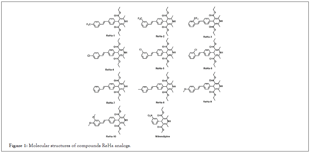
Figure 1: Molecular structures of compounds ReHa analogs.
3-(4,5-Dimethylthiazol-2-yl)-2,5-diphenyltetrazolium bromide (MTT) was purchased from Amresco, Inc. (Solon, OH, USA). Roswell Park Memorial Institute (RPMI)-1640 was obtained from Gibco. Propidium iodide (PI), RNase, 2′, 7′-dichlorofluorescein diacetate (DCFH-DA), dithiothreitol (DTT), catalase (CAT) and chloroquine (CQ) were obtained from Sigma-Aldrich ((St. Louis, MO, USA). 3-methyladenine (3-MA), Z-VAD-FMK (Z-VAD) and Necrostatin-1 (NEC) were from Selleck Chemicals. The primary antibodies against inducible LC3, p62, p-mTOR, p-p38, p-JNK, p-ERK1/2 and p-Akt were supplied from Cell Signaling Technology (Beverly, MA, USA) and antibodies against β-Actin was supplied from Santa Cruz Biotechnology (Santa Cruz, CA, USA). HRP-labeled secondary antibody was a product of Trans Gen Biotech (Beijing, China). All other chemicals were of analytical grade.
Synthesis of compounds ReHa analogs
The compounds ReHa analogs in Figure 1 were synthesized by the Wittig-Horner reaction as described in our previous papers [24,25]. The compounds ReHa analogs in Figure 1 were synthesized by the Wittig-Horner reaction between phosphonates and corresponding appropriate aldehydes (Figure 2). The detailed synthesis process, characterization of all the ReHa analogs, structural determinations of the compounds which based on 1H NMR, 13C NMR spectra and HRMS were included in the supplementary material.
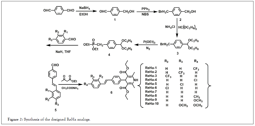
Figure 2: Synthesis of the designed ReHa analogs.
Cell culture
NCI-H460 cells were purchased from Shanghai Institute of Biochemistry and Cell Biology, Chinese Academy of Sciences and culture [26].
Cell viability assay
The cytotoxicity of ReHa analogs for 24 h against NCI-H460 cells was tested by the MTT assay as described previously [26]. In brief, NCI-H460 cells (3 × 10⁴ cells/mL) were treated with the ReHa analogs for 24 h, then replaced with fresh medium containing 0.5 mg/mL MTT and the medium with 100 µL DMSO for 4 h at 37°C. The absorbance was detected at 570 nm on a Bio-Rad M680 microplate reader. When necessary, the cells were pretreated with 3-MA (1 mM), zVAD (50 mM), Nec-1 (5 mM), DTT (400 mM) or CAT (0.5 mg/mL) for 1 h before ReHa-2 was added.
Assay for the ROS levels by a flow cytometry
The related procedure of production of ROS [27]. Briefly, NCI-H460 cells (3 × 10⁴ cells/mL) cells were cultured in 6-well plates and the cells were treated with the ReHa analogs and NIP for 6, 9 or 12 h. Then, the cells were washed with PBS and incubated with 3 μM DCFH-DA for 30 min at 37°C in dark. Finally, the cells were immediately analyzed with a FACSCanto flow cytometer.
Western blot analysis
Western blot analysis was carried out as described previously [28]. NCI-H460 cells (2 × 10⁶ cells/dish) were treated with the ReHa analogs for the indicated time. Then, the cells were lysed in a RIPA buffer with 1% PMSF for 5 min at 4°C and an equal amount of protein (40 μg) for each sample was resolved by 10% SDS-PAGE. Finally, chemiluminescent detection was performed using an ECL Western blot detection kit and Image Quant chemiluminescence system.
Statistical analysis
All experiments were repeated independently at least three times. Results are expressed as the mean values ± Standard Deviation (SD) and analyzed with SPSS 10.0 statistical software. Statistical analyses were performed by a one-way ANOVA followed by LSD test. A value of p<0.05 was considered to be statistically significant.
Synthesis of ReHa analogs with different substituents
ReHa analogs with different substituents were prepared by following the synthesis procedure shown in Figure 2. In brief, we used p-hydroxybenzaldehyde as raw material and then it was reduced to alcohol by sodium borohydride. The reactions were gone through bromination, Wittig-Horner reaction between appropriate aldehydes and corresponding phosphonates in the presence of NaH in tetrahydrofuran (for synthetic details and characterization of all the ReHa analogs.
Cytotoxicity of the ReHa analogs
As shown in the Table 1, MTT assay showed that among the ReHa analogs tested, comparison with the ReHa-7 (no substituent hybrid with resveratrol and hantzsch ester), the trifluoromethyl and Cl-substituent were more high toxicity than methoxyl groups and the meta-rtho trifluoromethyl (ReHa-2), Cl (ReHa-5)-substituent exhibited highest cytotoxicity on NCI-H460 cells, suggesting that the electron-withdrawing group were more toxic than electron-donating group. Because the electron pulling activity of trifluoromethyl is stronger than that of Cl-substituent, hence, ReHa-2 is the most cytotoxic among the compounds and its IC50 values was 9.90 μM in NCI-H460 cells. Therefore, we choose ReHa-2 to carry out the subsequent experiment (Figure 3).
| ReHa | IC50 (µM) | Comp. | IC50 (µM) |
|---|---|---|---|
| 1 | 46.1 ± 2.3 | 7 | 27.0 ± 1.8 |
| 2 | 9.9 ± 0.2 | 8 | 29.4 ± 0.7 |
| 3 | 27.6 ± 1.0 | 9 | >80 |
| 4 | 34.7 ± 3.7 | 10 | >80 |
| 5 | 14.4 ± 0.4 | Nitrendipine | 72 ± 4.9 |
| 6 | 27.4 ± 1.1 |
Table 1: Cytotoxicity of ReHa analogs against NCI-H460 cellsa. Note: aValues for IC50 (Concentrations to cause 50% of inhibition of cell viability after 48 hours of treatment) are presented as means ± SD for at least three independent experiments.
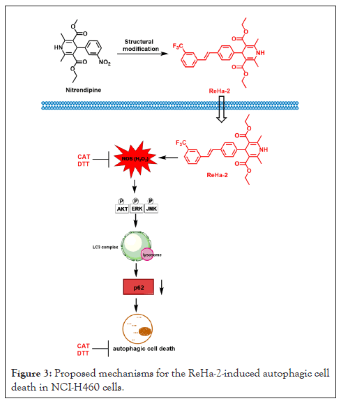
Figure 3: Proposed mechanisms for the ReHa-2-induced autophagic cell death in NCI-H460 cells.
Next, in order to assess the death models induced by ReHa-2 in NCI-H460 cells, some inhibitors on the cytotoxicity were examined. They contain 3-MA (an autophagy inhibitor), Z-VAD (an apoptotic inhibitor) and NEC (a necrotic inhibitor). As shown in Figure 4, the cells were pretreated with 3-MA (1 mM), zVAD (50 mM), Nec-1 (5 mM) for 1 h before ReHa-2 was added. 3-MA partally reversed the cytotoxicity, whereas Z-VAD or NEC further no affected the cytotoxicity. The above results suggested that the cytotoxicity of ReHa-2 depended partally on autophagy process rather than necrosis or apoptosis. In order to confirm the role of ROS in the cytotoxicity induced by ReHa-2, two redox modulators including CAT (a specific scavenger of H2O2) and DTT (a ROS scavenger) were used to conduct the experiment. The cells were pretreated with DTT (400 mM) and CAT (0.5 mg/mL) for 1 h before ReHa-2 was added. Figure 4 exhibited that both CAT and DTT partially reversed the cytotoxicity induced by ReHa-2 in NCI-H460 cells. These results suggested that the cytotoxicity of ReHa-2 were related to ROS generation.
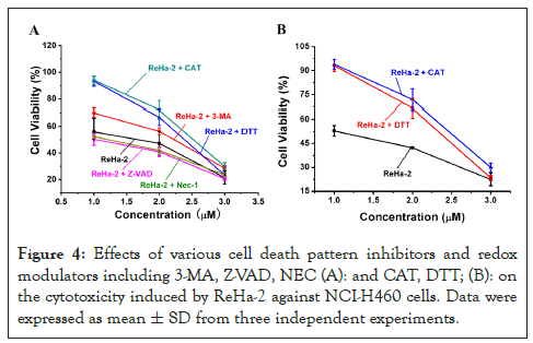
Figure 4: Effects of various cell death pattern inhibitors and redox modulators including 3-MA, Z-VAD, NEC (A): and CAT, DTT; (B): on the cytotoxicity induced by ReHa-2 against NCI-H460 cells. Data were expressed as mean ± SD from three independent experiments.
ReHa-2 induces ROS accumulation in NCI-H460 cells
Considering that ROS generation is essential for the cytotoxicity of ReHa-2 in NCI-H460 cells, we further used DCFH-CA, a widely used ROS fluorescent probe, to assay intracellular ROS levels in the cells. As shown in Figure 5, ReHa-2 induced an obvious intracellular ROS accumulation with 30 mM which increased 2.1-fold relative to the control at 9 h. In contrast, the cells treatment with diverse concentrations of NIP for 9 h induced negligible changes (Figure 5). The ROS accumulation induced by ReHa-2 is fully consistent with the design expectation.
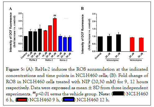
Figure 5: (A): ReHa-2 induces the ROS accumulation at the indicated concentrations and time points in NCI-H460 cells; (B): Fold change of ROS in NCI-H460 cells treated with NIP (20,30 mM) for 9, 12 hours respectively. Data were expressed as mean ± SD from three independent experiments. ##p<0.01 versus the vehicle group.

ReHa-2 induces autophagic cell death in NCI-H460 cells
Since ReHa-2 had no apoptotic and necrotic effect on NCI-H460 cells, we investigated the effects of ReHa-2 on autophagy by using Western blotting. During the formation of autophagosomes, the autophagy-associated protein LC3 in cytoplasm changed from LC3-I form to LC3-II form with autophagosomal membrane specific binding. Conversion of LC3-I to LC3-II is associated with the formation of autophagosomes. During the autophagy process, protein p62 can transport substrates to be degraded to autophagosomes and then be degraded together with autophagosomes under the action of lysosomes [29]. Therefore, changes in p62 content during autophagy can reflect the completion of autophagy flow. Moreover, mTOR activity is regulated by amino acids and glucose levels in mammalian cells. The inhibition of mTOR were induced by rapamycin or starvation, which the conditions induced autophagy [30]. Figure 6 exhibited that ReHa-2 triggered time dependently up-regulation of LC3-II as well as down-regulation of p62, p-mTOR and dose dependently up-regulation of LC3-II as well as down-regulation of p62. From these results we infer that ReHa-2 can induce enrichment of autophagosomes by increasing the formation of autophagosomes rather than blocking its flow. Ultimately, ReHa-2 resulted of autophagic cell death in NCI-H460 cells.
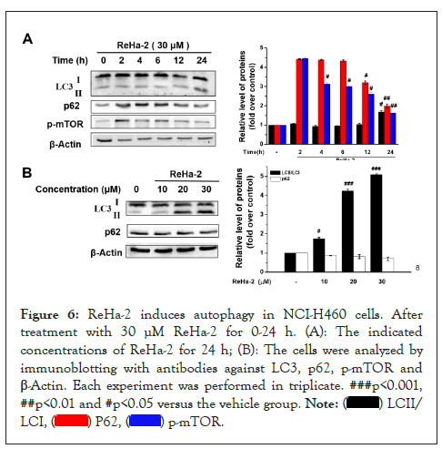
Figure 6: ReHa-2 induces autophagy in NCI-H460 cells. After treatment with 30 μM ReHa-2 for 0-24 h. (A): The indicated concentrations of ReHa-2 for 24 h; (B): The cells were analyzed by immunoblotting with antibodies against LC3, p62, p-mTOR and β-Actin. Each experiment was performed in triplicate. ###p<0.001, ##p<0.01 and #p<0.05 versus the vehicle group. 

ReHa-2 induces dysfunction of autophagy degradationautophagic and further leads to cell death in NCI-H460 cells
As an inhibitor of lysosome, chloroquine can inhibit the fusion of autophagy and lysosome. Thus, it is often used in the study of autophagy and autophagy flow [31]. We investigated the effects of CQ on LC3 expression level induced by ReHa-2. As shown in Figure 6, in this case of chloroquine, the expression levels of LC3-I and LC3-II were significantly enhanced with the increased of ReHa-2 concentration. In compare with parent molecule NIP, ReHa-2 exhibited down-regulation of p-mTOR expression. These results indicated that ReHa-2 activated autophagy by enhancing autophagy formation and then lead to autophagic death in NCI-H460 cells.
ReHa-2 activates MAPKs and Akt
MAPKs and Akt have also been confirmed to participate in the regulation of autophagy [32,33]. For example, in presence of Beclin complex, the apoptosis-related protein Bcl-2 enables the complex inhibits autophagy in vivo. Under starvation conditions, JNK can be activated and phosphorylation of Bcl-2 which weakens its binding ability with Beclin 1, then it activates autophagy [34]. Figure 7 shows that ReHa-2 induced a time-dependent phosphorylation of Akt, ERK and JNK. In brief, ReHa-2 induced the highest protein expression of phosphorylation of Akt, JNK and ERK at 30 and 60 min respectively. These results suggested that MAPKs and Akt have been participated in the ReHa-2-induced autophagic death in NCI-H460 cells.
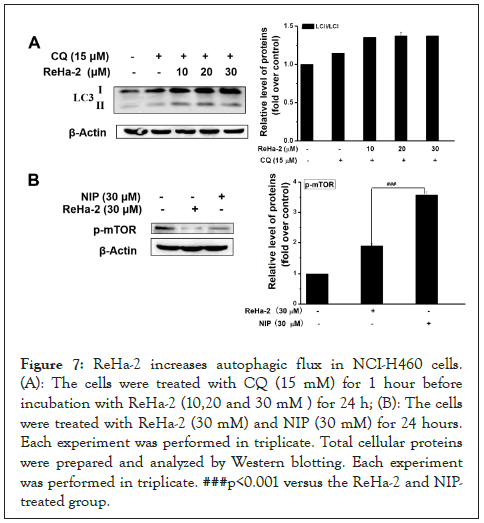
Figure 7: ReHa-2 increases autophagic flux in NCI-H460 cells. (A): The cells were treated with CQ (15 mM) for 1 hour before incubation with ReHa-2 (10,20 and 30 mM ) for 24 h; (B): The cells were treated with ReHa-2 (30 mM) and NIP (30 mM) for 24 hours. Each experiment was performed in triplicate. Total cellular proteins were prepared and analyzed by Western blotting. Each experiment was performed in triplicate. ###p<0.001 versus the ReHa-2 and NIP-treated group.
The cells were treated with indicated concentrations (30 mM) of ReHa-2 for 24 h. Total cellular proteins were prepared and analyzed by Western blotting. Each experiment was performed in triplicate. ##p<0.01 and #p<0.05 versus the vehicle group (Figure 8).
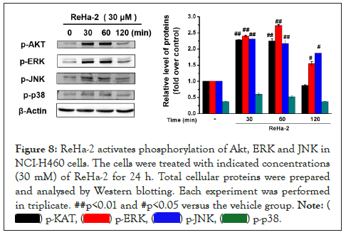
Figure 8: ReHa-2 activates phosphorylation of Akt, ERK and JNK in NCI-H460 cells. The cells were treated with indicated concentrations (30 mM) of ReHa-2 for 24 h. Total cellular proteins were prepared and analysed by Western blotting. Each experiment was performed in triplicate. ##p<0.01 and #p<0.05 versus the vehicle group. 

In summary, this work describes a novel autophagic cell death inducer ReHa-2, which based on hybrid with resveratrol and hantzsch ester derivatives in NCI-H460 cells. Mechanistic study focused on NCI-H460 cells reveals that this molecule compared with the parent NIP could more effectively increase of intracellular ROS (mainly including H2O2), resulting in the increasing protein LC3-II and reducing p62 in a time and dose-dependent manner. Moreover, ReHa-2 activated phosphorylation of Akt, JNK and ERK signal pathway.
In summary, this work describes a novel autophagic cell death inducer ReHa-2, which based on hybrid with resveratrol and hantzsch ester derivatives in NCI-H460 cells. Mechanistic study focused on NCI-H460 cells reveals that this molecule compared with the parent NIP could more effectively increase of intracellular ROS (mainly including H2O2), resulting in the increasing protein LC3-II and reducing p62 in a time and dose-dependent manner. Moreover, ReHa-2 activated phosphorylation of Akt, JNK and ERK signal pathway.
CRediT authorship contribution statement
Yu-Ting Du performed and designed the research, supervised the whole project and wrote the manuscript which was reviewed by all the authors; Hongliang Wang performed the research; San-Hu Zhao and Xiao-Juan Zhang contributed to the data analysis. All authors have read and approved this version of the article. The authors declare that all data were generated in-house and that no paper mill was used.
This work was supported by Scientific and Technological Innovation Programs of Higher Education Institutions in Shanxi (No. 2020L0545), 2021 Innovation and Entrepreneurship Training Program for College Students of Xinzhou Teachers University (56), Xinzhou Teachers University PhD startup fund (No. 00000460), the Fund for Shanxi “1331 Project” Key Subjects Construction and the Top Science and Technology Innovation Teams of Xinzhou Teachers University.
All items in the Submission Checklist have been provided and checked carefully. We declare no conflict of interest, and do not want color reproduction of figures in printed version but want color reproduction in wed version.
[Crossref][Google scholar][PubMed].
[Crossref][Google scholar][PubMed].
[Crossref][Google scholar][PubMed].
[Crossref][Google scholar][PubMed].
[Crossref][Google scholar][PubMed].
[Crossref][Google scholar][PubMed].
[Crossref][Google scholar][PubMed].
[Crossref][Google scholar][PubMed].
[Crossref][Google scholar][PubMed].
[Crossref][Google scholar][PubMed].
[Crossref][Google scholar][PubMed].
[Crossref][Google scholar][PubMed].
[Crossref][Google scholar][PubMed].
[Crossref][Google scholar][PubMed].
[Crossref][Google scholar][PubMed].
[Crossref][Google scholar][PubMed].
[Crossref][Google scholar][PubMed].
[Crossref][Google scholar][PubMed].
[Crossref][Google scholar][PubMed].
[Crossref][Google scholar][PubMed].
[Crossref][Google scholar][PubMed].
[Crossref][Google scholar][PubMed].
[Crossref][Google scholar][PubMed].
[Crossref][Google scholar][PubMed].
[Crossref][Google scholar][PubMed].
[Crossref][Google scholar][PubMed].
[Crossref][Google scholar][PubMed].
[Crossref][Google scholar][PubMed].
[Crossref][Google scholar][PubMed].
[Crossref][Google scholar][PubMed].
[Crossref][Google scholar][PubMed].
[Crossref][Google scholar][PubMed].
[Crossref][Google scholar][PubMed].
Citation: Du YT, Wang HL, Zhao SH, Zhang XJ (2022) Discovery of Novel Hybrid with Resveratrol and Hantzsch Ester Derivatives as Activators Induce Autophagic Cell Death in Tumoral NCI-H460 Cells through Production of ROS. Chemo Open Access. 10:161.
Received: 01-Sep-2022, Manuscript No. CMT-22-17858; Editor assigned: 05-Sep-2022, Pre QC No. CMT-22-17858 (PQ); Reviewed: 19-Sep-2022, QC No. CMT-22-17858; Revised: 27-Sep-2022, Manuscript No. CMT-22-17858 (R); Published: 04-Oct-2022 , DOI: 10.35248/2167-7700.22.10.161
Copyright: © 2022 Du YT, et al. This is an open-access article distributed under the terms of the Creative Commons Attribution License, which permits unrestricted use, distribution, and reproduction in any medium, provided the original author and source are credited.