Journal of Hematology & Thromboembolic Diseases
Open Access
ISSN: 2329-8790
ISSN: 2329-8790
Case Report - (2023)Volume 11, Issue 5
Only 10% of people diagnosed with multiple myeloma are under 50, and 2% of people are under the age of 40. Here we present the case of a 24-year-old female admitted to the hospital for recurrent genital infections, severe anemia and a normal phosphocalcique balance. The myelogram studies revealed normochromic normocytic non-regenerative anemia, as well as a 12% marrow infiltration by abnormal plasma cells. Immunological analysis by immunofixation revealed a homogeneous band in the gamma-globulin zone. After the 5th chemotherapy cycle, the 6-month follow-up assessment showed complete remission.
Multiple myeloma; Hemolytic anemia; Immunofixation; Young adult
Multiple Myeloma (MM) is a malignant hemopathy characterized by a malignant proliferation of plasma cells that produce Immunoglobulin (Ig) frequently of the IgG type, and rarely of the IgA type or lambda or kappa light chains [1]. It is the second most common blood cancer after non-Hodgkin's lymphoma, accounting for approximately 1% of all cancers and 2% of cancer mortality [2]. The average age of onset is 65-70 years, but it can occur in younger individuals, with 1-3% of cases occurring in patients younger than 40 years [3,4]. We present here a case of multiple myeloma in a 24-year-old female patient referred to the immunology department of the University Hospital of Annaba for investigation of severe anemia probably of autoimmune origin. This particular case underlines that MM can affect young subjects and that it can be the cause of severe anemia [5].
Patient information
The patient had no significant medical history and no family history of malignancies. She also had no known genetic history. The patient also reports several cases of neoplasia and chronic renal failure in her family. She reported being under pressure and stress from her family and their demands. Six months ago, she began experiencing generalized bone pain, fatigue, weight loss, and weakness. No intervention was performed.
This is a 24-year-old female patient originating from Eastern Algeria, single, unemployed, referred to the Immunology Department of CHU Annaba (Algeria) for evaluation of a severe anemia evolving since 6 months. The patient reports recurrent episodes of tonsillitis, recurrent genital infections, erosive Helicobacter pylori gastritis, lactose intolerance, and an episode of COVID-19 in February 2022 (moderated form). The patient also reports multiple cases of neoplasia and chronic renal failure in the family. The disease history goes back 6 years, marked by the onset of hair and eyelash loss, severe asthenia, generalized abdominal pain, bloating, anorexia, and oral ulcers.
The patient was conscious and oriented in time and space with an appropriate verbal response. Her body temperature was 37.2°C, heart rate was 80 beats per minute, blood pressure was 130/80 mmHg, and respiratory rate was 16 breaths per minute. Clinical examination of the head and neck revealed no significant abnormalities and the mucous membranes were pink and moist. Respiratory and heart sounds were normal and there were no rales or wheezes. The abdomen was slightly tender to palpation in the epigastric region. The liver and spleen were nonpalpable. Neurologic examination revealed normal muscle coordination and strength, and intact sensation. No evidence of neurologic deficit was detected. Examination of the extremities revealed diffuse bone pain with restricted joint motion, particularly in the proximal joints.
Diagnostic assessment and outcomes
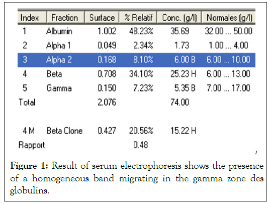
A blood count formula (NFS) was performed, revealing a hemoglobin of 7.3 g/dL, a normochromic normocytic regenerative anemia, a leucopenia of 1850/mm3, a neutropenia of 622/mm3 and a lymphopenia of 820/mm3. The renal function and erythrocyte sedimentation rate tests were normal, as well as the 24-hour proteinuria (0.9 g/L) and the calcium (2.80 mmol/L). The Immunofluorescent tests (IFI) for antinuclear (ANA) and cytoplasmic (ANCA) antibodies were negative. Radiological examinations did not reveal any suspicious bone lesions. The bone marrow analysis showed a normal cellular aspect, and the immunohistochemical study was positive for Syndecan-1 (CD138) in 30% of the cells. The electrophoretic analysis of serum proteins on agarose gel revealed a homogeneous narrow base band in the gamma globulin zone with a decrease in the other immunoglobulin classes (Figure 1). The 24-hour urine electrophoresis with a diuresis of 0.9 L showed no trace of protein with a 24-hour proteinuria of 0.08 mg/L. The results of an immunofixation of serum proteins on agarose gel revealed the presence of a monoclonal Immunoglobulin of IgG Lambda isotype and the search for Bence Jones protein was negative. The serum free Kappa and Lambda light chain dosages were normal and very increased respectively. The free Kappa/free Lambda ratio was 0.15 (norms: 0.26-1.65) (Figure 2). The diagnosis of monoclonal gammopathy was confirmed and the patient was put on a 6 VDT protocol with dexamethasone, thalidomide biphosphates, and bortezomib). After the 5th chemotherapy cycle, the 6-month follow-up assessment showed complete remission with a bone marrow plasmocyte rate of 1%, normal renal balance, calcium and blood count formula (Figure 3).

Figure 1: Result of serum electrophoresis shows the presence of a homogeneous band migrating in the gamma zone des globulins.
Figure 2: Serum immunofixation result shows IgG monoclonal immunoglobulin Lambda with clonal repression of polyclonal immunoglobulins.
Figure 3: Results of electrophoresis and serum immunofixation show the disappearance of monoclonal immunoglobulin after treatment.
Multiple myeloma is a rare disease in young people, characterized by an uncontrolled proliferation of plasma cells and the accumulation of malignant cells in the bone marrow. A retrospective study between 1976 and 1997 revealed that the incidence of monoclonal gammopathies in the adult population was 10.3 per 100,000 inhabitants [6-10]. Our patient presented atypical symptoms, such as profound anemia, recurrent genital infections and leukopenia and neutropenia. Other studies have shown that the frequency of multiple myeloma in patients under 30 years of age is 0.4%. The biomarkers for diagnosis are the presence of at least one defining myeloma event (EMD) and the presence of at least 10% clonal plasma cells in the bone marrow [11-13]. Serum protein electrophoresis and immunofixation should be performed to confirm the diagnosis [14-18]. Our patient had normal calcium, an IgG type monoclonal component and a very high serum free light chain, which was a good prognosis. The stigmas of an autoimmune disease were negative and the treatment aimed to use agents such as bortezomib, dexamethasone, cyclophosphamide and thalidomide [19,20].
In conclusion, multiple myeloma is a rare type of cancer that mostly affects older adults. However, this case report emphasizes that it can also affect young adults. Early diagnosis and timely treatment with chemotherapy can lead to complete remission, as demonstrated in this 24-year-old female patient's case. Healthcare professionals should be aware of the possibility of multiple myeloma in young patients presenting with unexplained symptoms.
All authors have read and approved the final version of the manuscript.
[CrossRef] [Google scholar] [PubMed]
[CrossRef] [Google scholar] [PubMed]
[CrossRef] [Google scholar] [PubMed]
[CrossRef] [Google scholar] [PubMed]
[CrossRef] [Google scholar] [PubMed]
[CrossRef] [Google scholar] [PubMed]
[Google scholar] [PubMed]
[CrossRef] [Google scholar] [PubMed]
[CrossRef] [Google scholar] [PubMed]
[CrossRef] [Google scholar] [PubMed]
[CrossRef] [Google scholar] [PubMed]
Citation: Meriche H, Gouri A, Bennouikes K, Aouadi F, Deridi A, Mahnaoui H, et al (2023) Early Onset Multiple Myeloma in a 24 Year-Old Female Revealed by Severe Anemia: A Case Report. J Hematol Thrombo Dis. 11:547.
Received: 03-Apr-2023, Manuscript No. JHTD-23-23480; Editor assigned: 07-Apr-2023, Pre QC No. JHTD-23-23480 (PQ); Reviewed: 21-Apr-2023, QC No. JHTD-23-23480; Revised: 28-Apr-2023, Manuscript No. JHTD-23-23480 (R); Published: 05-May-2023 , DOI: 10.35248/2329-8790.23.11.547
Copyright: © 2023 Meriche H, et al. This is an open-access article distributed under the terms of the Creative Commons Attribution License, which permits unrestricted use, distribution, and reproduction in any medium, provided the original author and source are credited.