Journal of Clinical and Experimental Ophthalmology
Open Access
ISSN: 2155-9570
ISSN: 2155-9570
Research Article - (2023)Volume 14, Issue 7
Addressing the biochemical and physical changes leading to development of cataract and reversal of this process is research target for ophthalmologists. Currently the only way to treat cataract is by surgical intervention which entails a big economic and logistic burden especially in underdeveloped nations and is not risk free. In this ex vivo experiment, we injected urea solution in a concentration of 96 mmol/L in hard cataractous nuclei removed by cataract surgery. Results showed that urea restored transparency of these opaque nuclei. Electron microscopic examination of those lenses before and after injection showed disorganization of lamellar bodies (hallmark sign of cataract) with restoration of the regular pattern of extracellular spaces within the lens and hence better light transmission. This observation may carry the potential for us to be able to develop a way to prevent and perhaps reverse cataract by simply administering urea eye drops.
Lamellar bodies; Urea; Electron microscope, Surgical intervention; Lamellar bodies
Cataract is the lamellar bodies, urea, electron microscope leading cause of blindness in the world [1]. Approximately 1% of Africa's population are blind and about half the blindness in Africa is due to cataract [2]. With shortage of ophthalmologists and progressive increase in the cataract prevalence, the idea about preventing or delaying development of age-related cataract has emerged.
Lens proteins are classified into urea soluble and urea insoluble proteins [3]. Also, some studies reported that in chronic renal failure, the occurrence of cataract is rare and the pathogenesis is unknown [4-6]. Thus increasing urea in these patient might be responsible for preventing lens opacity. Although many studies (in animals) mentioned to the existence of urea in aqueous humor by concentration equal to glucose and by concentration 7 fold of ascorbate and also by 80% concentration of blood urea, yet no study explained or mentioned the role of urea in the aqueous [7,8]. Multi-lamellar Bodies (MLBs) are lipid coated spheres (1 μm-4 μm in diameter) found with great frequency in the human age-related cataracts compared with human transparent lenses [9].
Aim of the work
To study the effect of urea solution on denatured lens protein of multi lamellar body in senile nuclear cataract.
18 cataractous brown nuclei, the degree of hardness of which was grade III in all of them. All of them extracted through extra capsular cataract extraction and all of them were subjected for electron microscopic examination (E/M). These nuclei were divided into 2 sections, one section used as a control and another section was injected by urea solution another two nuclei were injected at their center by the urea solution for ex vivo demonstration, to show the effect of urea on opacity of the nucleus.
Another two nuclear cataract, one immersed in BSS and other one immersed in urea solution for 12 weeks to see effect of urea on cataractous nucleus without injection.
One clear lens extracted from the anterior chamber (traumatic anterior dislocation) was subjected to E/M examination and used for calibration of success.
In ex vivo demonstration, after injecting the center of brown nucleus by the urea solution, the opaque nucleus which obstructs under print Figure 1 becomes transparent with clear under print (Figures 2-7).
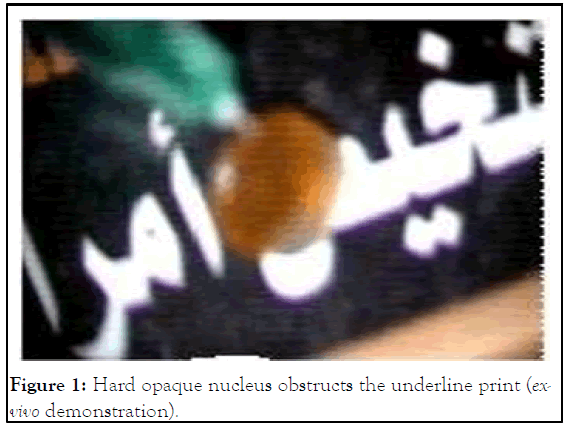
Figure 1: Hard opaque nucleus obstructs the underline print (exvivo demonstration).
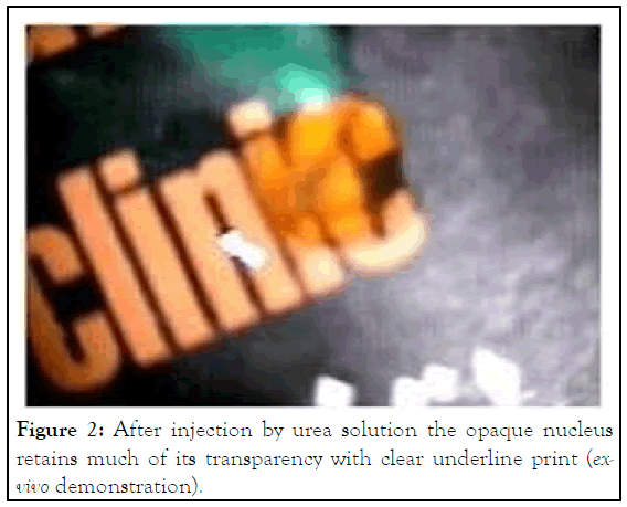
Figure 2: After injection by urea solution the opaque nucleus retains much of its transparency with clear underline print (exvivo demonstration).
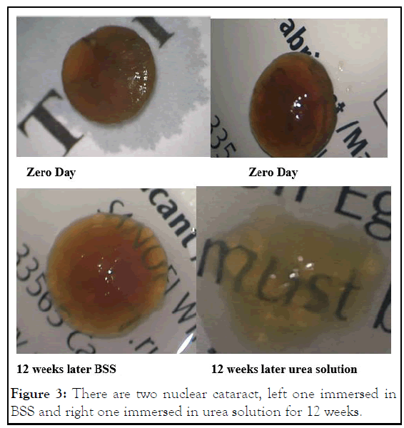
Figure 3: There are two nuclear cataract, left one immersed in BSS and right one immersed in urea solution for 12 weeks.
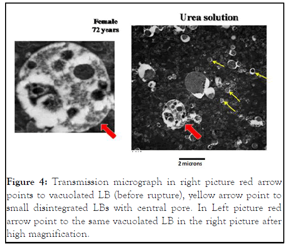
Figure 4: Transmission micrograph in right picture red arrow points to vacuolated LB (before rupture), yellow arrow point to small disintegrated LBs with central pore. In Left picture red arrow point to the same vacuolated LB in the right picture after high magnification.
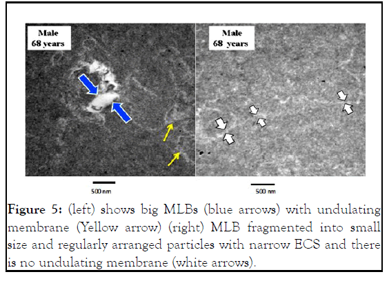
Figure 5: (left) shows big MLBs (blue arrows) with undulating membrane (Yellow arrow) (right) MLB fragmented into small size and regularly arranged particles with narrow ECS and there is no undulating membrane (white arrows).
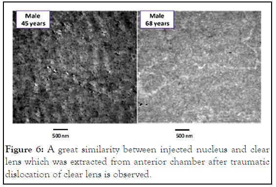
Figure 6: A great similarity between injected nucleus and clear lens which was extracted from anterior chamber after traumatic dislocation of clear lens is observed.
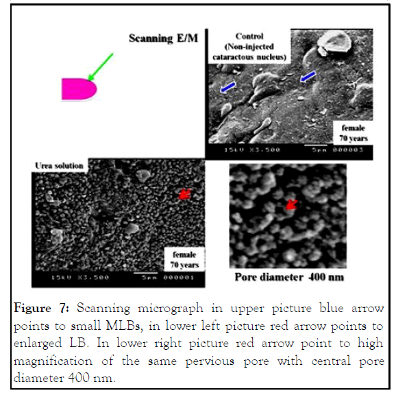
Figure 7: Scanning micrograph in upper picture blue arrow points to small MLBs, in lower left picture red arrow points to enlarged LB. In lower right picture red arrow point to high magnification of the same pervious pore with central pore diameter 400 nm.
The primary event in cataract opacification is the formation of intermolecular disulphide bonds between modified polypeptides as a result of increased oxidative stress [10]. The extensive intermolecular disulphide bonding would result in the formation of large insoluble complexes and these would be stabilized. Under the effect of urea solution with its strong electrostatic reaction and a chaotropic effect the cross linking between side chain of amino acid may be released and this help protein molecule to retain its native structure through protein folding machine (chaperonins) that assist the folding of a wide range of substrate proteins.
Under the effect of urea solution, each MLB increase in size then disintegrates into multiple small size MLBs and this may be due to sudden expansion in limited space. The later changes might render MLBs arranged in regular pattern with central pore diameter greater than wavelength of visible light, that help in restoration of lens transparency. So, extracellular space become smaller and get similar to normal Figure 4 and this will lead to decrease light scattering so, lines become transparent.
In ex-vivo demonstration, nuclear cataracts show great and rapid improvement by urea injection, this improvement proved by both E/M examination and clinical demonstration.
In Figure 3 there is much improvement in right picture than left picture. The nucleus become very soft and transparent with clear under print.
In injection method there is marked improvement at once, the same effect occurs in immersion method but after 12 weeks. In injection method urea solution diffuse to the whole lens, but in immersion method diffusion need long time to become effective due to strong adhesion between lens lamellae.
Many research trials focused on the role of the α-crystallins in preventing protein aggregation and precipitation has been demonstrated in experiments performed in vitro [11]. Also lanosterol study which identifies lanosterol as key molecule in the prevention of lens protein aggregation, and reverses protein aggregation in cataract. All these studies are still in the preclinical stage so, we hope in near future cataract will be a preventable disease [12].
Injection of urea solution, 0.96 mol/L, in human hard cataract nuclei in an ex vivo experiment leads to changes in the electron microscopic characteristics of cataractous lens mainly affecting lamellar bodies, namely via biochemical changes leading to changes in protein structure, which contributes to restoring lens transparency.
Better understanding of cellular biology, biophysics, and physiology of the lens, along with greater insight into the aging process, should provide strategies to prevent or significantly delay cataract formation in most individuals. We discovered that urea solution in very small concentration 96 mmol has very strong effect on multilamellar bodies which cause light scattering and decrease visual acuity in cataract. Under the effect of this solution each MLB increase in size then disintegrates into multiple small size MLBs, and this may be due to sudden expansion in limited space (ECS). MLBs then arranged in regular pattern with central pore diameter greater than wavelength of visible light. This leads to restoration of lens transparency. Each lens particle (MLB) acquires central pore (300 nm-400 nm) as amino acids may retain its original molecular configuration.
Sally A. Sayed, Mohamed G.A. Saleh, Mohamed Anwar, Ghada Hosny revised the manuscript.
Study was approved by the Faculty of Medicine, Medical Ethics Committee: IRB17300280, Assiut University, Egypt.
[Google Scholar] [PubMed]
[PubMed]
[Crossref] [Google Scholar] [PubMed]
[Crossref] [Google Scholar] [PubMed]
[Crossref] [Google Scholar] [PubMed]
[Crossref] [Google Scholar] [PubMed]
[Crossref] [Google Scholar] [PubMed]
[Crossref] [Google Scholar] [PubMed]
Citation: Fahmy HL, Sayed SA, Saleh MGA, Anwar M, Hosny G (2023) Effect of Urea on Lens Protein of Multi lamellar Bodies in Senile Nuclear Cataract. J Clin Exp Ophthalmol. 14:946.
Received: 23-Oct-2022, Manuscript No. JCEO-22-19808; Editor assigned: 25-Oct-2022, Pre QC No. JCEO-22-19808 (PQ); Reviewed: 08-Nov-2022, QC No. JCEO-22-19808; Revised: 30-Jan-2023, Manuscript No. JCEO-22-19808 (R); Published: 06-Feb-2023 , DOI: 10.35248/2155-9570.23.14.946
Copyright: © 2023 Fahmy HL, et al. This is an open-access article distributed under the terms of the Creative Commons Attribution License, which permits unrestricted use, distribution, and reproduction in any medium, provided the original author and source are credited.