Rheumatology: Current Research
Open Access
ISSN: 2161-1149 (Printed)
ISSN: 2161-1149 (Printed)
Research Article - (2022)Volume 12, Issue 2
Background: A conservative management strategy for knee osteoarthritis can include exercise therapy. Exercise therapy is hypothesized to reduce knee adduction. The effects of exercise therapy on knee adduction, along with other physical parameters, were assessed in a systematic review in people with knee osteoarthritis.
Methods: Searches performed on the following electronic databases: MEDLINE, Google Scholar, Cochrane Central, EMBASE, and OpenGrey. Study participants with knee osteoarthritis undergoing structured exercise therapy were randomized controlled trials. For every study, we conducted independent analyses to extract data and analyze the bias risks. We calculated the mean differences and 95% confidence intervals for each outcome.
Findings: In three studies that involved 233 participants, there were no significant differences in knee adduction moments between intervention and control groups. Two of the studies observed improvements in physical function after exercise therapy, and one of them demonstrated significant reductions in pain. All three trials favored the intervention group in terms of muscle strength and torque.
Interpretation: A change in knee adduction time was not associated with the therapeutic benefits of exercise therapy. Exercise therapy for knee osteoarthritis may not be effective if there is no momentary adduction. Dynamic joint loading may result from a shift in neuromuscular control after exercise therapy.
Neuromuscular; Osteoarthritis; Knee; Adduction; Joint
Exercise Therapy (ET); Knee; Knee Osteoarthritis (KOA); Knee Adduction Moment (KAM) (KAM); Adduction
The most common clinical presentations include pain, stiffness, and reduced physical ability, resulting in disability and activity limitations. The role of biomechanical variables in the development and progression of KOA has been studied recently [1]. When a person has KOA, the KAM is used more commonly as a replacement for the medial tibiofemoral contact force, reflecting the relative force distribution across the joint. KAM differs considerably between participants despite its close relationship to medial tibiofemoral contact forces. There is currently no link between structural disease and pain severity [2,3]. While several factors may explain the pain reduction and improved function seen in patients with KOA who undergo exercise training programs, exercise regimens like quadriceps and hip abductors and adductors strengthening and neuromuscular training which have been designed to reduce knee joint loading [4]. On the other hand, strengthening hip muscles will correct pelvic imbalances. It is possible for the KAM to increase as a result of the contralateral pelvis dropping and the center of mass shifting from the stance leg. Regardless of the specific ET technique used, the main mission is to restore the correct biomechanics of the lower limbs. Reduced KAM may be one of the reasons for the decrease in pain and impairment. Exercise training has observable clinical benefits, but it is unknown whether it affects the KAM. As a first step, we wanted to determine whether exercise therapy's clinical benefits relate to changes in KAM in people with KOA [5]. As there were so few studies that evaluated the required outcomes, only studies that measured pain scores and physical function were able to confirm ET's effects on KAM in patients with KOA, and a qualitative analysis of ET on these dimensions was conducted.
Protocol
A preferred reporting method for systematic reviews and meta- analyses that meets PRISMA standards, as well as the Cochrane Collaboration, are used to report systematic reviews.
Sources of data and search technique
We searched EMBASE, MEDLINE, and Cochrane CENTRAL from their creation until November 2020. To find potentially qualifying papers, the search was extended to include systematic reviews and citation monitoring methodologies [6]. The grey literature was found by using Google Scholar and OpenGrey, a specialized database of technical or research reports, conference papers, and official publications.
Eligibility for study
Randomised Controlled Trials (RCTs) were included if they examined physical function, pain, muscular strength and KAM in patients with KOA regardless of other outcomes [7].When diseases or injuries cause pain, physical exercise can be recommended for the relief of symptoms [8]. Exercise training is any type of training regardless of intensity, volume, or type of exercise (for example- Exercises that improve motor control and strength, like high-load and low-load strengthening exercises).We excluded a study that did not examine any of the three outcomes above, a study that only tested a single bout of exercise, and a study that used multi modal therapies (example- foot orthotics, manual therapy, and exercise therapy) [9].
Selection of studies and data extraction
Based on the eligibility criteria as shown in Table 1, we used a common screening checklist for each trial. Studies with titles or abstracts that did not meet the requirements were disqualified [10]. The reviewers discussed their differences regarding study eligibility. To obtain clarifications on studies where there was insufficient information to assess eligibility criteria, we contacted the authors via email. Insufficient information after this contact was excluded from the study. The review team decided to omit publications reporting results from the same population when more than one publication reported the same result [11]. Twice, authors were contacted by email whenever data was required for synthesis or to assess the quality of a study. Missing data estimation was conducted whenever possible. If insufficient data were present, the study was discarded.
| Patient | Intervention | Type of study |
|---|---|---|
| Osteoarthritis of the knee OR Osteoarthritis of the knee OR Osteoarthritis of the knee OR Osteoarthritis of the knee | Train in strength OR lift weights OR strengthen your body WITH weight-bearing exercises OR lift weights. | (randomized controlled trial [PT] OR controlled clinical trial [PT] OR randomized controlled trial [MH] OR random assignment [MH] |
| A lifting-weights strength-training program OR plyometric exercises OR cycling to stretch-shorten or stretching drill. | single-blind method [mh] OR clinical trial [PT] OR clinical trials [mh] OR (“clinical trials”[tw]) | |
| Exercise Therapy (ET) OR physical therapy OR physical training OR aerobic exercise OR stretch-shortening exercise. | The triple (or the tripple) AND the mask (or the blind)) OR the latin square (or the placebos [or the placebos [tw] or the randomness))) | |
| Isometric exercise, physical exercise, or a warm-up exercise. | Detailed design [Mh: experiment] OR comparative research [Mh] OR judgment research OR [Mh] review studies | |
| - | Study crossovers OR control studies (non-human) OR potential studies (non-human)) NOT (animal studies))) |
Table 1: PubMed database literature search strategy.
Evidence level and bias risk assessment
To assess bias risk the Cochrane Collaboration's method for measuring bias risk was used. In total, three types of bias were evaluated in the included studies: high, low, and unclear bias. In this case, a funnel plot was not appropriate due to the small number of studies examined [12]. Evidence is defined as the consistency of findings across several high quality trials or studies; evidence of moderate quality is consistent findings across multiple low-quality trials; evidence of limited quality is the consistency of findings among low-quality studies; and none (no trial evidence is available) [13]. According to the reviewer team, high-quality studies could only be considered if each of the five factors was present. The trials were deemed low quality when other biases were present. Consequently, the "unclear" classification was deemed harmful, and the evidence was lowered [14].
Measures of outcome
Kinematic and kinetic analysis is used to create KAM, and body weight is used as a normalization factor. The studies included in this review were conducted with subjects walking barefoot at their own pace. To assess pain, this study used the pain subscale of WOMAC, and to assess physical function, it used the physical function subscale. There was considerable variation in the numeric scales used for the physical function and pain subscales in trials, but there was no pooling, no modifications were necessary, so the data was reported in raw form [15].
Analysis of data
The biomechanical differences among the workouts included in the study precluded the pooling of data due to clinical heterogeneity [16-19]. The results were therefore analyzed qualitatively by author using original scale as described in Table 2.
| Authors | Knee Adduction Moment (KAM) | Clinical outcomes | Conclusion |
|---|---|---|---|
| Pagani CF, et al. [16] | MD 0.13 (95% CI -0.12 to 0.38) for KAM1 | The pain walking (0-10) scale has a MD of 1.37 (95%CI -2.16 to -0.59) | Increasing strength |
| Inflammatory pain (0-20)2: MD -2.40 (95% CI -3.25 to -1.54) | KAM was not affected by these symptoms. | ||
| The function (0-68)3 is 6.17 (95% CI -9.41 to -2.93) | |||
| Paterson K, et al. [17] | The md1 value for KAM1 is -0.12 (95% CI -0.36 to 0.82) | Inflammatory pain (0-20)2 : MD -0.67 (95% CI -2.03 to 0.69) | The high intensity resistance training did not work |
| MD -2.99 (95% CI -7.77 to 1.79) when function (0-68)3 is considered | KAM should be improved relative to controls. | ||
| MD 0.18 (95% CI -0.06 to 0.42) for KAM1malalignment | -1.6 MD (95 % CI -7.06 to 3.86) for pain (0-20)z maligned | Strengthening quadriceps had no significant effect | |
| Pereira LC, et al. [18] | The KAM1 alignment was -0.02 (95% credible interval -0.38 to 0.34) | MD -13.9 (95% CI -19.24 to -8.55) aligned for pain (0-20)z | Participants with either more or less KAM |
| Function (0-68)3 maligned: MD 2.20 (95% CI 4.39 to 11.07) | Aligned in a neutral or maligned direction | ||
| A function (0-68)3 aligned: MD -6.40 (95% CI -8.50 to 0.41). | |||
Table 2: The original scale is used for all values.
Study description
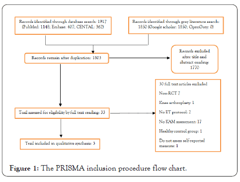
There were 1917 records produced by manual and automated searches from October to November 2020. A search of gray literature on Google Scholar found 1850 citations, but none on OpenGrey. These two databases contained no relevant articles, other than duplicates already in the list [20]. The 1803 registers were reviewed for title and abstract, with 1770 being rejected. A full-text assessment of the remaining 33 was conducted as shown in Figure 1. The qualitative analysis included 233 patients. In all other studies, except for the one that recruited only females, the age ranged from 60.8 to 67.2. The average Body Mass Index ranged from 28.5 to 34.2. Two studies used Kellgren-Lawrence classifications, while the third used Modified Outterbridge classifications.

Figure 1: The PRISMA inclusion procedure flow chart.
Throughout the included trials, training protocols varied. In a 12- week treatment, patients used ankle cuff weights and elastic bands five times/week to develop hip adductor and abductor muscles [21]. To achieve the goals of the study, patients were advised to carry out home workouts as well as directed to visit a physiotherapy clinic seven times to get instructions and measure their load progression.
In the exercise program, exercise physiologists focused on knee extension, hip adduction, hip abduction, leg press, and ankle flexion strengthening. As a control group, study participants did not undergo any ET procedures and were advised not to undergo additional treatments [22].
Intervention effects
Exercise's effect on KAM: The KAM did not differ between the strengthening and control groups during the 12-week study and 95% Confidence Interval (CI) is between 0.039 and 0.335 Nm/ BW × HT%=0.146. As compared to the control group, KAM in the strengthening group increased by 4.6 percent. No statistically significant differences were found between the strengthening and sham-exercise groups in KAM [23].
Effect of exercise on physical function and pain: Participants in the trial improved in pain and physical function after six months, with no significant differences between groups. Strengthening participants showed significant pain reduction when compared with controls in the neutrally aligned group [24].
Exercise's effects on muscle strength: The trial found that individuals undergoing a hip strengthening program had significantly higher hip joint torques as well as knee extension torques compared to patients in the control group. A similar set of findings were found in the strengthening group in the trial when compared with patients in the sham-exercise group in terms of knee extension strength, knee flexion, plantar flexion, hip adduction, and hip abduction [25]. In the study, both aligned and maligned individuals who participated in a strengthening program significantly increased their quadriceps strength compared to the control group.
Evaluation of bias and evidence
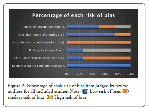
In general, the interventions in these studies did not perform blinding of therapists and patients [26]. ET positively affects pain, physical function, and muscle strength; however, ET does not have a meaningful effect on KAM in percentage as shown in Figure 2.

Figure 2: Percentage of each risk of bias item judged by review
authors for all included studies. 

According to the current systematic study, ET significantly reduces pain, increases athletic ability, and increases muscle strength, but it has minimal impact on KAM. Thus, clinical effectiveness of different Exercise therapy procedures did not result in a change in KAM in patients with KOA [27]. ET has been shown to have good clinical effects in several rigorous systematic studies and clinical guidelines; however, this is the first systematic evaluation to show that, even when ET had therapeutic improvements, its dynamic KAM remained unchanged. Only a few studies are included in this review, so conclusions should be interpreted with caution. On the other hand, the results of the study are supported by research that did not meet the inclusion criteria. After eight weeks of strengthening hip abductors, pain and strength improved, but no significant changes were detected in KAM. Data consistently demonstrate that the biomechanical principles underlying exercise efficacy owing to KAM reduction have no validity in the literature. In contrast, the KAM's balance is justified by the ability to induce abduction moments through quadriceps contraction.
Increasing quadriceps power decreases knee flexion, thus decreasing compression loads on the tibia and femur. Before they can be considered clinically as unloading factors in KOA patients, these pathways need to be studied further. Offloading is achieved by strengthening these muscles by shifting the center of mass away from the stance limb and toward the swing limb due to weak hip abductors on the stance limb. KAM is only affected by such processes when the hip abductors are weak and there is a contralateral hip drop. Only one study addressing this issue concluded that pelvic drop increased with age. A protective intervention in terms of joint loading was not examined in this review. According to any of the included studies, the KAM did not change significantly after ET, but other parameters should be assessed as well, including muscle strength and neuromuscular control, though each of these may contribute to illness progression. KOA is associated with high BMI levels, which were often observed in the studies included in the analysis. KOA is highly associated with a high BMI, according to previous research. In a cross-sectional study, higher BMI was associated with changes in knee biomechanical patterns during locomotion when KOA was moderate. Losing weight has many clinical benefits, including decreased pain and disability, improved walking speed, and improved knee function, but it may also cause joint degradation.
Over the course of a year, a 16-week weight loss program produced excellent clinical benefits despite an increase in joint stress, but no improvement in structural markers of disease development. Future research should investigate other mechanisms that explain ET's therapeutic success. In addition, the few studies we included could affect the generalizability of our findings. Because of clinical variability within ET regimens, data pooling was not possible. Due to the absence of control groups in randomized controlled trials, the available evidence may have been diminished. To test the effect of ET on dynamic knee stress, some specific types of biomechanical changes were required - for example, a greater trunk lean or reduced contralateral pelvic drop-rather than considering the entire KOA population. Researchers will be better able to control possible biases in future studies. Evidence quality was lowered by the absence of selective reporting bias and outcome assessor blinding in some of the included studies. ET did not reduce KAM, but it did improve physical function and pain. Aside from reducing dynamic joint load, there may be other mechanisms by which ET affects KOA.
Citation: Akhtar MW, Mahnoor M, Maqsood Q, Sumrin A, Alam MM, Hassan D (2022) Effects of KAM Adduction for Improvement of Knee Osteoarthritis. Rheumatology (Sunnyvale). 12:297.
Received: 25-Feb-2022, Manuscript No. RCR-22-46384; Editor assigned: 03-Mar-2022, Pre QC No. RCR-22-46384 (PQ); Reviewed: 18-Mar-2022, QC No. RCR-22-46384; Revised: 25-Mar-2022, Manuscript No. RCR-22-46384 (R); Published: 31-Mar-2022 , DOI: 10.35841/2161-1149.22.12.297
Copyright: © 2022 Akhtar MW, et al. This is an open-access article distributed under the terms of the Creative Commons Attribution License, which permits unrestricted use, distribution, and reproduction in any medium, provided the original author and source are credited.