Cell & Developmental Biology
Open Access
ISSN: 2168-9296
ISSN: 2168-9296
Research Article - (2013) Volume 2, Issue 1
After mating, the eggs were deposited, or oviposited, on setae of the pleopods of the female. The newly oviposited eggs were containing all the necessary material for synthetic processes associated with embryogenesis and morphogenesis and all the compounds required for oxidative metabolism and energy production. The fertilized eggs were opaque, greenish, round or oval in shape. The diameter of the egg was approximately 0.45 mm. As the development progresses, the greenish colour changed into light green, brownish-yellow and finally to dull whitish in colour about to hatch. The incubation periods varied from 12-14 days. The process of embryonic development includes nuclear division, cleavage (blastomeres), segmentation, formation of optic vesicle, eye pigment development and larva formation. At third minute after mating the sperm fused with the egg membrane and subsequently the male pronucleus entered the egg’s cytoplasm. The first and second nuclear divisions were completed without any corresponding division of the cell. Third division begun at 8 h and eight nuclei were formed after 9 h. Subsequent divisions of sixteen and thirty two nuclei stage took place at about 1 to 1.30 h interval and segmentation was completed at 18-20h. Embryonic development follows the normal blastula and gastrula stages, ending with the closing of the blastopore.
<Keywords: Embryonic development, M. idella idella, Hatching, Blastula, Gastula
Crustacean embryonic and larval systems offer a unique and valuable tool for furthering our understanding of both developmental processes and physiological regulatory mechanisms. Palaemonid females carry centrolecithal eggs in an external brood pouch during the development time [1]. This peculiarity of the palaemonids allows a systematic tracking of the embryonic development. Muller described the embryogenesis in M. carcinus and M. acanthurus [2,3]. Since there is no detailed study on the embryonic development of M. idella idella, the present study was carried out to know the information on the embryonic development of M. idella idella.
The berried females were kept in the fiber glass tanks (4530×37 cm). Optimum temperature (27-28°C) and dissolved oxygen (5 ppm) was maintained in the brood tank. The berried females were fed with commercial feed once in a day. Everyday the excess feed, excreta and shed out eggs were siphoned out. The development of the eggs was closely observed everyday. Daily colour changes of the eggs during incubation period could be noted. Eggs were sampled aseptically by gently removing a bunch of eggs from the brood pouch using sterilized forceps in random locations and separated with the help of a needle and forceps without damaging the eggs. After each sampling, brooders were given a 1-min prophylactic fungus dip treatment in malachite green (5mg L-1) before being returned to incubation tanks. All the developing embryos were examined with a light microscope to ensure that only viable embryos were sampled and the colour change corresponding to the development and length of the incubation period was noted. The time course of embryonic development, as indicated by the appearance of specific morphological features recorded from spawning time onwards. This includes from fertilization to hatching of first zoea. The gradual changes in the embryonic development and increase in the size of the eggs were recorded to understand the different developmental stages.
Hatching under laboratory condition
Once the first stage zoea inside the egg was fully developed, the larva was ready to come out of the eggshell to start active life. The process of hatching was studied through hand lens and compound microscope with the developing embryo removed from the brood sac. This slow process was accompanied by continuous vibration at the mouth of the larva, and stretching of its rolled body, forcing the egg shell to elongate gradually. Vibration at the mouth became more and more vigorous followed by further stretching of the body. About an hour later the thoracic appendages started to vibrate vigorously but intermittently for about a few minutes with increasing length of pereiopods vibration. This became very vigorous and continuous. The body continued to stretch the rostrum and telson, which was held like a mask covering and protecting the eyes and head, which started pushing outward. Suddenly the eggshell break and the telson thrashed out followed by the head, and with a forceful flex and stretch of the body the hatched zoea larvae started swimming actively in the water column.
During mating, the male placed its spermatophore on the thorax of a mature female, near the opening of the gonopores. As eggs were extruded from the oviduct, they passed across the spermatophore and were fertilized externally. Eggs were deposited, or oviposited, on setae of the pleopods of the female. The newly oviposited eggs were containing all the necessary material for synthetic processes associated with embryogenesis and morphogenesis and all the compounds required for oxidative metabolism and energy production. The eggs contain nutritive reserves in the form of proteinaceous yolk and lipid vesicles scattered throughout the cytoplasm. The fertilized eggs were opaque, greenish, round or oval in shape. The diameter of the egg was approximately 0.45 mm. As the development progresses the greenish colour changed into light green, brownish-yellow and finally to dull whitish in colour about to hatch. At this stage, the developing larvae were observed under microscope. During this period, there was considerable increase in size of the egg in long axis. Fecundity was ranged between 6,158 and 29,272 (60-92 mm total length of females). The incubation periods varied from13-14 days. The process of embryonic development includes nucler division, cleavage (blastomeres), segmentation, formation of optic vesicle, eye pigment development and larva formation (Figure 1).
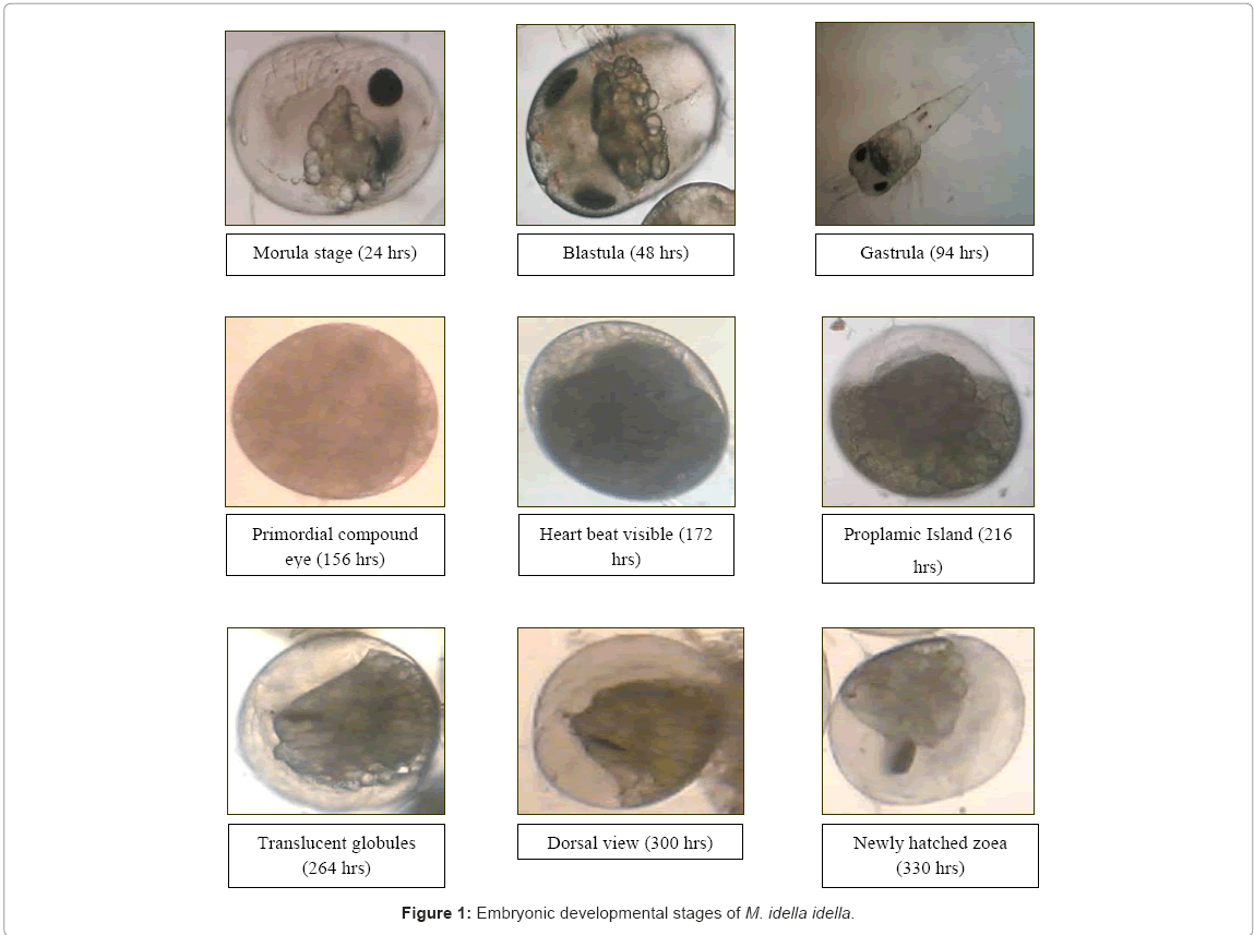
Figure 1: Embryonic developmental stages of M. idella idella.
Newly spawned egg and nuclear division
The attachment of sperm to the egg took place within the first minute of spawning. By the second minute of post-spawning, the egg became spherical and clear membrane was observed very beginning to envelop the egg. A third minute after mating the sperm fused with the egg membrane and subsequently the male pronucleus entered the egg’s cytoplasm. After about 2 hours, the stellate island of protoplasm containing the nucleus was discernable at the center of the egg and became clearly visible at about 1 h. Counting from the time of fertilization, the first nuclear division started at about 4 h and was completed within one hour.
Second nuclear division and segmentation
The second nuclear division started after 6 h and completed at 7:30- 8:00 h. Third division begun at 8 h and eight nuclei were formed after 9 h. Subsequent divisions of sixteen and thirty two nuclei stage took place at about 1 to 1:30 h interval and segmentation was completed at 22-24 h. In M. idella idella, the cleavage was superficial (meroblastic, i.e., a large mass of centrally located yolk confines cleavage to the cytoplasmic rim of the egg). The first and second nuclear divisions were completed without any corresponding division of the cell. Four cleavage furrows appeared when the third nuclear division was almost completed. They started from four subequidistant points on the surface and extend rapidly almost at right angles to each other to form four quadrants, or blostomeres, whose apexes become joined by a median furrow. The fourth nuclear division was holoblastic. Advanced segmentation stages showed distinct hexagonal markings on the surface up to this stage. The colour of the embryo was opaque, greenish, round or oval in shape. The diameter of the egg was approximately 0.47 mm.
Embryonic development follows the normal blastula and gastrula stages, ending with the closing of the blastopore, Increase in cell numbers was observed in the first 48 h and at 94-96 h a clear region at one pole of the embryonic mass was easily discernable (Figure 1). The clear region extended lengthwise forming the trunk of the growing embryo after 120-122h (Figure 1). The diameter of the egg was approximately found to be 0.57 mm translucent, light green in colour and oval in shape. As the clear region developed, the yolky mass lessened (Figure 1).
After 156h, small dark compound eyes appeared on the yolky mass and then at 170h, heartbeat was discernible (Figure 1). The colour was translucent, brownish-yellow in colour. The diameter of the egg was measured as 0.58 mm. In 216 h, the clear region which developed into trunk and caudal portion occupied about 2/3 of the embryo mass and the eye spots were enlarged and oval shaped (Figure 1). At 264 h, the appendages were formed beneath the clear trunk region, the eyes were enlarged and surrounded by striation and the translucent globules at the dorso-caudal portion of the yolky mass clearly exhibited rhythmic contraction. The diameter of the egg was approximately 0.62 mm and transparent, dull whitish in colour (Figure 1). At 300 h, the eyes were dark rounded and striation was observed, the translucent globules became enlarged and occupied most of the dorsal area of the yolky mass. The diameter of the egg was 0.65 mm and transparent dull whitish in colour (Figure 1). The newly hatched 1st zoeae were released at 330 h (Figure 1).
In certain decapod crustaceans, the females incubate their embryos on pleopods (swimmerettes) of the abdomen until hatching. During this period, the embryo’s investment coats (egg coats, egg envelopes) protect it from physical and chemical stresses and maintain the internal milieu. The outer investment coat, due to its immediate exposure to the aquatic environment, is of primary importance in this role. The outer coat has also been associated with the attachment of eggs to the maternal pleopods, selective permeability [4] and osmotic hatching [5], and it may serve as a substratum for aquatic microorganisms [6]. In the present study also, M. idella idella newly extruded eggs contain egg coats which helped the eggs to attach each other and to the pleopods. This helps to protect the eggs from physical and chemical damages.
During oviposition, the female stood upright and the eggs moved into a chamber formed by the pleopods and the lateral epirnera (pleura) on the underside of her abdomen. Eggs were attached to each other and to pleopodal setae by a connecting or adhesive material that formed the outer investment coat. This occurred for both fertilized and unfertilized eggs. At sites of attachment, the adhesive material took the shape of a flattened strand or a twisted stalk [7].
The externally brooding caridean P. macrodactylus exhibited a mechanism of egg attachment that differs from accounts of macruran and brachyuran egg attachment. A substance produced and stored in the female pleopods appeared to be released at moult to coat the external surfaces of the pleopods. Extruding eggs, fertilized or unfertilized, were connected to the pleopodal setae and to each other by the adhesive material, which simultaneously formed the outer investment coat of the eggs. This mechanism is unlike that suggested for Homarus [4] in that cement glands or ducts were not observed in P. macrodactylus, and secretion of adhesive material occurred before, rather than during, oviposition. It also differs from that proposed for Carcinus [8] in that fertilization was not necessary for attachment and the outer layer was formed by material secreted from the female pleopods, not from individual eggs. Attachment in Palaemonetes, described by Burkenroad [9] and Jefferies [10], was probably the same as that described here for P. macrodactylus, but the adhesive material escaped detection because of its close conformity with the external surfaces of the pleopods. In the present study also similar mechanism was observed.
The number of eggs produced by crustaceans varies widely [11]. According to Parson and Tucker [12], fecundity can vary seasonally, annually and between areas. In several crustaceans, there is a linear relationship between the number of eggs per brood and the size of the females. This has also been observed in M. lamarrei [13], the freshwater crayfishes Astacus leptodactylus [14] and P. (Austrocambarus) ilamasi [15]. According to Manush et al. [16], fecundity of M. rosenbergii varies from 40,000 to 60,000 eggs (body weight 100 g). In P. trituberculatus, [17] emphasized that oocyte number increased with increasing female’s body size and predicted estimates ranged between 0.8 and 4.5 million for the carapace width of 130-240 mm. Depending on the size of M. idea, it carries about 40-160 developing eggs [18]. In present study, M. idella idella fecundity were ranged between 6,158 and 29,272 (60 and 92 mm total length).
During the development in M. idella idella, the colour of the eggs changed through greenish opaque, light green, brownish-yellow and dull whitish in colour. At the time of development, the colour of the egg changed through brown to gray as the yolk is used up and the outline of the embryo becomes visible. The eyes and pigment spots appear first followed by the outlines of the abodomen and cephalothorax [19]. The colour change was caused by absorption of the yellow yolk and development of dark pigment in the eyes [20,21]. Extruded eggs of Macrobrachium species are of two colours either green, like in M. acanthurus [22] and M. amazonicum [23] or orange in colour as in M. heterochius, M. ohione [24], M. rosenbergii [25] and M. carcinus [26]. In Macrobrachium spp, eggs with embryos turn either grey or dark brown prior to exclusion [27]. Whereas in M. gangeticum, the colour of eggs is green yellow and become grey corresponding to embryonic development [28]. In M. lamarrei and M. lamarrei lamarrei, the eggs were green in colour [29,30]. Aubson and Patlan [31], Rodriguez (1977) [32] and Rodriguez (1985) [33] classified the eggs into four different developmental stages. However, Ajith Kumar [34] classified eggs into 4 stages based on the colour of the eggs in M. idella idella.
Many workers have divided the crustacean egg stages based on the appearance of distinctive morphological features such as eye, heart beat and appendages formation. However, such morphological characters only begin to appear mid-way during embryonic development. Cellular differentiation starts soon after gastrulation and requires enormous energy expenditure. Subramoniam [35] emphasized the importance of a detailed classification of early development of decapod crustaceans to understand the changes in the metabolic pathways involving interconversion of already stored substrates within the closed system of egg development.
The quantity and distribution of yolk in the eggs of different crustacean species is closely related to cleavage and embryonic development patterns [36,37]. Holoblastic or total cleavage usually occurs in eggs containing a small amount of yolk (oligolecithal eggs), in which the establishment of morphological characteristics occurs relatively fast, resulting in the development of the typical free nauplius larvae with three pairs of appendages [7,38]. This pattern is observed in most branchiopods and maxillopods, and in penaeids of the malacostraca [38]. Crustaceans with yolky eggs (centrolecithal eggs) present meroblastic or partial cleavage. The large amount of yolk triggers a delayed embryonic development that results in further structuring of the embryonized-nauplius (also called egg-nauplius), with the formation of paired appendages, growth of the caudal papilla and organization of appendages in the postnaupliar region [39,40]. This pattern is observed in most malacostracans in which the hatching form is the zoea [36].
The embryonic development in M. idella idella followed the general pattern of embroyogenesis described for other species that have centrolecithal eggs, as P. varians [41], M. carcinus [42], P. pugio [43] and M. acanthurus [44]. The formation of the zoea structures follows the organization of the basic body plan observed in the development of oligolecitic species eggs [45] where first larval phases correspond to the embryonized post-zoea in the meroblastic pattern [46].
The cleavage process observed in stage II indicates that development follows a holoblastic pattern, since cleavage furrows can be seen in the surface of the whole egg, individualizing the blastomeres. However, these cleavage furrows are shallow, and they do not reach the central yolk mass the subsequent organization of the germinal disc seen in stage III, followed by the organization of the embroyonized zoea and post-zoea, are typical of the meroblastic developmental pattern [47]. The recognition of both holoblastic and meroblstic developmental traits during the cleavage stage is common in most decapods species, due to the particular quantity and distribution of the yolk in the eggs [36]. The development of the M. idella idella shows that the initial morphogenesis is quite intense until organization of the embroyonized zoea. The zoea could be visualized due to the large size of the egg, superficial position of the embryo and colour contrast between embryonic cells and yolk mass.
In the present study, the egg size of the M. idella idella increased mainly in long axis during the embryonic development. These changes in egg size were also reported to most of the malacostracan specis, as the brachyuran Eriocheir japanicus [48] and the prawn, M. offersi [49]. According to Odinetz-Collart and Rabelo [50] and Narciso and Morats [51], in crustaceans the egg diameter tends to increase until hatching. Churchill [52] specified that egg diameter was not related in any way to female size and also the egg diameters increased at a relatively steady pace throughout ontogeny. Under constant environmental conditions, the variability in egg size and biomass has been attributed to variation in female size or age [53,54] and genetic factors [55]. The growth of egg size is associated, among other factors, to increase of water content in eggs, as the embryo develops [48]. The eggs of aquatic invertebrates range widely in size. Even within a single taxonomic group such as decapods or amphipods, egg size can vary enormously between species, and also within species. For example, echinoderm eggs vary in diameter from 50 to 1500 μm [56]. In general, of course, species with smaller eggs have higher fecundities than those with larger eggs, and the selective advantages of the different egg size have been discussed extensively in the literature of marine invertebrate reproduction [57-59]. The number of eggs containing embryos during development depends on the size of the mother shrimp, as is know for M. lamarrei [13], M. amazonicum [60], M. idae [61] and M. ohione [24]. Associated with these variations in egg size are differences in the time taken for the embryo to develop and hatch. This can vary from a few days in some tropical species to at least 18 months in some polar isopods [62,63].
In crustacean with yolky eggs, different developmental times are observed from spawning to hatching, such as 40 days for Cherax destructor [64] and 180 days for H. americanus [39]. The incubation of developing embryos by most female decapod crustacean may be one of the reasons for the success of this group. This ensures greater survival against predators and other adverse environmental conditions. Their different incubation times are related to the endogenous factors of development and can also be influenced by exogenous factors like water temperature, as described by Celada et al. [65]. In P. sanguinolentus embryonic development last for 8-11 days [66]. In M. rosenbergii embryonic development last for about 18.5 days [60]. The incubation time is 12-15 days for M. malcolmsonii and comparatively less duration of 12-13 days for in M. gangeticum. Whereas in giant freshwater prawn M. rosenbergii, longer period for incubation and embryonic development was reported at 18- 25 days [29]. However, in the present study the water temperature was controlled, suggesting that the endogenous factors, like egg size and the amount of yolk were decided to the variation of the development time. The embryonic developments of M. idella idella last for about 13-14 days.
A number of species belonging to the genus Macrobrachium are known to migrate from fresh water to brackishwater for breeding purposes [67]. Populations of M. idae inhabiting the rice fields along the west coast of south India are known to migrate into the backwaters during the breeding season. During the embryonic development, the eggs of M. idae increase their salt (ash) content from 4 to about 7% (dry body weight) by absorbing salts from the surrounding water. The gravid females of M. rosenbergii that were gradually exposed to salinities of 8 ppt during the last part of incubation had a higher number of larvae released. In the present study, the M. idella idella brooder were maintained at 4-5 ppt during incubation period [68].
The species of M. olfersi, P. pandaliformis and P. argentinus have similar sized eggs and have presented similar development times. On the other hand, the longest length of development was observed in M. potiuma, whose voluminous eggs allowed for a more prolonged embryogenesis, which results on a development of more complex structures [2]. According to Jalihal et al. [69], the species of Macrobrachium, which have larger eggs, tend to show a smaller fecundity, fewer larval stages, and a reduction of the larval period. These features have also been observed in other palaemonids, such as M. nattereri [69], M. iheringi [70], M. borellii [71] and M. jelskii [72,73].
A similarity of the length of the embryonic periods shows that a specific amount of time to the organization of embryonic features is necessary. The prezoea and zoea periods are faster due the organization of less complex embryonic structures. The post-zoea period is longer because the structures have to be finalized and to acquire functionality before hatching. In M. potiuna, the post-naupliar stage is even longer, because this specie hatches as more complex larvae [42]. In the present study also, prezoea and zoeal periods are faster than the post-zoeal period.