Medical & Surgical Urology
Open Access
ISSN: 2168-9857
ISSN: 2168-9857
Case Report - (2022)Volume 11, Issue 3
Emphysematous pyelonephritis is a rare infectious complication of kidney transplantation. It is defined by the presence of gas in the renal parenchyma, the excretory cavities and the perirenal space due to non-anaerobic gasforming bacteria. Its management is not codified nowadays, and this is due to its rarity and limited number of reported cases. During the past, nephrectomy was the rule. But nowadays, management is based on resuscitation measures and urinary tract’s diversion. In this article, authors present a case of emphysematous pyelonephritis in a 77-year-old patient with a transplanted kidney 29 years previously. His management was conservative with favorable ultimate evolution.
Renal transplant; Complication; Emphysematous pyelonephritis; Transplantectomy
Post-transplant infectious complications are a frequent and serious cause of morbidity and mortality, they occur mainly in the first three months. Urinary tract infections alone account for approximately 40%-50% of all infectious complications [1]. Emphysematous pyelonephritis is a rare infectious complication of kidney transplantation. It is defined by the presence of gas in the renal parenchyma, the excretory cavities and the perirenal space due to non-anaerobic gas-forming bacteria [2]. Previously transplantectomy was the rule. Currently, management consists of maintaining homeostasis by resuscitation measures and urinary drainage. We report a case of emphysematous pyelonephritis in transplanted kidney in a 77-year-old patient, which occurred 29 years after the transplant.
This is a 77-year-old patient followed for Biermer's disease and having had a kidney transplanted in the left iliac fossa 29 years ago for end-stage renal failure of undetermined cause. The baseline post-transplantation creatinine varied around 100 mmol/l. The patient was on iron supplementation, immunosuppressants, corticosteroids and antibiotics, once a week. The patient presented to the emergency department for asthenia associated with fever for 48 hours.
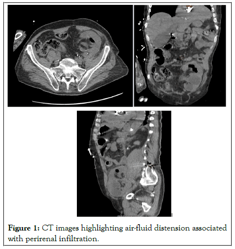
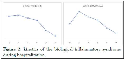
An episode of abdominal pain associated with diarrhoea, dysuria and burning during urination for few days were reported. On admission, a performans status of 2, a temperature of 38.7°C, a heart rate of 94 bpm and a blood pressure of 103/71 mmHg were noted. Biologically, hemoglobin was 8 g/dl, white blood cells were 19000 elements and C reactive protein was 199 and creatinine was 8.7 mg/l. The abdominal and pelvic CT-scan performed in an emergency noted air-fluid distension of the upper calicial group of the renal graft with fat infiltration around the renal graft (Figure 1).

Figure 1: CT images highlighting air-fluid distension associated with perirenal infiltration.
After conditioning, the patient is taken to the operating room for a JJ probe placement. During cystoscopy, the ureteral meatus was impossible to intubate. Decision to puncture the kidney with a Chiba needle by direct approach was token, filiform passage of the contrast product to the bladder and then descent of a JJ probe on a hydrophilic thread. From 24 hours after the diversion, the fever and the kinetic of the inflammatory syndrome began to decrease. Pseudomonas aeruginosa was the isolated germ in the urine culture recovered during the puncture. A probabilistic antibiotic therapy based on meropenem was instituted. The ultimate evolution was favorable marked by the amendment of the fever and clinical signs, reduction of the inflammatory syndrome (Figure 2).

Figure 2: kinetics of the biological inflammatory syndrome during hospitalization.
The pathogenesis of emphysematous pyelonephritis involves the association of three factors: the presence of gas-producing bacteria, a high local glucose concentration, and tissue hypoperfusion and ischemia. The risk factors for occurrence in a transplanted kidney are the same as those reported for native kidneys: diabetes, immunosuppression, urinary obstruction. A particularity is added in transplant patients: the absence of Gerota's fascia. The time to onset of complications in a transplanted kidney is variable, between 2 weeks and 12 years after the transplant [3]. The particularity in our patient is the very late onset of this infectious complication (29 years). The only obstructive risk factor is stenosis of the ureteral meatus preventing the rise of the hydrophilic thread. As reported in the literature, our patient was on immunosuppressants and corticosteroids. Biermer's disease can be considered as a risk factor for the occurrence of this pathology in a transplanted kidney. In renal transplant, the infection’s clinical picture may not be complete. Thus, positive diagnosis is based on imaging. The unprepared X-ray of the urinary tract and ultrasound may show suggestive signs. However, the abdominopelvic computed tomography is the reference examination. The presence of air bubbles is the pathognomonic sign. The CT-scan also makes it possible to specify the size, the localization of the lesions, and the delimitation of the gases, to appreciate the perirenal extension and to guide the therapeutic conduct with follow-up of the patient [4]. In 2010 Al-Geizawi, et al. [5] proposed the classification of pyelonephritis on transplanted kidney,
Stage 1: Gases in the collection system.
Stage 2: Gas replacing <50% of renal parenchyma, with minimal spread to surrounding tissues and rapid control of sepsis.
Stage 3: Gas replacing >50% of renal parenchyma; or widespread spread of infection in the perinephric region; or patient with evidence of multiple organ failure, uncontrolled sepsis, or shock unresponsive to medical management.
Based on the classification proposed by Al-Geizawi, et al. our case cannot be classified: presence of gas just at the level of the collector system and presence of sepsis quickly controlled. The bacteriological spectrum causing emphysematous pyelonephritis is often shared for native kidney and transplanted kidney. E. coli is the most prevalent pathogen germ (62.7% of cases). Other incriminated bacteria are K pneumoniae, Acinetobacter, Proteus mirabilis, Pseudomonas and Anaerobes. Fungal agents can be isolated. Pseudomonas aeruginosa was the isolated germ in our case. Emphysematous pyelonephritis management in kidney transplant recipients remains a subject of controversy. In the past, transplantectomy was the reference treatment, but data from the literature show that currently, several cases have been successfully treated only with conservative treatment. According to the staging system proposed by Al-Geizawi, et al. treatment at stage 1 requires aggressive medical management combined with urethral catheterization and upper urinary tract diversion. At stage 2, they recommend aggressive medical management with the placement of percutaneous drainage, as well as regular follow-up by imaging. In patients with poor course or presented at stage 3, nephrectomy of the transplanted kidney may be warranted. Our attitude was the adoption of intensive resuscitation measures associated with drainage of the urinary tract by descent of a JJ probe after an attempt to climb from JJ doomed to failure.
Emphysematous pyelonephritis on transplanted kidney can occur several years after the transplant. The clinical presentation is atypical. It should be considered in presence of any infectious syndrome in a transplant recipient with burning or any urinary signs. The abdominopelvic CT-scan is the exam of reference. The conservative treatment is preferable and consists of intensive and resuscitation measures with urinary diversion. In case of failure of conservative treatment, a transplantectomy can be discussed.
[Crossref] [Google Scholar] [PubMed]
[Crossref] [Google Scholar] [PubMed]
[Crossref] [Google Scholar] [PubMed]
[Crossref] [Google Scholar] [PubMed]
[Crossref] [Google Scholar] [PubMed]
Citation: Saleh NA, Deogratias N, Mahamat MA, Mostapha A, Amine M, Mohamed D, et al. (2022) Emphysematous Pyelonephritis on Renal Transplant Due to Stenosis of Uretero Vesical Anastomosis: Observation and Initial Intervention (Case Report). Med Surg Urol. 11: 298.
Received: 11-Nov-2022, Manuscript No. MSU-22-20075; Editor assigned: 14-Nov-2022, Pre QC No. MSU-22-20075 (PQ); Reviewed: 30-Nov-2022, QC No. MSU-22-20075; Revised: 06-Dec-2022, Manuscript No. MSU-22-20075 (R); Published: 14-Dec-2022 , DOI: 10.35248/2168-9857.22.11.298
Copyright: © 2022 Saleh NA, et al. This is an open-access article distributed under the terms of the Creative Commons Attribution License, which permits unrestricted use, distribution, and reproduction in any medium, provided the original author and source are credited.