Journal of Clinical and Experimental Ophthalmology
Open Access
ISSN: 2155-9570
ISSN: 2155-9570
Research Article - (2015) Volume 6, Issue 4
Purpose: To assess the impact of laminin and collagen IV on the proliferation of human lens epithelial cells (LECs) and the performance of two hydrophilic and one hydrophilic IOL with a hydrophobic surface coated with erufosine on their properties to inhibit LEC proliferation and migration in a well-established in vitro anterior chamber model of PCO.
Setting: Research Laboratory for Experimental Ophthalmology, Ludwig-Maximilians-University, Munich, Germany.
Design: Experimental study.
Methods: Three IOLs were selected after pre-evaluation for their properties to inhibit LEC proliferation in vitro. All three IOLs were then coated with erufosine while uncoated IOLs of the same type and lot served as controls. Twelve well cell culture inserts were coated with either laminin or collagen IV and erufosine coated or uncoated IOLs were placed on these inserts into the wells. The wells were inserted into 12 well plates and kept under standard cell culture conditions for 6 days. After removal of the IOLs, the cell culture inserts were analyzed for LEC proliferation and migration.
Conclusions: Collagen IV has a stronger impact on LEC proliferation in vitro than laminin has. All tested erufosine coated IOLs were able to significantly decrease the formation of PCO in an in vitro anterior chamber model.
Keywords: Intraocular lenses; Erufosine; Cataract surgery
Cataract surgery is truly a success story of modern medicine. Today, the exchange of the opaque natural lens by an artificial intraocular lens (IOL) is usually facilitated in an outpatient setting and the WHO estimates that the number of cataract surgeries will rise to around 32 million by the year 2020 [1]. Cataract is overall still the most common reason for visual impairment in the world. However, only 10% of these cases are amongst industrialized countries, reflecting the better access to surgical intervention and earlier treatment ages in those regions [2-4]. The demand for cataract surgeries does not only increase due to the ageing population but also because elderly people in developed countries are more active than earlier generations, have higher expectations towards their visual quality and often want to reduce or avoid the need of glasses.
The rising number of implanted premium IOLs reflects the trend to increased refractive considerations prior to cataract surgery today with large numbers of different multifocal, bi- and trifocal and astigmatism neutralizing IOLs on the market.
Cataract surgery is a safe and proven treatment modality today with a relatively low complication rate. The major reason for secondary visual deterioration after successful in-the-bag cataract surgery however, remains posterior capsule opacification (PCO) with an estimated 40% of individuals developing PCO within a few years after surgery [5,6]. Although neodymium:YAG capsulotomy, the treatment of choice in PCO, is well tolerated with good results, the high number of interventions places a significant financial burden on the health care system and complications can occur [7,8]. There have been tremendous efforts to overcome this problem from several approaches. From these, the strongest impact on the prevention of PCO seems to arise from modifications to the IOL design, favoring sharp edged IOLs [9,10].
A simple approach to address this problem and the issue of PCO in general without adding additional surgical steps or surgical devices seems to be a coating of the IOL with a suitable active pharmaceutical component. A major limiting factor so far however, has been the fact that from the myriad of pharmaceutical agents investigated in the context of PCO, only very few show good biocompatibility and promising properties as a PCO inhibiting drug at the same time [11-13]. Studies that have been carried out to evaluate coated IOLs for this purpose include rapamycin and heparin coated as well as surface modified IOLs [14,15]. Rapamycin coated IOLs showed good results in terms of PCO inhibition for the first 6 months in a rabbit model, but increased efforts are needed to coat the IOL due to the necessity of polyglycolic lactic acid (PGLA) and chloroform as a linker molecule [16]. Heparin coated IOLs were well tolerated by the patients but did not show sufficient effects on PCO inhibition [17].
We showed previously that Alkylphosphocholines (APC; namely erufosine) are capable to inhibit major lens epithelial cell properties that are associated with PCO formation such as cell proliferation, adhesion, migration and contraction by attenuating the PI3K/Akt pathway [18-20]. Furthermore are APCs well tolerated by cells of the anterior and posterior part of the eye [21-24]. APCs are composed of a long carbon chain and an alkyl chain giving them amphiphilic that is hydrophilic as well as lipophilic, characteristics. These chemical properties would theoretically result in an accumulation of APCs in the water compartment of hydrophilic IOLs leading to a soaking or loading to them [25]. We recently demonstrated that hydrophilic IOLs, even with a hydrophobic surface, can be coated with APCs and without the need of additional linker molecules [24].
Derived from these data, the present study aimed at investigating whether different commonly used hydrophilic IOLs, coated with APCs are capable of inhibiting PCO formation in an in vitro model of posterior capsule opacification and whether the extracellular matrix (ECM) components laminin and collagen IV, associated with the formation of PCO, would alter the behavior of LEC proliferation with APC coated IOLs.
Human lens epithelial cell culture
The human lens epithelial cell (LEC) line HLE-B3 was obtained from ATCC, Rockville, Maryland, USA, and cultured on tissue culture flasks and Nunc plates (Thermo Fisher Scientific, Inc.) in Eagle’s modified essential medium (Biochrom AG) supplemented with 10% fetal calf serum, 50 IU penicillin/mL, and 50 μg streptomycin/mL at 37°C in an atmosphere of 5% carbon dioxide. HLE-B3 cells are a well-established in vitro model for PCO research [23-25]. The medium was changed every third day. Versene with 2.5% trypsin (Invitrogen-Gibco, Life Technologies Corp.) was used for sub culturing cells on confluence. Cellular growth was observed daily under a Leica phase-contrast microscope.
Alkylphosphocholines
Erufosine was of analytical grade. It was synthesized and provided by Hansjoerg Eibl, PhD, Max-Planck-Institute for Biophysical Chemistry, Goettingen Germany [26]. Erufosine was dissolved in phosphate-buffered saline (PBS) and stored at 4°C. A stock solution was prepared to obtain a final concentration of 10 mM in PBS. This solution was used as the coating solution in further experiments. Equal volumes of PBS without addition of erufosine were used for the control IOL of the same lot.
Intraocular lens coating with alkylphosphocholines
Six different IOLs were initially tested for their properties to lessen the degree of LEC proliferation when they are coated with APC compared to an uncoated IOL of the same type. All six IOLs are commonly used for cataract surgery and commercially available. Out of these, we identified three which were particularly prone for PCO formation that we used for further experiments.
These three hydrophilic foldable IOLs, one with a hydrophobic surface, were selected for coating with erufosine without linker molecules, such as PGLA. The optic diameter of the IOLs tested were 6.0 – 6.15 mm, further IOL properties can be found in Table 1. All IOLs had a square-edged design as indicated by the manufacturer.
| IOL 1 | IOL 2 | IOL 3 | |
|---|---|---|---|
| IOL Design | 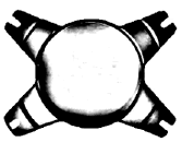 |
 |
 |
| Material | Hydrophilic acrylic | Hydrophilic acrylic | Hydrophilic acrylic, Hydrophobic surface |
| Water Content | 26 % | 26 % | 25 % |
| Total Diameter | 11.0 mm | 10.8 mm | 11.0 mm |
| Optic Diameter | 6.0 mm | 6.15 mm | 6.0 mm |
| Incision Size | 1.8 mm | 2.8 mm | 2.2 mm |
| Haptic Angulation | 10° | 6° | 0° |
Table 1: Design and characteristics of the three tested IOLs.
Using a 12-well plate, 2 IOLs were coated separately in 500 μl APC coating solution for 12 hours at 4°C as described previously [25]. In brief, APC as amphiphilic compounds are able to form a monomolecular film on acrylic IOLs. Because they can permeate through cell membranes and can pass the blood–brain barrier, it can be postulated that they will diffuse into the capsular bag after IOL implantation. Two other IOLs of the same lot were incubated in PBS and served as controls. Consecutively, all IOLs were allowed to dry for 4 hours at 4°C, followed by rehydration with PBS for 1 hour under standard cell-culture conditions. The IOLs were controlled for transparency under a phase-contrast microscope (Axioplan ×100, Carl Zeiss Meditec AG).
Cell proliferation assay with or without laminin and collagen IV
In order to test for the best suited IOL for our further experiments, we tested six different IOLs for their performance on the proliferation of LECs. A tetrazolium dye-reduction assay (MTT, 3-[4,5 dimethylthiazol- 2-yl]-2,5-diphenyltetrazolium bromide) was used to determine the cell proliferation. In this, HLE-B3 cells (500 ml/well at a density of 5 × 10⁴ cells/well) were seeded in 12 well plates to grow for 48 to 72 hours. Uncoated (control) or APC coated IOLs were then placed onto these human LECs for 5 days under standard cell-culture conditions. A cell-culture weight was placed on top of each IOL to ensure proper positioning of the IOL on the cell-culture plate.
For the assessment of laminin or collagen IV on their influence on LEC adhesion and proliferation respectively, the ground of 12 well plates were coated with laminin (10 µg/ml) or collagen IV (50 µg/ml). Five hundred microliters of either component was placed in a well and incubated at room temperature for one hour before the solution was aspirated and the well carefully rinsed with PBS. An APC coated IOL was then placed into the well as described before followed by an MTT test, performed as described by Mosmann with some modifications. After removal of the cell-culture weight, the IOL, and the cell-culture medium, the cells were washed with PBS and the MTT solution was added. The cells were incubated at 37°C for 30 minutes. After 3 washes with PBS, the insoluble formazan crystals were dissolved in dimethyl sulfoxide. The optical density was determined using a microplate reader at 550 nm (Molecular Probes, Life Technologies Corp., NY, USA). Results were expressed as the mean percentage of the control. Experiments were performed in duplicate and repeated at least 4 times (minimum of 8 IOLs per IOL type).
In vitro PCO model for the assessment of cell migration and proliferation
The in vitro PCO model was performed as described by Gotoh et al. [27]. In brief: All three square-edged acrylic IOLs coated with APC as described before, were placed on a cell culture insert coated with 5 μg/cm2 type IV collagen (BD Biosciences, San Jose, CA, USA) in a 12-well culture plate to simulate the formation of PCO in cataract surgery and implantation of an IOL (Figure 1). Analog to the in vivo situation, lens epithelial cells tend to proliferate and migrate beneath the IOL forming PCO after implantation of the artificial lens. In this in vitro model for PCO, a tiny weight (0.85 g) was placed on top of the IOL to assure IOL contact to the underlying membrane. Cells (4 × 10⁴) were placed into each culture well, and the cells were incubated in 700 μl 10% FBS/MEM within the upper chamber and in 1 mL of 10% FBS/MEM in the lower chamber. After 6 days, cells were stained with 0.1% crystal violet and digitally documented with a light microscope and an attached digital camera (Leica GmbH, Wetzlar, Germany). For further evaluation, representative fields underneath the IOL were selected and marked with a square to count cell numbers and to quantify the area covered with LECs using software (Leica GmbH, Wetzlar, Germany). Each experiment was carried out in duplicate (n=2) and repeated 12 times per IOL (24 IOLs per IOL type), summarizing to a total of 72 IOLs investigated.

Figure 1: In order to find IOLs that would display the best results in combination with APC, six different IOLs were selected and evaluated in regard of their ability to inhibit LEC proliferation. Out of these six, two showed a significant decrease of LEC proliferation (IOL 2 and 3; p<0.0001) while a third IOL (IOL 1) showed results that were barely significant (p=0.0532). These three IOLs were therefore chosen for further evaluation.
Assessment of the influence of collagen I and laminin on the proliferation of LEC
Similar to the description of the cell proliferation assay, we investigated the influence of the ECM components collagen I and laminin.
Twelve well plates were coated with laminin or collagen I for 24 h or remained uncoated before HLE-B3 cells (500 ml/well at a density of 5 × 10⁴ cells/well) were seeded in these plates to grow for 48 to 72 hours. APC coated IOLs were then placed onto these human LECs for 5 days under standard cell-culture conditions. A cell-culture weight was placed on top of each IOL to ensure a proper position of the IOL on the cell-culture plate. As described before, an MTT assay was done after the removal of the weight, IOL and cell medium. Experiments were performed in duplicate and repeated at least eight times (minimum of 16 IOLs per IOL type).
Statistical analysis
Results for cell proliferation and the area covered by cells beneath the IOL were expressed as the percentage of the control where the control was set to 100% and the mean was calculated for each experimental group. The analysis of remaining cells beneath the IOL was expressed as the actual number. The influence of ECM components is displayed as the x-fold increase of proliferating cells compared to the control which was arbitrarily set to 1. The error is expressed as the standard error of the mean for all experiments. All differences were tested with a Mann-Whitney U test. For discrimination of two univariate groups an analysis of variance was applied and a post hoc Bonferroni test to determine differences between each tested group was calculated afterwards.
All statistical testing was performed using Graphpad Prism 4 for Macintosh. For all analyses a p value below 0.05 was considered significant with a confidence interval (CI) of 95%.
LEC proliferation
Six IOLs that are commonly used in the operating theater were selected and tested for their properties on LEC proliferation in APC coated or uncoated conditions. We compared APC coated IOLs with an uncoated version of the same IOL as a control. IOLs 4, 5 and 6 did not show evidence to reduce LEC proliferation promising enough to be further evaluated. IOLs 2 and 3 however, displayed very strong and statistically persuading results to be further used for our experiments in the in vitro PCO model and the evaluation of ECM components. IOL 2 in combination with APC reduced the number of proliferated cells by 79.5% compared to the uncoated IOL (p<0.0001) while IOL 3 decreased the amount of proliferated cells by 73.5% (p<0.0001). Although statistically barely not significant (p=0.0532), we also included IOL 1 for further studies. Coated with an APC, it displayed the third strongest impact on the reduction of LEC proliferation out of the six tested IOLs (Figure 1).
Influence of ECM components on LEC proliferation
Our results clearly show, that a difference between uncoated, laminin and collagen I coated inserts exists: All three IOLs have been tested and the amount of proliferated LECs was calculated. In order to simplify the reading of results, all results were normalized to the uncoated insert, which was arbitrarily set to 1. The results are therefore expressed as the fold increase of LEC proliferation compared to the uncoated insert. IOL 1 showed a significant increase in LEC proliferation for both laminin as well as collagen I coated inserts compared to the uncoated insert (uncoated vs laminin: p<0.05; 95% CI -1.843 to -0.05362; uncoated vs collagen I: p<0.001; 95% CI -3.212 to -1.423). We could also appreciate a significant difference between laminin and collagen I. (p<0.01; 95% CI -2.349 to -0.3894). IOL 2 also showed a trend towards stronger proliferation for LECs under the influence of collagen I, no statistically significant difference could be observed between any of the three groups (p>0.05). The difference between collagen I and uncoated inserts for IOL 3 on the other hand was statistically significant (p<0.01; 95% CI -2.191 to -0.3793). No significance could be appreciated for uncoated vs. laminin coated wells (p>0.05) while a significant difference exists between laminin and collagen I (p<0.05; 95% CI -1.926 to -0.1146) in this IOL though.
Influence of APC coated IOLs on collagen I coated Inserts in the PCO Model
Derived from our previous results, collagen I seem to have a stronger influence on the attachment and proliferation of LECs. The membrane that we used for this PCO model was therefore coated with collagen I. Both foldable hydrophilic IOLs as well as the IOL with a hydrophilic core and a hydrophobic surface showed a significant capability of reducing human lens epithelial cell growth compared to the uncoated equivalents which served as controls. IOL 1 was able to reduce the area of cell spreading to 22.3% (p<0.001; 95% CI 16.5-28.2%), IOL 2 reduced the area with covered LECs to 32.4% (p<0.001; 95% CI 24.4-40.3%) and the hybrid IOL 3 reduced the distribution of cells beneath the IOL to 21.6% (p<0.001; 95% CI 16.2-27.03%). Although slight differences in the range of inhibition of cell distribution underneath the IOL between the three tested IOLs can be observed, no statistically significant differences could be appreciated (p>0.05). We also counted and measured the absolute number of cells under the IOLs. They match the results of the correspondent areas covered by LECs: We found a reduction in the number of cells compared to uncoated IOLs down to 14.3% (p<0.001; 95% CI 11-17.7%) for IOL 1, IOL 2 reduced the number to 31.5% (p<0.001; 95% CI 28.3-34.7%) and IOL 3 to 23.3% (p<0.001; 95% CI 19.4-27.3%) compared to the control while all results are shown as the actual number of cells counted. And as for cell area measurements, no significant difference in the number of cell reduction between all three lenses could be appreciated (p>0.05).
Posterior capsule opacification is still an unresolved problem in the tremendously evolving field of refractive lens surgery resulting in secondary visual deterioration and the need of additional interventions.
Our results suggest that the coating of hydrophilic IOLs can be done by bathing an IOL in APC, even if the surface of the IOL is hydrophobic.
In a first approach to identify IOLs that would work well when coated with APC, we tested six different commonly used IOLs for their potential to reduce LEC proliferation (Figure 1). We identified three IOLs that showed promising results and were therefore used for further experiments (Table 1).
The idea of placing an IOL into the capsular bag that carries a pharmaceutical component that would inhibit the formation of PCO is obvious, yet no coated IOLs for this purpose are commercially available.
The sandwich theory that was proposed by Linnola implied a specific role of extracellular matrix (ECM) proteins in the development of PCO [28]. In this, proteins such as fibronectin are believed to act as a biologic glue to connect the capsule with the IOL [29-31]. The firm adhesion of the capsule and the IOL would not allow LECs to migrate into the optical zone and therefore reduce the occurrence of PCO. While fibronectin may therefore be of particular importance to form a barrier with the IOL against LECs, other ECM components such as laminin or collagen IV have also been shown to be expressed in PCO and may act as attractants for LECs. We therefore investigated the influence of laminin and collagen IV in combination with IOLs and APC coated IOLs. Our results show that collagen IV more likely than laminin has a strong effect on the adhesion and proliferation of LECs in a model of LEC proliferation why we chose to use collagen IV for further evaluation in an in vitro model of PCO (Figure 2).

Figure 2: ECM components have been shown to have an important function in the formation of PCO but also in its inhibition. Laminin and collagen IV have therefore been tested on their impact on LEC adhesion and proliferation with all three APC coated IOLs. Both components showed a trend towards more proliferated LECs in all three IOLs. However, collagen IV showed a significant increase in LEC proliferation for IOL 1 and 3 while laminin increased LEC proliferation only slightly in IOL 1. Collagen IV was therefore chosen for further evaluation. (*p<0.05;**p<0.01;***p<0.001).
While in vitro studies have obvious disadvantages over animal and particularly clinical studies, they allow the investigation of PCO formation and inhibition from different approaches in an almost infinite number. In order to mimic the in vivo situation better, Gotoh et al. developed an in vitro model that would allow studying PCO and its inhibition with an IOL without the need to sacrifice animals (Figure 3).

Figure 3: Schematic of the PCO model. A cell culture insert is placed into a well of a 12 well plate. The ground of the insert consists of a PET membrane with 1 μm pores allowing flow between the upper and lower chamber. The used PET membrane was coated with type IV collagen and one of the three IOLs was then placed on it and pushed down with a small weight. The upper chamber contained LECs. LECs were cultured for six days before the membrane was removed and stained for determination of cell proliferation and migration.
Drawn from our results of the laminin and collagen IV tests, we coated the membrane between the upper and lower chamber with collagen IV. The best result of the tested IOLs were achieved by one hydrophilic (IOL 1) and the hydrophilic IOL with the hydrophobic surface (IOL 3) although the results for all IOLs were very good and differences between all three IOLs were rather small and statistically not significant (Figures 4 and 5). We believe that IOL 1 performed somewhat better because of its slightly bigger size which may have led to a larger contact area on the membrane, leading to more contact of the LECs to the IOL and therefore to APC. Hydrophilic or hydrophobic acrylic materials are flexible and therefore especially appropriate for small incision cataract surgery. Hydrophobic IOLs have been proposed to perform better in terms of PCO inhibition though [32-34]. Hydrophilic IOLs on the other hand are meant to have a better uveal biocompatibility, leading to fewer inflammation and cellular response [35]. They are also more flexible than hydrophobic IOLs and therefore even better suited for minimal small incision cataract surgery compared to hydrophobic IOLs. As mentioned, these IOLs seem however, to be associated with higher PCO rates [36,37], possibly due to the hydrophilic material which is more difficult to handle resulting in less accurate manufacture of the IOL´s sharp edge [38]. It therefore seems intriguing to develop a hybrid IOL that would have both hydrophilic as well as hydrophobic properties at the same time to overcome this problem.

Figure 4: Examples of measured PET membranes are shown. The area as well as the number of cells was calculated using Leica´s digital microscope software. Every cell covered area and each single cell under the IOL was marked and the total area or total number of cells was calculated by the software.

Figure 5: Area under the IOL covered by lens epithelial cells. IOLs were coated with APCs or served as uncoated controls. The area was measured in μm2 and the results normalized and expressed as percent of control. All three IOLs showed a strong significant reduction in the amount of migrated cells underneath the IOL (p<0.001). No statistical difference was seen between the three IOLs however (p>0.05).
The results of IOL 1 (hydrophilic) and 3 (hybrid) are very similar and also both IOL total diameters are equal suggesting that the APC coating of the hydrophilic IOL without hydrophobic properties is sufficient to gain results that were not inferior to the hybrid lens with hydrophobic parts. We also did not see major differences in the absolute number of migrated cells underneath the IOLs when compared to each other matching the results that we received by measuring the area of cell distribution but they are significantly lower than for the uncoated IOLs for all tested IOLs (Figure 6). This may seem obvious, but it also shows that the actual amount of cells that migrated underneath the IOLs is significantly lower than in the case of uncoated compared to APC coated IOLs, implying that not only cell spreading is reduced but also cell proliferation and migration, confirming our previous results that were carried out in a cell culture model [19,20].

Figure 6: The number of migrated cells beneath the IOLs were counted and the actual number of cells underneath the IOL in a defined, representative field is shown. All three IOLs showed a significant reduction in the number of LECs under the IOL (p<0.001) but no statistical difference could be appreciated when comparing the three IOLs to each other (p>0.05).
The amphiphilic character of APC make them particularly well compatible with hydrophilic IOLs as they can get soaked into the water compartment of the IOL as shown before [24].
We could assert that not all LECs were completely eradicated by any of the coated IOLs. Particularly in the peripheral parts, were no contact of the IOL to the membrane was achieved, some LECs could be appreciated. It may, however even of an advantage to have some LECs after cataract surgery left, in order to have a better stability of the IOL within the capsular bag. It is also comprehensible, that the contraction of the capsular bag by the remaining LECs and the expression of fibronectin may provide a higher pressure of the posterior capsule to the edge of the IOL providing better mechanical prevention of LECs growing into the center of the IOL. Thus, a situation in which most LECs in the central part of the IOL are inhibited while most LECs peripherally are not as affected may, considering the sharp edge of the IOL, together with the APC coated surface of the IOL even be beneficial in terms of PCO prevention.
In our first experiments we determined nontoxic concentrations for APC when used with LECs [18]. We furthermore showed that APC can inhibit proliferation, migration, attachment and spreading of LECs, being the most important cellular features in the formation of PCO development [20]. In addition to that have we been able to prove that APC inhibit the contraction of LECs by inhibiting the Pi3K/Akt pathway [19].
Further efforts are needed to investigate the performance of APC coated IOLs in an in vivo situation. We do believe however, that the substantial preliminary work so far make APC a promising candidate as an IOL coating agent for the future.
What was known
• Cellular properties such as migration and proliferation are pivotal for the development of PCO and APC are well suited to inhibit these qualities in concentrations proven to be biocompatible to all tested ocular cells and donor corneas.
• Both hydrophilic as well as hydrophobic IOLs can be successfully coated with APC and may therefore serve as drug carrier to prevent PCO occurrence.
What this paper adds
• Collagen IV, an important ECM component, mediates stronger LEC adhesion and proliferation than laminin with an IOL in an in vitro setting.
• All tested APC coated IOLs were able to significantly inhibit the proliferation and in vitro formation of PCO even with collagen IV as an additional ECM component.
• APC coated hydrophilic IOLs perform just as well as APC coated IOLs with a hydrophobic surface and confirm all prior in vitro investigations in a sophisticated in vitro model of PCO.
Dr. Eibl-Lindner holds the patent for “Intraocular lenses treated with Alkyl-phosphocholines for pharmacological after cataract prophylaxis,” international application no. PCT/EP2010/051490.
No other author has a financial or proprietary interest in any material or method mentioned.