Journal of Research and Development
Open Access
ISSN: 2311-3278
ISSN: 2311-3278
Research Article - (2015) Volume 3, Issue 1
The aim of this study was to evaluate the etiology of ascites ultrasonographically using laboratory paracentesis results as a reference. A total of 53 patients with ascites were examined ultrasonographically using 3.5 MHz probe, during the period from 2012 up to 2013. The ascites volume, texture, total protein, total albumin, and Serum Ascites Albumin Gradient (SAAG) levels were evaluated and correlated with the underlying causes. The result of this study showed that the etiology of ascites was liver cirrhosis 23 (43.4%) followed by cancers 10 (18.9%), inflammations 8 (15.1%), renal diseases 7 (13.2%), heart diseases 3 (5.7%), portal hypertension 1.0 (0.53%) and urinary bladder Schistosomiasis 1.0 (0.53%). The ascites was detected in sub hepatic area in one patient, in the hepato renal in 9 patients, in the vesico ureteric in16 patients and occupying the intraperitoneal space with fully distention was found in 27 patients. Association between ascites characters, patient diagnostic etiology and paracentesis results were found to be significant at p<0.05. In conclusion the volume of ascites and etiology were directly associated with the SAAG therefore ultrasound can be suggested as a safe noninvasive imaging method for diagnosis the etiological factors of ascites.
<Normally, about 50 ml of fluid is present in the peritoneal cavity and ascites become clinically obvious when at least 1500 ml of fluid has to accumulate [1]. As little as 10 ml of free fluid can be detected [2]. The International Ascites Club had proposed a grading system for ascites [3]. The causes of ascites are: liver cirrhosis, portal hyper tension, heart failure, hepatic venous occlusion, pericarditis, cancers, tuberculosis, pancreatitis, renal infections and other different causes [4]. Ascites formation in malignancies of the abdomen and pelvis generally has been attributed to increased rates of formation intra peritoneal fluid and decreased rates of removal [5]. Assessment of the volume of ascites is necessary in monitoring the progress of the disease and in selecting appropriate methods of treatment [6]. In recent years the uses of ultrasound was found to be increased in evaluating ascites [7] and determining its location [8]. Transvaginal is highly sensitive in the detection of free fluid in the pelvis [9-11]. Computerized Tomography evaluation of the abdomen is considered of high sensitivity in detecting as little as 100 ml of ascitic fluid, Magnetic Resonance Imaging (MRI) may reveal diseases causes [12,13]. History and physical examination findings are of less sensitivity yields varying results [14]. Abdominal paracentesis is crucial for determining the cause of a patient's ascites [15]. This study took place in order to evaluate the etiology of ascites ultrasonographically using laboratory paracentesis results as a reference.
Sample, area and duration
A total of fifty three patients in both genders who were admitted to the Military Hospital, Alia Specialist Hospital, Wad- Medani Teaching Hospital, National Cancer Institute-Wad-Madani and Omdurman Military Hospital during the period from 2012 to 2013 to determine the etiology of ascites or those in whom ascites were determined during the course of other investigations were included in the study.
Scanning technique of abdominal and pelvic ultrasound
Scanning was obtained according to the international scanning guidelines and protocols [16] using Aloka SSD-500 with frequency (3.5 MHz) convex probe, and Honda SSD-500 with frequency (3.5 MHz) convex probe. The following potential spaces have been evaluated including Hepatic recesses and around the peripheral hepatic borders, Splenic recesses and around the peripheral splenic borders, right subphrenic space. Left sub-phrenic space, Sub-hepatic space (Morrison`s pouch). No patients preparation was done. Patients were scanned in supine position in right lateral decubitus and in left lateral decubitus. For ethical issue: permission of department and patients consent were taken.
Measuring the ascites volume using the ultrasound machine
During scanning the abdomen (similar to the assessment of the amniotic fluid) the largest ascites pockets or pools were located, the ultrasound images in both transverse and longitudinal planes were taken, then the volume of ascites were estimated by applying the ultrasound volume function 2-axis (A and B) or 3-axis (L,W, and H) to determine the distances, using tract ball and set knob the volume value immediately display in ultrasound screen representing pool 1, then measured the second largest pocket representing pool 2 and the third largest pocket representing pool 3 and so on. Lastly the summation of (pool1, pool2, pool3…) representing the total estimated abdominal ascites (TEAA) is recorded. This was similar Szkodziak et al. in measuring the amniotic fluid [17]. The volume was classified as follows:-If TEAA recorded (<200-600) ml the Ascites is of grade 1 (mild) and (>600-800) ml it is of grade 2 (moderate), (>800-1000) ml is grade 3 (severe/gross). IF TEAA recorded (>1000-2000) ml is grade 4 massive. Data were collected and analyzed using SPSS software version 16.
Paracentesis parameters
Total ascites protein, serum Ascites albumin, Serum Ascites Albumin Gradient (SAAG: the Serum Albumin Concentration minus the ascitic fluid albumin concentration) were evaluated. Ascites class as Transudate (low in total protein concentration) <30 g/dl, Excaudate (high in total protein concentration)>30 g/dl were also considered.
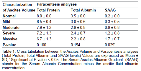
The sample size was classified into groups according to the ascites grading:-Mild (grade 1) (<200-600) ml constituting 4 (7.5%), Moderate (grade 2) (>600-800) ml were 9 (17.0%) Severe/gross (grade 3) (>800- 1000) ml were 29 (54.7%) Massive (grade 4) (>1000-2000) ml were 10 (18.9%) and Normal 1 (1.9%). Ages for each group were registered, and it was found that the patients mean ages were 51.3 ± 19.2, 60.0 ± 12.8, 51.6 ± 16.0, and 54.9 ± 18.3 years old for mild ascites, moderate, severe, massive ascites respectively. After ultrasound scanning, the Ascites was detected in different regions: Sub hepatic area in one patient and in the hepato-renal in 9 patients then in the vesico- ureteric in16 patients and Ascites occupying the intraperitoneal space with fully distention was found in 27 patients.
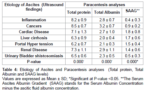
Assessment of ascites is one of the clinical indications of abdominal ultrasound which is able to differentiate between fluids and solid tissues [18]. In our study we used ultrasound as an imaging noninvasive tool. All the internal organs in the abdomen and pelvis of the ascetic patients had been evaluated including the liver, spleen kidneys, ovaries; pelvic organs as well as ascites echo texture and volume. Abdominal Ultrasound can accurately estimating the volume of intra-abdominal ascites or any other fluids collections [19] with a sensitivity of 97% [20]. It can detect amounts as small as 100 mL and is considered the gold standard for the diagnosis of ascites [5]. Abdominal ultrasound is also useful in locating the optimal site to perform a paracentesis, particularly in patients with a small amount of ascites or in those with loculated ascites. The simplest and most inexpensive way to orient the diagnostic workup of a patient with ascites, is through the analysis of ascitic fluid [21]. A diagnostic paracentesis was the test performed in our patients who were affected with ascites. Table 1 characterized the ascites according to the volume and correlated the characteristics with the laboratory analyses of the total protein, total albumin and SAAG levels and showed a significant relation between the characteristics of the ascites and SAAG Level. These laboratory tests taken together are the most useful in determining the etiology of ascites and thereby in further directing the workup of patients with ascites [21]. The results revealed that the most common causes of ascites in Sudanese patients was the liver cirrhosis followed by cancer integrated cancers of breast, prostate, colon, ovaries, lymphomas, hepatocellular carcinoma and cystoadeno carcinoma, inflammations including: pelvis inflammatory diseases, hepatitis ,abdomen tuberculosis(TB), followed by renal disease, heart diseases ,portal hypertension and urinary bladder Schistosomiasis .Similarly; another studies had mentioned that the most common cause of ascites in patients from Western Europe and North America is cirrhosis, which accounts for approximately 80% of cases [22,23]. It is reported that approximately 50% of patients with cirrhosis develop ascites [24], then malignancies (10%) and cardiac failure (5%) and (TB). The malignant Ascites is of critical predictive significance with survival in patients with ovarian cancer [25]. Table 2 presented the cross tabulation between the character of Ascites as volume with the etiology of ascites, it showed a significant relation at p=0.002. Although the underlying cause of ascites was noticed from the history and clinical examinations of the patients, however, it is important to exclude other causes of ascites. Therefore, investigations were directed at diagnosing the etiology of ascites. The essential test which was done was the diagnostic paracentesis as well as the abdominal ultrasound scan to evaluate the appearance of the liver, pancreas, lymph nodes, and presence of splenomegaly, which may signify portal hypertension which is considered as one of the main cause of ascites [26]. Characterization of Ascites according to the etiology was presented in Table 3 these were done by evaluating the abdominal and pelvis organs, total protein, total albumin and SAAG levels, ascites type as transudate or exudates, and were correlated with ascites etiology as regards the ultrasonographic findings. P-value was found to be <0.05; therefore the statistical relationship is highly significant between the etiology of ascites and ascites echo texture and paracentesis results as transudate and exudate. The sensitivity and true positive results of the ultrasound diagnosis when compared with the paracentesis results ;were found to be 100% in cardiac disease, liver cirrhosis, renal disease, portal hyper tension urinary bladder Schistosomiasis cases and 87.5% in inflammatory cases and 77.8% in patients with cancers. Table 4 showed a significant relation between the ascites etiology and paracentesis analyses of total protein, total albumen and SAAG levels. SAAG was found to be high in the cases of cardiac disease, liver cirrhosis, portal hyper tension and renal disease. This was similar to what was mentioned previously [27] who showed that SAAG is considered a best reflector for the elevated hydrostatic pressure, contributing significantly to the development of portal hypertensive ascites, even in patients with a low serum albumin concentration, and reported SAAG to be particularly helpful in distinguishing congestive heart failure with high total protein from malignant ascites without liver metastases ,so our reflection of the significances between the ascites etiology as diagnosed by ultrasonography and the paracentesis results will be of great value in predicting the laboratory analyses results convincing us to consider the ultrasonography as one of the excellent non- invasive imaging methods done without complications. The priority to establish the cause of ascites will determine its management [21]. Scanning by ultrasound and measuring the ascites volume may reflected the etiology and the issue appears to affect the spread of intraperitoneal fluid including peritoneal compartments, intraperitoneal pressure, area from which the fluid originates, rapidity with which fluid accumulates, the presence or absence of adhesions, ligamentous attachments, cancers, inflammations or other findings. In addition we believe the density, texture and character of the fluid with respect to other abdominal organs is important: liver, spleen, ovaries and pelvic organs have densities greater than that of the average ascitic fluid, this may conclude the ascites cause as cirrhosis, splenomegaly, or other pathological findings. The pliability of the abdomen was accountable for ascitic fluid localization. To localize the ascites in our sample, the two methods were obtained, physically and ultrasonographically. Regarding the results, this study recommended that ultrasound should take pride of place in diagnosis the ascites by measuring the ascites volume and evaluating its texture as routine imaging investigations, because ultrasonography can suggest the underlying cause, differentiates between the transudates and exudates ascites collections with appreciated sensitivity. It was proved to be reliable, valuable method of investigation, and will often prevent the necessity for more laborious and hazardous procedures as it is noninvasive, cheap cost and available.


