Journal of Nutrition & Food Sciences
Open Access
ISSN: 2155-9600
ISSN: 2155-9600
Research Article - (2016) Volume 6, Issue 1
In the present work, we investigated the protective effects of Guangdong Herbal Tea (GHT) on liver and kidney damage induced by restraint stress in mice, and we explored the relationship between these effects and the Nrf2/ARE signaling pathway. Pretreatment with GHT prior to the exposure to restraint stress significantly prevented an increase in the serum levels of hepatic and kidney functional markers (ALT, AST and BUN), lipid peroxidation (MDA) and alleviated oxidative stress, as determined by glutathione content (GSH), Catalase (CAT) activity and Total Antioxidant Capacity (T-AOC) in the liver and kidney. Meanwhile, GHT-induced Nrf2/ARE signaling pathway activation was evidenced by Nrf2 translocation into the nucleus. Moreover, GHT up-regulated heme oxygenase-1 (HO-1). Taken together, these results suggest that the protective effects of GHT against liver and kidney damage induced by restraint stress may be due to its antioxidant capacity and the up-regulation of HO-1 via the Nrf2/ARE signaling pathway.
Keywords: Guangdong herbal tea; Restraint stress; Liver and kidney damage; Nrf2/ARE signaling pathway
Stress plays an important role in the occurrence and development of many diseases [1,2]. Animal models are basic tools for the study of stress. Restraint stress animal model is a widely employed model [3]. Through limiting animals’ movement, researchers can induce a state of oxidative stress [4,5]. Oxidative stress is the result of an imbalance between the free radical generation and antioxidant defenses. It has been shown to be related with the occurrence of many diseases by damaging lipid, protein, DNA and inactivating the antioxidant enzyme [6-9].
The Nrf2/ARE signaling pathway, which is important to the modulation of cellular defense mechanisms against oxidative stress, is a hot point in research [10-12]. Under normal conditions, the nucleus factor erythroid related factor 2 (Nrf2) is captured in the cytoplasm by the Kelch-like ECH associating protein 1 (Keap1), which degrades Nrf2 through the ubiquitin–proteasome pathway [13-16]. Under oxidative stress, electrophiles and chemopreventive agents disrupt the reactive residues in Keap1. As a result, Nrf2 is dissociated from Keap1 and translocates into the nucleus, where it binds the antioxidant response element (ARE) [17] and initiate the transcription of the phase II enzyme genes and antioxidative stress protein genes [18-23]. Many studies showed antioxidants can induce the expression of many antioxidant enzymes, such as HO-1, by activating Nrf2 [24-26].
Herbal tea is a kind of beverage made from natural plants. Previous studies reported herbal tea could be used to treat many diseases because of its capabilities to clear away heat, moistening lungs, detoxication, alleviating pain, dewetting, and modulating immunity [27-30]. Guangdong Herbal Tea (GHT), originated from Guangdong province, has been used for approximately two centuries. Currently, GHT is commercialized by being produced and sold as a boxed or canned product and is widely popular in Mainland China, Hong Kong and many other regions in Asia. GHT as a daily beverage is drunk by many people for health purposes. Although recent studies have demonstrated that GHT plays a critical role in attenuating dysfunctions through relieving oxidative stress damage induced by restraint stress [31-35], little is known concerning the underlying mechanisms. These facts led to our focus on the correlation between the Nrf2/ARE signaling pathway and the effects of GHT on restraint stress damage in the liver and kidney. Therefore, this study may provide a scientific basis for developing GHT as promising antioxidant therapy for inhibiting the progression of restraint stress-associated diseases and promoting good health.
Ethics statement
This study was carried out in strict accordance with the recommendations in the Guide for the Care and Use of Laboratory Animals of the National Institutes of Health. The protocol was approved by the Committee on the Ethics of Animal Experiments of the Ethics Committee of Guangdong Provincial Centre for Disease Control and Prevention. All surgeries were performed under sodium pentobarbital anesthesia, and all efforts were made to minimize suffering.
Preparation of GHT extract
Guangdong Herbal Tea (GHT) extract was obtained from Guangzhou Wanglaoji Pharmaceutical Co., Ltd (Guangzhou, China) (batch No: 120301). It was extracted from raw herbs, mainly including Mesona, Plumeria Rubra, Lonicera Japonica, Microcos paniculata, Chrysanthemum, Prunella Vulgaris and Liquorice, through water extraction followed by alcohol precipitation and concentration. One hundred grams of raw herbs typically generates 18.5 g of extract, so 1 g of extract is equivalent to 5.4 g of raw herbs. The extraction was performed at 4°C.
Based on the Professional Standard and Chinese Pharmacopoeia, the main bioactive components of Guangdong Herbal Tea (GHT) extract were identified and measured by using gas chromatography and mass spectrometry (GC-MS) system, spectrophotometric method. The results were showed that the contents of flavonoids, triterpenoids, polysaccharides, alkaloids, volatile oil and anthraquinone were 9.0, 1.0, 0.18, 0.025, 0.0095, 0.0081(w/w,%) respectively.
Reagents
Rutin, ursolic acid and glucose were provided by the National Institutes for Food and Drug Control (China). Dendrobine was purchased from Dalian Meilun Biotech Co., Ltd (China). Reagent kits for alanine aminotransferase, aspartate aminotransferase, blood urea nitrogen and creatinine (ALT, AST, BUN and CREA) were bought from BioSino Bio-technology and Science Inc. (Shanghai, China). Assay kits for Lipid Peroxidation (MDA), GSH and GSSG, Catalase (CAT), Total Superoxide Dismutase (SOD) and Total Antioxidant Capacity (T-AOC) were purchased from the Beyotime Institute of Biotechnology (Jiangsu, China). NE-PER Nuclear and Cytoplasmic Extraction Reagents and a super signaling West Pico Trial Kit were purchased from Thermo Fisher Scientific Company Inc. (Rockford, USA). A Bovine serum Albumin (BSA) Standard Set was obtained from Bio-RAD Company (California, USA). Primary antibodies against GAPDH (14C10) Rabbit mAb, Lamin B1 Antibody, HO-1 (P249) Rabbit mAb and nuclear Nrf2 (D1C9) Rabbit mAb were brought from Cell signaling Technology Inc (Boston, USA). The HRP AffiniPure Goat Anti-Rabbit IgG (H+L) (the secondary antibody) was obtained from EarthOx, LLC (San Francisco, USA). Transfer membranes (0.45 nm) were purchased from Millipore Corporation (Billerica Ma, USA).
Determination of active compounds
Flavonoid content was measured with rutin as a reference by nitritealuminum nitrate colorimetry at 510nm [36]. The triterpenoid concentration was detected with ursolic acid as the standard product by vanillin colorimetry at 546nm [37]. The polysaccharide content was measured using the phenol–sulfuric acid method at 490nm based on a standard curve for glucose [38].
Animals and experimental design
Forty male BALB/c mice (6 weeks old, 20 ± 2 g body weight) were purchased from Guangdong Medicine Laboratory Animal Center (Foshan, China). All mice were housed in a specific pathogen-free room under controlled temperature (22 ± 2°C) and humidity (65 ± 5%) with a 12 h day-night cycle (lights on at 06:00). They were fed a pelleted basic diet (provided by Guangdong Medicine Laboratory Animal Center) with clear water ad libitum. After a quarantine period of 7 days, all of the mice were randomly divided into five groups: control group, restraint stress group and three GHT groups. The control group and the restraint stress group received distilled water and the GHT groups received GHT extract at 0.48 g/kg, 1.44 g/kg or 4.32 g/kg, which was equivalent to 2.60 g/kg, 7.78 g/kg or 23.33 g/kg of raw herbs, respectively. To prepare the test substances for the three treatment groups, 0.48 g, 1.44 g or 4.32 g GHT extracts were dissolved in water to a final volume of 10 ml. The test substances were administered orally (0.1 ml/10 g) daily for 5 consecutive days. One hour after the last administration, mice of restraint stress group and three GHT groups were exposed to acute restraint stress. According to the method described by Satoh E et al. and Bao L et al, mice were placed in well-ventilated 50 mL polypropylene tubes (2.8-cm diameter × 11.5-cm length) for 2 h. There were fifteen 0.8-cm-diameter holes drilled on the sides and one 1-cm-diameter hole drilled on the bottom of the tubes for mice to breathe[39,40]. Those mice were not physically compressed and did not experience pain. At the same time, mice in control group were kept in cages with no food or water to avoid differences of food and water intake between the restraint and control groups.
After restraint for 2 h, the mice were weighed and anesthetics via intraperitoneally injection of 0.6% pentobarbital sodium (0.1 ml/10 gBW). Then, all mice were extracted blood from the abdominal vein and removed livers and kidneys. To ensure death in all animals, cervical dislocation was used to euthanize mice.
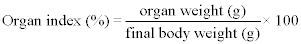
Calculation of the organ index
The weights of livers and kidneys relative to the final body weights (organ indexes) were calculated. The livers and kidneys were stored immediately at -80°C for later biochemical assays.
Analysis of serum biochemistry
Blood was centrifuged at 2500 r/min for 15 min at 4°C by refrigerated centrifuge to obtain serum. Serum levels of alanine aminotransferase, aspartate aminotransferase, blood urea nitrogen and creatinine (ALT, AST, BUN and CREA) were determined using an Auto Chemistry Analyzer (Automatic Analyzer 7600-010; HITACHI, Tokyo, Japan).
Tissue homogenization
Liver and kidney samples were homogenized in chilled distilled water using a homogenizer (IKA ULTRA® TURRAX T18 basic, GmbH, Staufen, Germany) at 10000 r/min for 30s in an ice bath, then centrifuged at 10000 × g for 10 min at 4°C. Liver and kidney homogenates were used to determine biochemical parameters.
MDA-based determination of lipid peroxidation levels
Lipid peroxidation levels in livers and kidneys were measured by the method of TBA-reactive substances. Briefly, in acidic medium, MDA reacted with thiobarbituric acid (TBA) upon boiling, and generated pink MDA-TBA adducts, which were measured by a Varioskan Flash (Thermo Fisher Scientific Company Inc, Rockford, USA) at 532 nm. Peroxidation was determined from a standard curve using a standard MDA concentration series and expressed in terms of nmol/mg protein.
Determination of glutathione content (GSH)
Total glutathione (GSH) was determined in tissue homogenates using yeast glutathione reductase, 5,5’ dithio-bis-(2-nitrobenzoic acid) and NADPH. The yellow color developed by the reaction of GSH with DTNB was read at 412 nm. Oxidized glutathione (GSSG) was determined by the same method in the presence of 2-vinylpyridine. GSH was calculated from the difference between total glutathione and GSSG.
Determination of catalase (CAT)
CAT activity was measured by following the decrease in absorbance at 520 nm due to the decomposition of hydrogen peroxide (H2O2). One enzyme unit was defined as the amount of enzyme decomposing 1 mM H2O2 per minute at 25°C.
Determination of superoxide dismutase (SOD)
SOD activity was assayed using the nitro blue tetrazolium (NBT) method according to a SOD assay kit following the manufacturer’s protocol. NBT was reduced to blue formazan by superoxide, which has strong absorbance at 560 nm. One unit (U) of SOD was defined as the amount of protein that inhibits the NBT reduction rate by 50%. SOD activity was expressed as U/mg protein.
Determination of total antioxidant capacity (T-AOC)
T-AOC levels were based on the reduction of Ferrictripyridyltriazine (Fe3+-TPTZ) to Ferrous-tripyridyltriazine (Fe2+- TPTZ) according to the FRAP method, and the blue adducts were measured at 593 nm and expressed in terms of nmol/mg protein.
Nuclear and cytoplasmic protein extraction and western blot analysis
Dissected liver and kidney tissues (50 mg) were homogenized using a homogenizer (IKA ULTRA® TURRAX T18 basic, Staufen, Germany) in 500 μL ice-cold CERI at 10000 r/min for 30s in an ice bath. Then, 27.5 μL of ice-cold CER II was added to each sample and centrifuged at a speed of 12,000 × g for 5 min at 0°C. Supernatants (cytoplasmic protein extraction) were immediately transferred to clean pre-chilled tubes. The remaining pellets were suspended in 250 μL ice-cold NER and vortexed at 3000 r/min for 30min at 0°C using a vortex mixer (ABSON, USA) before they were centrifuged at 12,000 × g for 10 min at 0°C. Finally, the supernatants were subjected to nuclear protein extraction. All extracts were immediately stored at -80°C until use.
The protein concentrations of the nuclear and cytoplasmic extracts were determined using the Quick Start Bovine Serum Albumin (BSA) Standard Set. The proteins (30 μg/sample) in sodium dodecyl sulfatepolyacrylamide gel electrophoresis (SDS- PAGE) sample loading buffer were boiled for 5min, separated by 10% SDS-polyacrylamide gel electrophoresis, and transferred to PVDF membranes at 300 mA for 2h. Each membrane was blocked with 5% skim milk-TBST (containing 0.1% Tween 20) at room temperature for 2 h. The bands were incubated with nuclear Nrf2 (1:500), HO-1(1:500), GAPDH (1:1000) and LaminB1 (1:1000) antibodies overnight at 4°C. After incubation, these bands were incubated with HRP AffiniPure Goat Anti-Rabbit IgG (H+L) secondary antibody (1:10000) at room temperature for 2 h. The immunoreactive bands were visualized using enhanced chemiluminescence reagents according to the manufacturer’s instructions and recorded on film using an analysis system for gel imaging (Vilber Lourmat Company, France).
Statistical Analysis
SPSS 16.0 software was used for statistical analysis, and all data were expressed as the mean ± standard deviation (SD). The statistical significance of differences between groups was analyzed using a oneway analysis of variance (ANOVA). A difference of P<0.05 was considered statistically significant.
The contents of active compounds
The flavonoid content was determined as 9.0% with rutin as a reference. The triterpenoid concentration was measured at 1.0% with ursolic acid as the standard product. The polysaccharide content was 0.18% based on glucose as the reference.
The effect of GHT on the final weight, liver weight and kidney weight in restrained mice
In this study, no significant differences could be observed for the final weight or the liver and kidney weights among the different experimental groups (P>0.05). There were also no obvious differences between the liver and heart indexes among the different experimental groups (P>0.05) (Table 1).
| Groups | Final weight(g) | Liver weight(g) | Kidney weight (g) | Liver index (%) | Kidney index (%) |
|---|---|---|---|---|---|
| control | 21.80 ± 1.41 | 1.33 ± 0.16 | 0.33 ± 0.04 | 6.13 ± 0.46 | 1.52 ± 0.17 |
| RS | 22.00 ± 1.59 | 1.27 ± 0.13 | 0.34 ± 0.03 | 5.82 ± 0.73 | 1.56 ± 0.21 |
| 0.48 g/kg | 21.41 ± 1.76 | 1.30 ± 0.11 | 0.32 ± 0.02 | 5.55 ± 0.98 | 1.52 ± 0.13 |
| 1.44 g/kg | 21.14 ± 1.39 | 1.18 ± 0.12 | 0.32 ± 0.05 | 5.59 ± 0.33 | 1.54 ± 0.13 |
| 4.32 g/kg | 20.74 ± 1.40 | 1.15 ± 0.07 | 0.32 ± 0.02 | 5.55 ± 0.91 | 1.55 ± 0.23 |
Table 1: The effect of Guangdong Herbal Tea (GHT) on the final weight, liver weight, and kidney weight in restrained mice. The results are shown by mean ± SD.
The effect of GHT on serum AST, ALT, BUN and CREA levels in restrained mice
Mice subjected to restraint stress for 2 h exhibited severe liver and kidney injuries, as evidenced by obvious increases in the levels of serum ALT, AST and BUN (P<0.01 or P<0.05) compared to the control group. The CREA content was also elevated in the model group, but no significant difference was found (P>0.05). However, GHT treatment obviously attenuated this status. Depletions of ALT levels were observed in all treatment groups (P<0.01). Simultaneously, the increases in AST and BUN levels induced by restraint stress were significantly decreased by 4.32 g/kg in the GHT treatment (P<0.01). Furthermore, the CREA levels were ameliorated in a dose-dependent manner (P>0.05) (Table 2).
| Groups | ALT (U/L) | AST (U/L) | BUN (mmol/L) | CREA(mmol/L) |
|---|---|---|---|---|
| Control | 59.50±8.28 | 108.90 ± 21.84 | 5.30 ± 0.36 | 31.88 ± 3.00 |
| RS | 80.78 ± 19.58## | 127.00 ± 18.76# | 8.25 ± 1.42## | 33.90 ± 3.07 |
| 0.48g/kg | 63.80 ± 12.76** | 119.60 ± 16.60 | 8.08 ± 0.67 | 34.10 ± 5.07 |
| 1.44g/kg | 63.78 ± 8.27** | 119.90 ± 18.19 | 7.67 ± 0.60 | 33.22 ± 3.15 |
| 4.32g/kg | 60.30 ± 7.56** | 105.40 ± 5.78** | 6.06 ± 1.16** | 31.40 ± 2.41 |
Table 2: The effect of Guangdong Herbal Tea (GHT) on serum AST,ALT, BUN and CREA levels in restrained mice. The results are shownby mean ± SD. #P<0.05 and ##P<0.01 vs Control group, **P<0.01 vs RS group.
Effects of GHT on oxidative stress of the liver in restrained mice
Restraint stress significantly increased the production of MDA in the liver (P<0.01). Meanwhile, obvious decreases in GSH and T-AOC contents in the liver were found when compared to the control group (P<0.01). The results also illustrated that restraint stress inhibited the activity of CAT (P<0.01). Although there was an evident reduction in SOD activity, no significant difference was found (P>0.05). Interestingly, treatment with 4.32 g/kg GHT significantly attenuated the production of MDA and the depletion of GSH and the T-AOC content induced by restraint stress in the liver (P<0.01). Additionally, the present study shows that treatment with 1.44 g/kg or 4.32 g/kg GHT elevated CAT activity (P<0.01). However, GHT treatment failed to change the SOD activity significantly, despite a dose-dependent effect (P>0.05) (Table 3).
| Groups | MDA (nmol/mg prot) | GSH (nmol/mg prot) | CAT (U/mg prot) | SOD (U/mg prot) | T-AOC (nmol/mg prot) |
|---|---|---|---|---|---|
| Control | 2.82± 0.26 | 20.65± 4.28 | 39.34± 1.03 | 85.82± 5.79 | 34.87± 2.98 |
| RS | 3.95± 0.27## | 12.91± 3.54## | 34.64± 1.67## | 74.82± 7.81 | 26.33± 2.12## |
| 0.48g/kg | 3.72± 0.16 | 13.84± 2.24 | 35.12± 1.54 | 75.82± 7.28 | 27.72± 2.17 |
| 1.44g/kg | 3.55± 0.31 | 15.16± 1.86 | 37.46± 1.32** | 82.17± 7.34 | 29.41± 4.56 |
| 4.32g/kg | 2.93± 0.36** | 19.41± 3.94** | 38.75± 1.28** | 85.13± 10.49 | 33.36± 4.45** |
Table 3: The effect of Guangdong Herbal Tea (GHT) on MDA, GSH, CAT, SOD and T-AOC levels in livers of restrained mice. The results are shown by mean ± SD. ##P<0.01 vs Control group; **P<0.01 vs RS group.
Effects of GHT on oxidative stress in the kidney in restrained mice
Furthermore, to ascertain whether GHT influenced kidney damage induced by restraint stress, we measured the levels of MDA, GSH, TAOC and the activities of SOD and CAT in the kidney. The results showed that restraint stress led to a significant elevation in the MDA level (P<0.01) compared to the control group. Simultaneously, evident reductions of GSH, CAT and T-AOC content were also observed in the restrained mice (P<0.01). However, after treatment with 4.32 g/kg GHT, MDA generation was attenuated and the concentrations of GSH, CAT and T-AOC were increased significantly (P<0.01). Neither restraint stress nor GHT treatment had significant effects on SOD activity in the kidney (P>0.05) (Table 4).
| Groups | MDA (nmol/mg prot) | GSH(nmol/mg prot) | CAT (U/mg prot) | SOD (U/mg prot) | T-AOC (nmol/mg prot) |
|---|---|---|---|---|---|
| Control | 2.74 ± 0.24 | 17.90 ± 1.73 | 27.52 ± 2.66 | 83.16 ± 8.07 | 32.58 ± 3.02 |
| RS | 3.44 ± 0.21## | 12.72 ± 1.78## | 22.52 ± 2.07## | 71.20 ± 5.87 | 26.20 ± 3.28## |
| 0.48g/kg | 3.29 ± 0.35 | 13.16 ± 1.11 | 23.10 ± 1.74 | 77.62 ± 11.42 | 26.85 ± 2.99 |
| 1.44g/kg | 3.14 ± 0.29 | 14.71 ± 2.23 | 24.95 ± 1.34 | 80.78 ± 7.69 | 29.70 ± 2.85 |
| 4.32g/kg | 2.83 ± 0.39** | 17.12 ± 2.51** | 27.13 ± 1.92** | 81.68 ± 12.26 | 31.85 ± 1.52** |
Table 4: The effect of Guangdong Herbal Tea (GHT) on MDA, GSH, CAT, SOD and T-AOC levels in kidneys of restrained mice. The results are shown by mean ± SD. #P<0.05 and ##P<0.01 vs Control group; *P<0.05 and **P<0.01 vs RS group.
Effect of GHT on the expression of HO-1 and nuclear Nrf2 proteins in the Nrf2/ARE signaling pathway in the liver of restrained mice
To investigate whether the observed protective effects of GHT were related to the Nrf2/ARE signaling pathway, the expression levels of HO-1 and nuclear Nrf2 protein in the liver were determined. The results indicated that the expression of HO-1 and nuclear Nrf2 protein was up-regulated in the model group (P<0.01) (Figure 1), while significant effects of increased up-regulation of HO-1 protein expression in the 4.32 g/kg GHT group (P<0.01) (Figure 1A) and nuclear Nrf2 expression in the 1.44 g/kg GHT group and the 4.32 g/kg GHT group (P<0.05 and P<0.01) were found (Figure 1B).

Figure 1: The effect of Guangdong Herbal Tea (GHT) on expression of relative liver proteins in Nrf2/ARE signaling pathway. (A) The expression of HO-1 in liver and the concentration of HO-1in liver. (B) The expression of Nrf2 in liver and the concentration of Nrf2 in liver. Mice were pretreated with GHT (0.48, 1.44 or 4.32 g/kg), while mice in Control group and RS group were given pure water for 5 consecutive days. 1 h after the last administration, all mice except in Control group were exposed to restraint stress for 2 h. ##P<0.01 vs Control group; *P<0.05 and **P<0.01 vs RS group, n=3.
Effect of GHT on protein expression of HO-1 and nuclear Nrf2 in the Nrf2/ARE signaling pathway in the kidney of restrained mice
In this study, exposure of the kidney to restraint stress led to obvious induction of HO-1 and nuclear Nrf2 expression compared to the control group (P<0.01) (Figure 2). However, the 4.32 g/kg GHT treatment also had a greater effect on the induction of the upregulation of HO-1 and nuclear Nrf2 expression (P<0.01).

Figure 2: The effect of Guangdong Herbal Tea (GHT) on expression of relative kidney proteins in Nrf2/ARE signaling pathway. (A) The expression of HO-1 in kidney and the concentration of HO-1in kidney (B)The expression of Nrf2 in kidney and the concentration of Nrf2 in kidney. Mice were pretreated with GHT (0.48, 1.44 or 4.32 g/kg), while mice in Control group and RS group were given pure water for 5 consecutive days. 1 h after the last administration, all mice except in Control group were exposed to restraint stress for 2 h. ##P<0.01 vs Control group; **P<0.01 vs RS group, n=3.
In this study, the restraint stress model was established by placing male BALB/c mice into 50 ml polypropylene plastic centrifuge tubes as restraint cages. Mice subjected to restraint stress for 2 h showed remarkable increases in serum ALT and AST levels, which are two important markers of liver damage. As an end product of lipid peroxidation, MDA can be used as an indicator of the extent of oxidative stress damage [41]. This study clarified that a substantial increase in hepatic lipid peroxidation was evident from the elevated MDA level. Antioxidant enzymes, such as GSH, CAT and SOD, are important in the protecting cells against oxidative stress and keeping the redox status balanced [42-44]. Therefore, GSH, CAT and SOD as oxidative stress biomarkers are also considered to be measured in our study. However, it was found that restraint stress failed to inhibit the activity of SOD in the liver of the restraint-stressed mice. Our results are consistent with the results reported by H. G. Kim, who found after receiving restraint stress for 6 h, the serum AST and ALT levels of mice increased significantly, the ROS and MDA levels in liver tissues also increased, however SOD activity had no significant change [45]. Meanwhile, our study shows the CAT activity and GSH level in liver tissues remarkably decreased. In Zaidi et al. research, restraint stress for 6 h decreased the activities of CAT and GSH in liver tissues [46]. BUN and CREA are two critical biomarkers of kidney damage. This study showed that the BUN level was obviously elevated in mice exposed to restraint stress. However, no significant increase in the CREA level induced by restraint stress was found. This result may be attributed to the time of restraint duration. It was also found that the production of MDA was aggravated when mice were exposed to restraint stress. Meanwhile, the content of GSH and T-AOC was reduced and accompanied by the inhibition of CAT activity in the kidney. The above results clearly suggest that restraint stress induced an imbalance between oxidants and antioxidants, which contributed to liver and kidney oxidative stress damage.
A plenty of stressors insult human self-defense systems. When the stress burden exceeds self-defense capacity, severe damages and toxicities can be induced, which even are occasionally lethal. When excessive free radical generation overwhelms the antioxidant defense capacity, the Nrf2/ARE system can be activated to elevate detoxifying activity. The present study identified that the expression of nuclear Nrf2 protein was up-regulated in restraint-stressed mice, demonstrating that restraint stress may promote the activation of the Nrf2/ARE signaling pathway. It was also found that the process of endogenous antioxidant/Phase II detoxification was initiated to protect against cellular and tissue injury, as evidenced by the expression of heme oxygenase-1 (HO-1) in liver and kidney tissues. In previous studies, the rats subjected to restraint stress for 3 h exhibited a significant increase in HO-1 expression in stomach [47] and the exposure of acute restraint stress for 6 h caused Nrf2 and HO-1 in rat’s hippocampus remarkably increased [48].
Herbal and natural plants have been used for hundred and thousand years for their preventive and/or therapeutic characters throughout the world. Considerable researches have been carried out to study the biological benefits of these remedies. GHT is a popular beverage consumed by people in China and other regions for health purposes. In the present study, damage in the liver and kidney provoked by restraint stress was obviously alleviated by GHT treatment, as reflected by the recovery of ALT, AST and BUN levels in the serum. Further investigation indicated that the protective effect of GHT in the liver and kidney was related to the improvement of the oxidative stress status because of its antioxidant activity, as evidenced by reduced MDA production and elevated CAT activity, and GSH and T-AOC levels in the liver and kidney. These results are partly consistent with another study, in which GHT decreased the MDA level and elevated the GSH content in the liver [49-52]. Retrospectively, the present study also showed that treatment with GHT significantly activated the Nrf2/ARE signaling pathway by enhancing up-regulation of nuclear Nrf2 and HO-1 protein expression induced by restraint stress, accompanied by the reduction of MDA production and the elevation of CAT and GSH levels in the liver and kidney. These results suggested that the induction of the Nrf2/ARE signaling pathway may be one of the underlying mechanisms for the effects of GHT treatment on restraint stress. However, the results also showed that neither restraint stress nor GHT had obvious effects on the activities of SOD in the liver and kidney. One explanation may be that the nuclear Nrf2 protein expression induced by restraint stress and GHT are not sufficient to activate the SOD genes.
In previous studies, researchers have identified several classes of chemo preventive agents with the capability of protecting cells against different damages [49]. In our present work, it was shown that the content of flavonoids, triterpenoids and polysaccharides was 9.0%, 1.0% and 0.18%, respectively. Flavonoids, which are abundant in many plants, consist of a large group of Polyphenolic compounds. Recently, most researchers and scientists have focused on the effects of flavonoids on health. Flavonoids have been proved to have antioxidant activity, free radical scavenging capacity, hepatoprotective and antiinflammatory properties [50]. A past study showed the ability of flavonoid to induce that expression of Nrf2, which plays an important role in regulating various cytoprotective target genes [51]. A plenty of triterpenoids have been found to have several biological activities, including anti-inflammatory, cytotoxic and anticancer activities [52]. Polysaccharides, which are composed of more than 100 monosaccharides, possess various biological activities [53]. The various biological activities of these phytochemicals, including antioxidant activities, immune-modulating, antitumor, anti-inflammatory have drawn much attention [54].
Therefore, we hypothesize that the active compounds responsible for the effects of GHT on oxidative stress induced by restraint stress may be flavonoids, triterpenoids or polysaccharides, but not necessarily limited to these chemical classes. In addition, a great number of studies shown that some plant compounds in GHT extracts could alleviate antioxidative stress through the activation of Nrf2/ARE signaling pathway. It was revealed Prunella vulgaris could up-regulate HO-1 by activating Nrf2, subsequently alleviate vascular inflammation [55]. Past studies showed Tea Chrysanthemum extract could protect cells against oxidative stress and inflammatory through inhibiting NF- κB-mediated inflammatory and activating the Nrf2/ARE signaling pathways [56]. Liquiritigenin and Dehydroglyasperin C extracted from Liquorice were found to possess activation effects of Nrf2, GSH and HO-1, which would contribute to their alleviating oxidative injury effects [57,58]. Moreover, it was revealed Lonicera japonica extract could increase GSH level, SOD activity and CAT activity [59]. In light of the above facts, we propose that GHT may contain a variety of phytochemicals besides flavonoids and triterpenoids that might affect antioxidative stress via the activation of the Nrf2/ARE signaling pathway. Meanwhile, the interactions of these phytochemicals are complicated and not clear. The many questions regarding GHT may be addressed in future studies.
In summary, the present study demonstrated that oral administration of Guangdong Herbal Tea could attenuate restraint stress-provoked liver and kidney damage in mice and that the protection can be explained not only on the basis of non-enzymatic action but also because of its effect on the enzymatic action of the Nrf2/ARE signaling pathway. Flavonoids and triterpenoids may be the active compounds responsible for the effects of GHT on oxidative stress induced by restraint stress, but these effects are not necessarily limited to these chemical classes. Therefore, these findings provide novel insight into the mechanisms of protection afforded by Guangdong Herbal Tea against restraint stress-induced oxidative stress. However, further studies are needed to clarify the interactions of GHT phytochemicals. From these observations, it is concluded that Guangdong Herbal Tea may be a potential dietary supplement for the prevention of some diseases induced by oxidative stress.
This work was financially sponsored by National High Technology Research and Development Program 863 (2010AA023001), the Guangdong Science and Technology Plan Project (2013B010404033) the Guangdong Natural Science Foundation Project (S2013010013289), the Guangzhou Science and Technology Plan Project (201300000161) and the Guangdong Provincial Bureau of Chinese Medicine Project (20132108 and 20121275).
Min Zhao, Xingfen Yang designed the research; Mengjiao Zhang, Guiyuan Ji, Jianbin Tan, Rui Huang, Jiao Tang conducted the studies; Mengjiao Zhang, Junming Huang, Rongbo Zheng, Xiaodan Huang analyzed the data and Mengjiao Zhang, Lingjie Zhou, prepared the manuscript; all authors read and approved the manuscript.
The authors declare no competing financial interests.