Journal of Biomedical Engineering and Medical Devices
Open Access
ISSN: 2475-7586
ISSN: 2475-7586
Review Article - (2022)Volume 7, Issue 9
BCI is a type of communication system that allows humans to communicate with their environment by using control signals produced from EEG activity, without the involvement of peripheral nerves and muscles. A BCI is characterized as a tool that measures brain or central nervous system activity and transforms these signals into artificial output. Robotics and advanced mechatronics have played a significant role in the development of assistive technologies to help people overcome a large range of disabilities. There are several techniques available and currently being experimented upon in various research and development setups for obtaining signals from the brain for further processing and utilizing with appropriate BCIs for multiple applications in the biomedical field, however, the most suitable in terms of cost effectiveness, safety protocols and Information Classification: General acceptable resolution is the EEG. This article identifies various artefacts observed in an EEG signal which need further filtration before it can be used for a certain application. In addition to this identification, this article elaborates the techniques and that can be used for this purpose.
Brain Computer Interface (BCI); EEG; Signal pre-processing; Artefacts; Independent Component Analysis (ICA); Peripheral nerves
BCI has always been an attractive area for researchers. More recently, it has become an interesting area of scientific investigation and a potential means of demonstrating a direct link between the brain and technology. It is also one of the fastest growing areas of research. Many scientists have tried and applied various methods of communication between humans and computers using BCI in various forms. However, from a simple concept in the early days of digital technology, today it has evolved into highly complex signal detection, recording and analysis techniques. Hans [1] recorded for the first time an electroencephalogram (EEG) [2] showing the electrical activity of the brain measured through the scalp of the human brain. The author tried it on a boy with a brain tumour. Since then, EEG signals have been used clinically to identify brain disorders. The concept of combining the brain and technology has always been of interest to the scientists, and recent advances in neurology and technology have made it a reality, paving the way for repairing and possibly enhancing human physical and mental capabilities. The sector flourishing the most based on BCI is considered the medical application sector. Cochlear implants [3] for the deaf and deep brain stimulation for Parkinson’s illness are examples of medical uses becoming more prevalent. A BCI can be used for a variety of purposes, and the application dictates the design of the BCI. Robotics and advanced mechatronics have played a significant role in the development of assistive technologies to help people overcome a large range of disabilities. BCI's immediate purpose is to offer control and power to persons with extreme disabilities or people with partial or complete body paralysis. Various neurological neuromuscular disorders, such as amyotrophic lateral sclerosis, brain stem stroke, or spinal cord injury can be the cause of these disabilities. Such an interface would increase their quality of life and would minimize the expense of intensive care at the same time. The majority of BCI integration and research has focused on medical applications, and many BCIs aim to replace or restore CNS function lost due to disease or accident. Other BCIs are narrower. In diagnostic applications, BCIs are also used for biological and emotional purposes, in CNS disease and post-traumatic therapy, and in motor rehabilitation. Biomedical technologies and applications can minimize the longevity of disease and empower people with reduced mobility to care, protect and support rehabilitation. The need to develop precise techniques that can process abnormal brain responses that can occur due to diseases such as stroke is a key challenge in developing such platforms [4]. BCIs are as accurate as the input provide to the system after reading a certain signal from the brain. The more a signal is void of unwanted and unnecessary information in the form of noises and artefacts better will be the functioning of the BCI system. This paper is aimed at identifying various artefacts commonly encountered while using EEG for signal acquisition and further elaborates the methods and techniques that can be used for removal of such unwanted components from the obtained signals. Furthermore, this review will provide a quick guide and a learning opportunity to the new researchers entering the field of biomedical engineering for prosthesis and rehabilitation developments.
Structure of BCI system
To better understand the requirement of signal pre-processing as the paper proceeds, there is a need for a basic understanding of the structure of a basic BCI. The BCI system works in a closed loop system. A certain amount of feedback is returned for each user action. For example, movements of an imaginary hand can lead to commands to move a robotic arm. This simple arm movement requires a lot of process. In this regard, BCI is characterized as a tool that measures brain or central nervous system activity and transforms these signals into artificial output. As an artificial intelligence system BCI can identify a certain set of patterns in brain signals and can control the actuator after four consecutive phases. These phases include signal acquisition, signal pre-processing and Feature Extraction, classification, and control interface. The stage of signal acquisition collects the signals from the brain and can also minimize noise and process artifacts. The preprocessing stage prepares the signals for further processing in a suitable manner. This part of the BCI is the highlight of this research. The feature extraction stage recognizes discriminative information in the recorded brain signals. The stage of classification classifies the signals considering the vectors of the function. The choice of a good classifier is necessary to achieve efficient pattern recognition in order to decipher the intentions of the user. Finally, the control interface step converts the classified signals into practical commands, for an actuator like a wheelchair, walker, or a computer. Basic structure of a BCI system is shown in Figure 1.
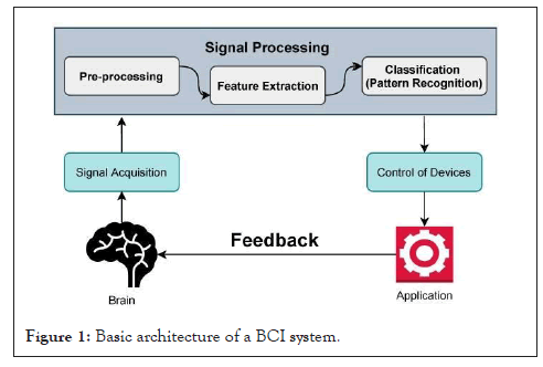
Figure 1: Basic architecture of a BCI system.
Signal acquisition techniques
The human brain generates electrical signals known as EEG signals. These signals can be acquired through invasive and non-invasive techniques. While choosing between the invasive and non-invasive methods of signal acquisition, the resolution needed for proper translation and the feasibility of obtaining the signals are considered. This research is focused upon the non-invasive technique of Electroencephalography (EEG) for acquiring the required signals from the brain. Other non-invasive techniques include Positron Emission Tomography (PET), Magnetoencephalography (MEG), functional Near IR Spectroscopy (fNIRS), and functional Magnetic Resonance Imaging (fMRI). Keeping in view the scope of research, only EEG technique is elaborated in detail.
Electroencephalography (EEG)
EEG monitors electrical activity in the scalp produced by neurons in the brain. Multiple electrodes implanted directly into the scalp, primarily the cortex, are often used to rapidly record these electrical activities. Due to its excellent temporal resolution, ease of use, safety and affordability, EEG is the most widely used technique for capturing brain activity. Active and passive electrodes are two types of electrodes that can be used. Active electrodes typically have a built-in amplifier, whereas passive electrodes require an external amplifier to amplify the detected signal. The main purpose of implementing a built- in or external amplifier is to reduce the effects of background noise and other interfering signals caused by cable movement. One problem with EEG is the need to use gels or saline to reduce the resistance of skin electrode contacts. Additionally, the signal quality is poor and varies with background noise. The International 10-20 system of electrodes placement [5] is commonly used to implant electrodes on the scalp surface for recording purposes. Electrical activity across different frequency bands is commonly used to describe EEG.
Emotiv EPOC headset
Emotiv EPOC is a multi-channel EEG research framework that supports a wide range of applications, including neuro-therapy, biofeedback, and an interface between the brain and computers. The Emotiv EEG headset is a high resolution, multi-channel, wireless, and portable EEG system. It has a 128 Hz sampling rate and communicates with the PC through a wireless USB dongle link. To moisturize its electrodes, this headset requires a hydrating solution and it is ready to use with almost no set-up time needed. It has 14 EEG channels. The locations of these channels are based on the International 10-20 system.10-20 System EEG electrodes are placed on the surface of human skull through a cap or a headset. These electrodes are portable and are placed on skull surface using 10-20 system. International 10-20 system of electrodes placement is shown in Figure 2. In a 10-20 system, electrodes are positioned at 10% and 20% points along lines of longitude and latitude. The name of the electrode starts with one or two letters indicating the general area or lobes of the brain where the electrode is located (Fp=frontopolar; F=frontal; C=central; P=parietal; O=occipital; T=temporal). Each name of the electrode terminates with a number or letter indicating the distance from the midline. Odd numbers are used in the left hemisphere and for the right hemisphere, even numbers are used. Electrodes placed at the midline are labeled with a ‘z’ for zero. Electrodes can easily capture physiological signals from the brain of a person and transmit them to data acquiring systems of applications.
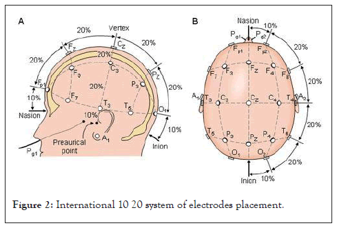
Figure 2: International 10 20 system of electrodes placement.
Signal pre-processing
Pre-processing is the procedure of transforming raw data into a format that is more suitable for further analysis and interpretable for the user. In the case of EEG data, pre-processing usually refers to removing noise from the data to get closer to the true neural signals. The signals that are picked up from the scalp are not necessarily an accurate representation of the signals originating from the brain, as the spatial information gets lost. Secondly, EEG data tends to contain a lot of noise which can obscure weaker EEG signals. Artifacts such as blinking or muscle movement can contaminate the data and distort the picture. Finally, the separation of the relevant neural signals from random neural activity that occurs during EEG recordings is carried out. A number of strategies are available to deal with noise effectively both at the time of EEG recording as well as during pre-processing of recorded data. A comparison of filtered vs unfiltered EEG signal is shown in Figure 3.
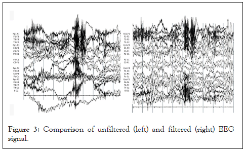
Figure 3: Comparison of unfiltered (left) and filtered (right) EEG signal.
Artefacts
One of the biggest challenges in using EEG is the very small signalto- noise ratio of the brain signals, coupled with the wide variety of noise sources. The factors like a heartbeat, the number of electrodes, placement of electrodes, subject’s movement, scalp preparation, any external distractions, and many more play an important role in either obtaining a proper recording or increasing the ratio of noise in the EEG data, in turn, causing poor recording session. Some of the most common artifacts along with their properties are listed in the Table 1. There are different techniques to remove noise from the data. Some sources of noise can be relatively easy to remove; others present more of a challenge and can introduce unwanted consequences. These artifacts can be divided into two types.
| Artefact | Source/Cause | Frequency range | Amplitude | Morphology |
|---|---|---|---|---|
| Cardiac | Heart | > 1 Hz | 1-10 mV | Epilepsy |
| Transmission line nose | Transmission line | 50-60 Hz | Low | Beta or Gamma Wave |
| Muscle artefact | Body muscle | ≥ 35 Hz | Low | Beta frequency |
| EOG | Eye | 0.5-3 Hz | 100 mV | Tumour, Delta Wave |
| Phone artefacts | Mobile and land line phones | High | High | Morphology different from actual EEG |
| Electrode artefact | Electrode | Very low | High | Morphology different form actual EEG |
Table 1: Some of the most common artifacts along with their properties are listed.
External artefacts
The noise arising from any external source like the number of electrodes, placement of electrodes AC power lines, lighting, and a large array of electronic equipment (from computers, displays, and TVs to wireless routers, notebooks, and mobile phones) is called external noise or external artifacts. The most common sources of this external noise are the ambient electrical current in any room with electrical wiring, either 50 Hz or 60 Hz, and any other electrical equipment in close proximity to the EEG sensors.
Internal artefacts
Internal Artifacts are defined as undesired signals caused by the internal sources of physiologic activities and movement of the subject during the recording of data that may introduce changes in the measurements and affect the signal of interest. EEG can be contaminated in frequency or time domain by artifacts. The most important internal artifacts in a typical EEG recording are ocular artifacts or EOG, and muscular artifacts (EMG). Electrical potentials which are due to eye movements and blinks propagate over the scalp and create hostile electrooculography (EOG) artifacts in the recorded electroencephalogram (EEG). Eye movements are a major source of contamination of EEG. Figure 4 shows the eye blink artefact in the window. Electrical activity on the body surface due to the contracting muscles is recorded via Electromyogram (EMG). Since independent myogenic activities of the head, face, and neck muscles are conducted through the entire scalp, it can be monitored in the EMG. Figure 5 shows the muscle artefact due to contraction of face muscles.
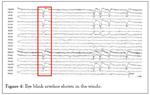
Figure 4: Eye blink arteface shown in the windo.
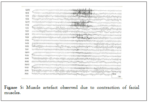
Figure 5: Muscle artefact observed due to contraction of facial muscles.
Techniques for removal of artefacts
Removal of noise: The easiest sources of noise to deal with are external and environmental sources of noise, such as AC power lines, lighting, and a large array of electronic equipment (from computers, displays, and TVs to wireless routers, notebooks, and mobile phones). The most basic steps in dealing with environmental noise are removing any unnecessary sources of electromagnetic (EM) noise from the recording room and its immediate vicinity, and, where possible, replacing equipment using alternate current with equipment using direct current (such as direct current lighting). A more advanced and costly measure is to insulate the recording room from EM noise by use of a Faraday cage. While being very effective in eliminating most of the environmental EM noise, EM insulation requires either planning or costly rebuilding work.
Signal averaging
Possibly the simplest way to deal with noise in the recorded data is signal averaging. The key assumption in signal averaging is that the noise in the signal is random, or at least occurs with a random phase about the event of interest, whereas the signal of interest is stable. If the EEG signal is recorded over several occasions, noise at each time point will increase at some points; reduce on others, but on average cancel itself out, leaving the stable EEG response of the event of interest. Signal averaging is a simple and powerful way of dealing with noise, but it has several limitations and caveats. For these reasons, signal averaging is used as a last resort for truly unavoidable noise sources only and uses other strategies to prevent noise before recording and remove it after recording.
Filtering: One of the most common and easiest ways to remove noise from the raw data is filtering. To be able to filter it, the noise needs to fall within one of the three categories: the frequency of the noise needs to be either below the frequency of the phenomena we are trying to observe, above it, or it needs to fall within a very narrow well-specified range.
High-pass filtering: It’s a type of filtering that allows only the signal varying above the selected cut-off frequency to pass and is routinely used already during acquisition itself. Several factors such as sweating and drifts in electrode impedance can lead to slow changes in the measured voltage, which can, in turn, lead to saturation of the amplifier and loss of data during recording, as well as to significant distortions in the averaged event-related time course. For those reasons, it is often recommended to filter the frequencies below 0.01 Hz.
Low-pass filtering: It is a type of filtering that allows only the signal varying below the selected cutoff frequency to pass and is used to remove noise at the other end of the spectrum of frequencies that one is interested in. Contraction of muscles typically leads to a strong signal with frequencies above 100 Hz, so suppression of frequencies above 100 Hz would (to a large extent) remove movement artifacts in the acquired signal. Additionally, sampling of the data itself can lead to aliasing-a phenomenon in which frequencies higher than the sampling frequency can appear as lowfrequency artifacts in the sampled data. To eliminate aliasing, the data should be low-pass filtered at frequencies at least one third lower than the sampling rate.
Notch filter: It is a type of filter that can be used with a frequency band of the artifact, to remove the particular artifact. A 50 Hz notch filter can use for removal of the transmission line frequency. Using a notch filter is very effective to remove noise but it can be very risky at times. It can also result in the rejection of some meaningful data that falls within the band of the notch filter. While filtering data, one needs to be aware that filtering depending on the nature of the filter used–can also significantly affect the data in the nonfiltered frequency ranges, thus affecting estimates of onsets and/or amplitude of observed ERP waves as well as introducing artefactual oscillations. So for those types of artifacts whose frequency falls within the frequency range of useful data, techniques like ICA are used.
Independent component analysis (ICA): Independent Component Analysis (ICA) is a statistical and computational technique for revealing hidden factors that underlie sets of random variables, measurements, or signals. ICA defines a generative model for the observed multivariate data, which is typically given as a large database of samples. In the model, the data variables are assumed to be linear mixtures of some unknown latent variables, and the mixing system is also unknown. The latent variables are assumed non gaussian and mutually independent and they are called the independent components of the observed data. These independent components, also called sources or factors, can be found by ICA. To understand how non gaussianity relates to statistical independence, it is necessary to understand the central limit theory, which states that the sum of many independent processes tends towards a Gaussian distribution. Therefore, if S(t) is assumed to be a set of truly independent sources, the observed mixed-signal X(t) will be more Gaussian by the central limit theory. A single estimated source ˆsi(t) is a linear mixture of X(t) given by the weights in the spatial filter wi. The with at maximizes the non- Gaussianity of ˆsi(t) is used to find the closest approximation to the true independent source si(t). Therefore, the optimization criteria are to find the unmixing matrix that maximizes non-Gaussianity in all of the source components. ICA provides extraction of the eyerelated signals present in the EOG, and removal of this information or artifact, rather than the complete EOG which still has some brain activity. However, detection and removal of transient artifacts such as head and neck muscle contractions and movements are difficult with ICA.
BCI as a biomedical technology has vast scope and potential with regards to contributing towards improving the lives of countless people who are suffering from diseases and disorders related to CNS functionality. A great deal of research is being carried out in various multidisciplinary settings around the globe to enhance the usage of the technology in cost effective and efficient ways. To make the technology available to all spheres of public around the world, it has to be designed and perfected at low costs so that people from all walks of life can afford it. In this vein, EEG is the most suitable option for acquiring signals from the brain due to several reasons including safety offered by the tools and gadgets used and its low cost. However, the signal that is picked from the brain using this simple option is not free from various distortions and disturbances which need to be removed to make it useful. Identification of the abnormalities is the first and the foremost task in this process and types of noises and abnormalities that are generally encountered have been mentioned in this article which shall serve as a guideline for the new researchers in this field. Depending upon the type of noise encountered, appropriate method can be adopted for its cancellation and pre-processing of the acquired signal. The more noise free and clean a signal will be, better information will be provide to the BCI and this will eventually improve the functionality of the entire system that is aimed at improving the life style of its user.
The author reports no conflicts of interest in this work.
[Crossref][Google Scholar][PubMed].
[Crossref][Google Scholar][PubMed].
[Crossref][Google Scholar][PubMed].
[Crossref][Google Scholar][PubMed].
Citation: Qammar S, Khan AK (2022) Identification of Artefacts Observed in EEG Signals Obtained from Human Brain Using Emotiv EPOC Headset and Artefacts: Removing Techniques in Signal Pre-Processing Stage of Brain Computer Interface. J Biomed Eng Med Dev. 07:236. DOI: 10.35248/2475-7586.22.07:236.
Received: 19-Sep-2022, Manuscript No. BEMD-22-19255; Editor assigned: 22-Sep-2022, Pre QC No. BEMD-22-19255 (PQ); Reviewed: 06-Oct-2022, QC No. BEMD-22-19255; Revised: 13-Oct-2022, Manuscript No. BEMD-22-19255 (R); Published: 20-Oct-2022 , DOI: 10.35248/2475- 7586.22.07.236
Copyright: © Qammar S, et al. This is an open-access article distributed under the terms of the Creative Commons Attribution License, which permits unrestricted use, distribution, and reproduction in any medium, provided the original author and source are credited.
Competing interests: The authors have declared that no competing interests exist.