
Journal of Clinical Chemistry and Laboratory Medicine
Open Access
ISSN: 2736-6588

ISSN: 2736-6588
Review Article - (2023)Volume 6, Issue 1
Key biomarkers such as BDNF (Brain-derived Neurotrophic Factor) and NfL (Neurofilament Light Chain) play important roles in the development and progression of many neurological diseases, including multiple sclerosis, Alzheimer's disease and Parkinson's disease. In these clinical conditions, the underlying biomarker processes are markedly heterogeneous. In this review, robust biomarker discovery is of critical importance for screening, early detection, and monitoring of neurological diseases. The difficulty of directly identifying biochemical processes in the Central Nervous System (CNS) is challenging. In recent years, biomarkers of CNS inflammatory response have been identified in various body fluids such as blood, cerebrospinal fluid, and tears. Furthermore, biotechnology and nanotechnology have facilitated the development of biosensor platforms capable of real-time detection of multiple biomarkers in clinically relevant samples. Biosensing technology is approaching maturity and will be deployed in communities, at which point screening programs and personalized medicine will become a reality. In this multidisciplinary review, our goal is to highlight clinical and current technological advances in the development of multiplex-based solutions for effective diagnosis and monitoring of neuroinflammatory and neurodegenerative diseases.
Biosensors; Brain; Ultra-sensitive; BDNF; NfL; CNS; Neurological diseases
BDNF: Brain-derived Neurotrophic Factor, ELISA: Enzyme-Linked Immunosorbent Assay, IMR: Immuno Magnetic Reduction, EIS: Electrochemical Impedance Spectroscopy, IDE: Inter Digitated Electrode, MWCNTs: Multi-Walled Carbon Nanotubes, LOD: Limit of Detection, LOQ: Limit of Quantitation, CV: Cycling Voltammetry, DPV: Differential Pulse Voltammetry, SPE: Screen-Printed Electrode, NfL: Neurofilament Light Chain, SMA: Single-Molecule Array, MB’s: Magnetic Microbeads, PEC: Photo-Electro Chemical, PtWP: Woodpile- like Pt nanowire, EGOFET: Electrolyte-Gated Organic Field-Effect Transistor, IDE: Inter Digitated Electrode, 3,5-DClPDT: 3,5-Di-Chlorophenyl Diazonium Tetrafluoroborate, Na-DEDTC: Sodium Diethyldithiocarbamate.
Brain-Derived Neurotrophic Factor (BDNF) belongs to a family of neurotrophins that play imperative roles in the survival and differentiation of developing neuronal populations [1,2]. In the adult brain, BDNF maintains high levels of expression and regulates both inhibitory synaptic transmission and excitatory and activity-dependent plasticity [2]. It profoundly affects neuronal physiology and morphology, promotes synaptic stabilization and neurite germination, and increases long-term potentiation. BDNF synthesis and release are regulated by neural activity and are consistent with BDNF's role as the main mediator of activity- dependent neuroplasticity [3]. Recent information recommends that BDNF may also be associated with spontaneous, activityindependent transmission [1,3]. BDNF is a neurotrophic signaling molecule related with synaptic plasticity, neuronal growth, synapse maturation during development, and axonal targeting [4]. BDNF is related to various of mental disorders, including: bipolar disorder, deep depression, anxiety disorders, and other psychiatric disorders associated with exposure to stressful conditions [5]. Therefore, alterations in BDNF blood levels due to genetic and environmental factors are increasingly associated with several psychiatric disorders [6]. Decreased serum BDNF occurs in depression, bipolar disorder, eating disorders, and schizophrenia, and there is evidence that drug treatment for these disorders upregulates BDNF levels. Low serum BDNF levels may also be associated with depression-related personality traits in healthy subjects, suggesting their potential role as risk markers. Additionally, Neurofilament Light Chain Protein (NfL, approximately 68 kDa) is one of many proteins in the neuronal cytoskeleton [7]. The protein is released upon axonal damage in the Central Nervous System (CNS) and can be detected in serum, Cerebro-Spinal Fluid (CSF), and EDTA-plasma after passage through the blood-brain barrier. NfL levels in serum/ plasma are several times lower than in CSF, but the correlation is strong. Plasma levels are lower than serum levels (approximately 10 percent lower). NfL levels in serum/plasma can thus be used as a marker of CNS neuronal degeneration. NfL levels are an unspecific marker of neuronal degeneration that does not reveal the underlying cause [8]. The level of NfL is extremely useful in monitoring the progression of demyelinating diseases. The reference ranges are broad because the NfL level varies greatly between individuals in the general population. Therefore, precise monitoring of BDNF and Nfl in Cerebro-Spinal Fluid (CSF) and other physiological fluids has the potential to discover important clues for development of therapeutic and diagnostic approaches for psychiatric diseases and neurodegenerative diseases such as Parkinson’s disease and Alzheimer’s disease. To evaluate the physiological significance of human salivary BDNF, a research work has optimized a sensitive and specific Enzyme-Linked Immunosorbent Assay (ELISA) (Table 1).
| Techniques | Biomarker | LOD/sensitivity | Findings and significances |
|---|---|---|---|
| ELISA | BDNF | ~15 pg/μl | Developed system was based on colorimetric detection. Also, the potential utilization of chemiluminescence based detection technique was approved successfully. |
| ELISA | BDNF | 618 pg/mL | There was no association between salivary BDNF levels and the other biological characteristics examined. |
| Western blotting | BDNF | _ | _ |
| Western blotting | BDNF | 10 μg | Levels of mBDNF proteins in mBDNF-GFP transfected cells (both the pro- and mature forms) were detectable. |
| Western blotting | BDNF | _ | _ |
| ELISA | NfL | 78.0 pg/mL | Finding indicated SMA to be more sensitive than ELISA or the ECL assay |
| ELISA | NfL | 10 µg/L | ELISA, which quantifies NfL from human CSF, has revealed the difficulty of preparing accurate and consistent standards. |
| ELISA | NfL | 15 nM | These results show a noble analytical performance of the novel ELISA for quantification of NfL concentrations in the CSF. |
| ELISA | NfL | 1000 pg/ml | Developed method was promising not only for screening dementia but also for differential diagnosis. |
| IMR | NfL | 0.18 fg/ml | _ |
| SMA | NfL | 14 ng/L | Plasma NFL is associated with AD diagnosis and with cognitive, biochemical, and imaging hallmarks of the disease. |
Table 1: Methods in the detection of BDNF and NfL.
Plasma levels of BDNF and NfL
In healthy volunteers, mean plasma BDNF levels ranged from 92.5 pg/mL (8.0 to 927.0 pg/mL). It was higher in women and decreased with age in both men and women [9]. BDNF is spread over numerous brain areas and helps neurons survive, support, and function. Other sources of BDNF include the gastrointestinal tract, lungs, heart, spleen, and liver. Apart from that, BDNF is known to be expressed in vascular smooth muscle cells, fibroblasts, and thymic stroma. The results from the review confirmed that BDNF levels in the colon, bladder, and lungs were higher than those in the skin and brain. Positive associations have been reported between blood levels of BDNF and Low-Density Lipoprotein (LDL) cholesterol, diastolic blood pressure, total cholesterol, body weight index, adipose tissue mass, and triglyceride. It has been recommended that women with low plasma BDNF levels are at increased risk of death. The observation supports that a significant decrease in plasma levels of BDNF in women was associated with age and weight gain. Plasma NfL levels were positively associated with age (r=0.355, p<0.001) and negatively with global cognition (r=-0.355, p<0.001) in all subjects.
Methods in the detection of BDNF and NfL
Given the above references to those different roles, there is increasing interest in achieving precise and quantitative detection of different BDNF protein morphologies. Because BDNF is a secretory protein, it is found both intracellularly and extracellularly. BDNF is a diversity of astrocytes (primarily due to trans-cytosis), neurons, microglia, vascular endothelial cells, monocytes, male submandibular glands, smooth and striatum muscles, peripheral pericytes, etc [10]. The Enzyme-Linked Immunosorbent Assay (ELISA) is an immunological assay frequently used to quantity proteins, antibodies, antigens, and glycoproteins in biological samples. Some examples include pregnancy tests, diagnosis of HIV infection, and quantity of cytokines or soluble receptors in cell supernatant or serum. The working principle of ELISA is presented in Figures 1-4.
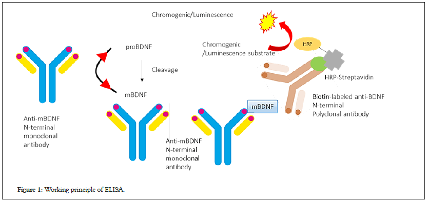
Figure 1: Working principle of ELISA.

Figure 2: Drug discovery in biosensor methodology.
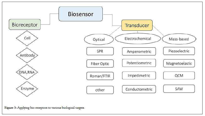
Figure 3: Applying bio receptors to various biological targets.

Figure 4: Developed immunosensor for detection of BDNF. 
To evaluate the physiological significance of human salivary BDNF, a specific and sensitive ELISA was developed appropriately. To determine the relationship between salivary BDNF levels and other biological characteristics, an ELISA method was examined. Results showed no association between BDNF levels in saliva and other biological characteristics. Western blotting showed BDNF levels were reduced in treated animals compared to control animals [11]. Western blotting is an experimental technique for detecting specific proteins in blood or tissue samples. This method uses gel electrophoresis to separate the proteins in the sample. The separated protein is transferred from the gel to the surface of the membrane. In an original study, western blotting evaluated the secretion of met BDNF and BDNF in serum. Finding revealed equal amounts of proteins were loaded in each lane so, mBDNF and vBDNF proteins are expressed at an equal level and treated in the same method. Targeting histone deacetylation for recovery of maternal deprivation-induced variations in AKAP150 expression and BDNF in the Ventral Tegmental Area (VTA) were determined by the western blotting (Table 2).
| Platform | Technique | Matrix | NPs | Model | Linear range | LOD |
|---|---|---|---|---|---|---|
| Electrochemical/Immunosensor | DPV | Plasma | AuNPs | Human | 0.1-2.0 ng / mL | 0.2 ng/mL |
| Electrochemical/Immunosensor | Impedance spectra | Brain | _ | Mice | 10 fg/mL to 10 ng/mL | _ |
| Electrochemical/Immunosensor | EIS | Cancer cells | AuNPs | Human serum | 100 to 450 μg/ml | 1.81 ± 0.012 pg/ml |
| Electrochemical/Immunosensor | _ | Glaucoma | _ | Human serum | 0.41 to 250 ng/mL | 0.2 ng/mL |
| Electrochemical/Immunosensor | CV, DPV | _ | MWCNTs | Human serum | 0.01 ng/mL to 100 ng/mL | 5 pg/mL |
| Electrochemical/Immunosensor | MIP, CVs, DPV | Neurological diseases | SPE | Human serum | 0.01 to 0.06 ng/mL | 6 pg/mL |
| Electrochemical/Immunosensor |
Table 2: Developed biosensors for detection of BDNF.
Biosensor methodology
The term "biosensor" is an abbreviation for "biological sensor". The device consists of biological elements such as enzymes, antibodies, nucleic acids, and a transducer. The biological element interacts with the analyte under test, and the transducer converts the biological response into an electrical signal. Depending on the application, biosensors are also known as immunosensors, genosensor. The commonly cited definition of the biosensor is "a chemical sensing device in which biologically obtained detections are combined with a transducer to enable the quantitative development of several complex biochemical parameters. Figure 1, presents the biosensor methodology. Mandatory use of biosensors has become of paramount importance in biomedical drug discovery, food safety standards, and environmental monitoring. Biosensors have been classified based upon numerous outlooks; however, the most generally used classification relies on two factors: the transducer and the bioreceptor [12].
Developed biosensors for BDNF detection
The Point of Care device was manufactured for use with a small amount of blood that can be collected from a finger puncture wound to detect BDNF. Specifically, the presented device is a polymer-based chip with a nanoporous wrinkled gold film that acts as an electrode/detection layer, enabling electrochemical detection of BDNF in plasma. Increasing the BDNF concentration (0.1- 2.0 ng/mL) resulted in a significant difference in redox current.
Biosensors are ideal for generating signal displays in seconds and achieving multiplexing. An Interdigital Micro-Electrode (IME) immunosensor coated with anti-BDNF antibody and anti-BDNF in a Polydimethylsiloxane (PDMS)-based microfluidic channel chip was developed to detect BDNF in the brain of mice. The platform detected BDNF from microliter liquid samples, even at femtogram/ mL concentrations, demonstrating higher selectivity than other growth factors. An ultrasensitive dual probe immunosensor was proposed to monitor of nicotine induced-brain derived neurotrophic factor released from cancer cells. Created biosensor showed acceptable linearity and high sensitivity at optimized conditions. BDNF dysregulation is generally associated with several neurological and psychiatric disorders such as schizophrenia, and the available evidence is that BDNF is also strongly associated with Parkinson's disease and Alzheimer's disease. The BDNF target sequence was detected with a capture probe attached to an aluminum micro comb electrode on the surface of a silicon wafer. A capture target reporter sandwich assay was performed to improve the detection of BDNF targets (Table 3).
| Platform | Technique | Matrix | NPs | Model | Linear range | LOD |
|---|---|---|---|---|---|---|
| Electrochemical/Immunosensor | CVs, EIS | Parkinson | GMA | Human | 1 to 50 µg/L | 5.21 ng/L |
| Electrochemical/Immunosensor | CVs, EIS | Brain | MBs | Plasma | 10−5000 pg mL−1 | 3.0 pg mL−1 |
| PEC | photochemical | Blood | Pt | Human | 1 ng/ mL | 38.2 fg/mL |
| Electrochemical/Immunosensor | CVs, DPV | Plasma | ZrO2@La2O3 | Human | 0.05 ~ 200 ng·mL-1 | 0.012 ng·mL−1 |
| PEC | _ | Serum | Fe3O4/AuNPs | Human | 5 pg/mL to 10 ng/mL | 1 ng/mL |
| EGOFET | Adsorption | Biological | Gold gate | Serum | 100 fm to 10 nm | 30 fm |
| Electrochemical/Immunosensor | EIS | Plasma | CNTs | Human | 5–10 pg ml−1 | 10 pg ml−1 |
| Electrochemical/Immunosensor | Spectophotometry | Serum | AuNPs | Human | 100 fM to 1 nM | 100 fM |
Table 3: Developed biosensors for detection of NfL.
A 3D aptersensor based on Bio-Layer Interferometry (BLI) technology for early detection of glaucoma, an irretrievable disease that causes blindness, followed by rapid and sensitive detection of BDNF was settled recently. The developed 3D aptersensor has made it possible to batch convert a small amount of biomarker BDNF binding into an optical interference signal in one step. An innovative immune microelectrode array was developed as a label-free electrochemical biosensor for the detection of BDNF in human serum. The examination results of performance, Figure 5 showed that the engineered immune-platform had a good selectivity, stability, and the limit of detections for BDNF measurement and was applicable in neuroscience A MIP-based synthetic receptor selectively bind to a clinically relevant protein, such as BDNF. BDNF-MIP was produced directly on the Screen- Printed Electrode (SPE) by surface initiation control/living radical photopolymerization. The resulting BDNF-MIP/SPE electrochemical sensor was able to detect BDNF-acceptable LODs in the presence of interfering HSA proteins and identify BDNF from structural analogs such as the neurotrophic factors CDNF and MANF [13]. Molecularly Imprinted Polymers (MIPs), which are entirely synthetic materials with antibody-like ability to bind and discriminate between molecules, exhibit enhanced stability and reduced fabrication cost compared to biological receptors. MIP based immunosensor Figure 6, was advanced for sensitive determination of BDNF directly on a Screen-Printed Electrode (SPE). The developed approach may be a promising method for developing innovative PoC diagnostic devices for early diagnosis and/or therapeutic monitoring of neurological diseases.
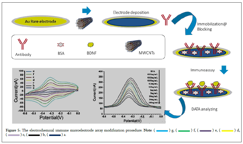
Figure 5: The electrochemical immune microelectrode array modification procedure. 

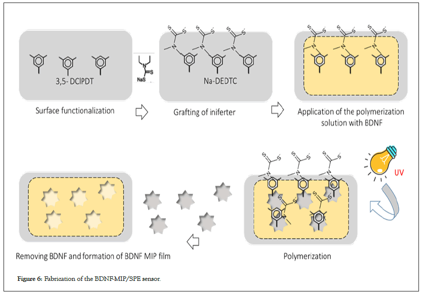
Figure 6: Fabrication of the BDNF-MIP/SPE sensor.
Neurofilament light chains are intra-axonal structural proteins released into both CSF and blood fluids upon axonal injury, regardless of the cause. It is vague how blood levels of NFL correlate with neurodegeneration, but it is one of the most consistent plasma biomarkers of neurodegeneration. A rapid and specific NfL diagnosis is therefore of great value. In the following, some advanced biosensors for its detection are reviewed and introduced (Figure 7).
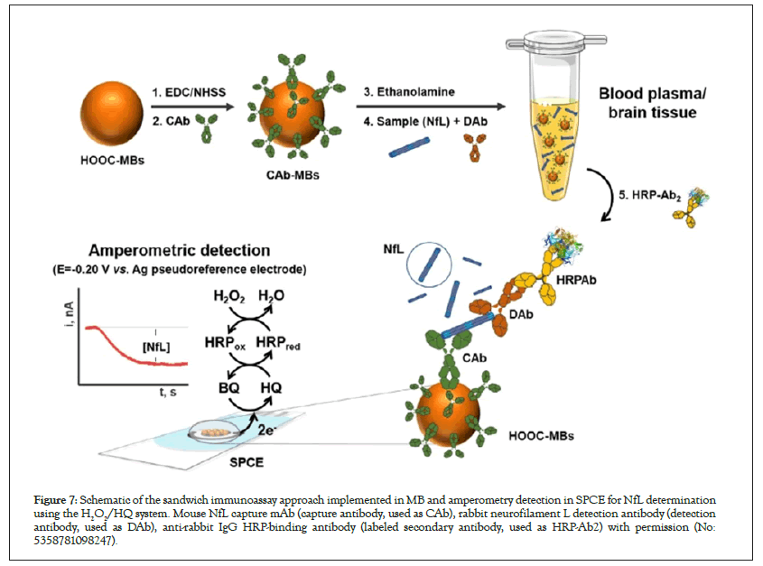
Figure 7: Schematic of the sandwich immunoassay approach implemented in MB and amperometry detection in SPCE for NfL determination using the H2O2/HQ system. Mouse NfL capture mAb (capture antibody, used as CAb), rabbit neurofilament L detection antibody (detection antibody, used as DAb), anti-rabbit IgG HRP-binding antibody (labeled secondary antibody, used as HRP-Ab2) with permission (No: 5358781098247).
Glycidyl Methacrylate (GMA) efficient monomer was applied to form thin polymeric films on the electrode surface and the anti-NfL antibody was immobilized via covalent linkage with GMA as a biorecognition agent. The advantage of a developed immunosystem involves in its high recognition ability and simplicity [14]. Magnetic microbeads (MBs)-based electrochemical bioplatform established for the detection of NfL-based on a sandwich type immunoassay including an HRP-secondary antibody to enzymatically label the detector antibody on the surface of commercial MBs functionalized with carboxylic acids. Observed analytical performance is suitable for determining NfL in brain tissues and plasma from AD patients using fast and straightforward methods and needing low samples. To assemble a bias-free PEC detection system, water-oxidizing with an iron oxyhydroxide-deposited bismuth vanadate DA2-PtWP biocathode (FeOOH/BiVO4) photoanode. Proposed biosensor was employed for determination of NfL in blood samples appropriately. A lanthanide-doped MOFs (La/UiO-66-NH2) material is pyrolyzed and synthesized as an ancestor to obtain a novel MOFs-derived material (ZrO2@La2O3) with exceptional electrochemical properties and effective immobilization of antibodies. ZrO2@La2O3 is employed as an electrode modification material to build a label-free electrochemical immunosensor. The system created was cheap, easy to manufacture, and easy to see. This could meet her future NfL assessment needs and become an efficient platform that would be of great help in the clinical diagnosis and treatment of neurodegenerative diseases. Organic semiconductor molecule Y6 was utilized in the construction of a novel immunosensor for ultra-sensitive detection of NfL. The effective demonstration of Y6 in this research for PEC recognition of NFL paved an original approach for the application of organic semiconductors in PEC sensing field [15]. An Electrolyte-gated Organic Field-Effect Transistor (EGOFET) was settled for sensitive detection of NfL in sub-pM levels. The selective, labelfree, and fast response makes this EGOFET biosensor a suitable device for monitoring and diagnosing neuronal injuries through detecting NF-L in physio-pathological ranges. An innovative electrochemical immuneplatform for the determining of NfL was advanced based on sandwich immunocomplexes labeled with HRP (b) and amperometric technique at SPCEs. Planned biosensor showed excellent selectivity and sensitivity and was usable for the determination of some biomarkers such as NfL, Tau, and p-Tau in human plasma. Gold-nanourchin complexed silicon dioxide-probe on gap-fingered IDE surface was established for NfL determining using current–volt measurement. Created biosensor, demonstrated excellent analytical performance and was applicable to quantity the level of NfL and leads to determine the condition with PD (Figures 8 and 9).
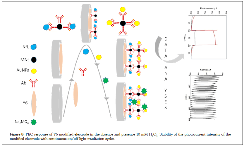
Figure 8: PEC response of Y6 modified electrode in the absence and presence 10 mM H2O2. Stability of the photocurrent intensity of the modified electrode with continuous on/off light irradiation cycles.
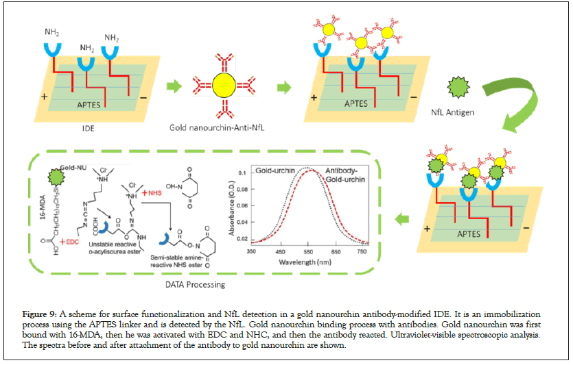
Figure 9: A scheme for surface functionalization and NfL detection in a gold nanourchin antibody-modified IDE. It is an immobilization process using the APTES linker and is detected by the NfL. Gold nanourchin binding process with antibodies. Gold nanourchin was first bound with 16-MDA, then he was activated with EDC and NHC, and then the antibody reacted. Ultraviolet-visible spectroscopic analysis. The spectra before and after attachment of the antibody to gold nanourchin are shown.
This review tried to collect all recent trends in advanced biosensors for the detection of BDNF and NfL. Presented biosensors have ultrasensitive detection limits, wide range linearity, and acceptable selectivity and stability in some cases. The main challenge in biosensor expansion needs to keep improving these technologies. For instance, the selection of enzymes, electrodes, and nanomaterial’s are important factors in the development of the biosensor because they must have similar operating conditions (concentration, temperature, pH,). Future work should focus on understanding the mechanism of interaction between the nanomaterials and enzymes and on the fabrication of new materials with electrode surface that could be applied in clinical diagnosis. As a result, it is recommended that the following items be considered in future studies. (A) Development of biosensors capable of detecting different critical neuroscience-related biomarkers such as BDNF, NfL, Tau protein, and Aβ. (B) Improving biosensor functional stability in various laboratory conditions. (C) Dedicated effort to develop more affordable, simple, and user-friendly biosensors with a higher sensitivity approach, less sample volume, and fewer pre-treatment steps. (D) Concentrate on miniaturization, such as microfluidic device fabrication and the integration of nano-materials with other biosensor components for in-situ heavy metal detection. (E) Using nanotechnology to integrate artificial intelligence and biosensors to continue developing biomarker detection processes that are scalable, sensitive, and multifunctional.
Citation: Charsouei S, Mobed A, Yazdani Y, Gargari MK, Ahmadalipour A, Sadremousavi SR, et al. (2023) Identification of Electrochemical Immunosensors for Neurological Diseases by BDNF and NfL. J Clin Chem Lab Med. 6:264.
Received: 27-Feb-2023, Manuscript No. JCCLM-22-20693; Editor assigned: 01-Mar-2023, Pre QC No. JCCLM-22-20693 (PQ); Reviewed: 15-Mar-2023, QC No. JCCLM-22-20693; Revised: 22-Mar-2023, Manuscript No. JCCLM-22-20693 (R); Published: 29-Mar-2023 , DOI: 10.35248/JCCLM.23.6.264
Copyright: © 2023 Charsouei S, et al. This is an open-access article distributed under the terms of the Creative Commons Attribution License, which permits unrestricted use, distribution, and reproduction in any medium, provided the original author and source are credited.