Journal of Medical Diagnostic Methods
Open Access
ISSN: 2168-9784
+44 1300 500008
ISSN: 2168-9784
+44 1300 500008
Short Communication - (2023)Volume 12, Issue 2
Localization and tracking of miniaturized wireless sensors in the Gastrointestinal Tract (GI) with high spatiotemporal accuracy is of high clinical value. It can enable continuous monitoring and transit-time evaluation of the GI tract, which is essential for accurate diagnosis, treatment, and management of GI motility disorders such as gastro paresis, ileus or constipation. GI motility disorders are also increasingly associated with a variety of metabolic and inflammatory disorders such as diabetes mellitus and inflammatory bowel disease. Together, these GI disorders affect more than one-third of the population globally and impose a considerable burden on healthcare systems. High resolution and real-time tracking of wireless sensors in the GI tract can also benefit anatomically-targeted sensing and therapy, localized drug delivery, medication adherence monitoring, selective electrical stimulation, disease localization for surgery, 3D mapping of GI anatomy for pre-operative planning, and minimally invasive GI procedures [1].
The current gold-standard solutions for these procedures include invasive techniques such as endoscopy and manometry, or procedures that require repeated use of potentially harmful X-ray radiation such as Computerized Tomography (CT) and scintigraphy. These techniques also require repeated evaluation in a hospital setting, which can confound observations given the recognized variability in motility and activity. Alternative approaches-including video capsule endoscopy and wireless motility capsules allow monitoring of the GI tract in real-world settings without interruption to daily activities. However, these methods lack direct measurement of the capsule’s location in the GI tract and have limited acquisition time. Commercial systems using electromagnetic-based tracking of sensors have also been developed but fail to simultaneously achieve a high Field-Of-View (FOV), high spatiotemporal resolution, fully wireless operation and miniaturization of the sensing devices, and system scalability with the number of devices [2].
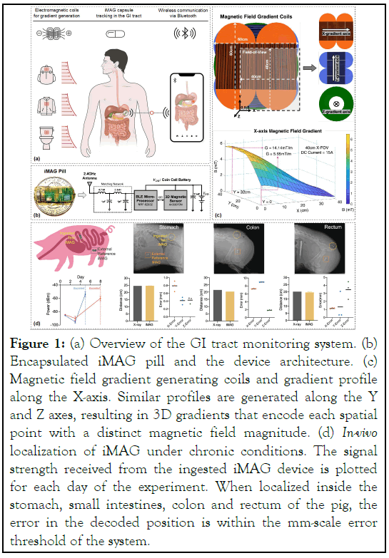
In our Nature Electronics article, we report a platform for localizing and tracking miniaturized wireless sensors inside the GI tract in real time and in non-clinical settings, with millimeterscale spatial resolution, and without using any harmful X-ray radiation [3]. Inspired by Magnetic Resonance Imaging (MRI), we generate monotonically varying magnetic fields in three orthogonal directions, resulting in gradients that encode each spatial point uniquely (Figures 1a and 1b). We designed highefficiency planar electromagnetic coils to generate the 3D magnetic field gradients in a FOV spanning the entire GI tract. For sensing the magnetic field, we designed highly miniaturized and wireless devices-termed ingestible Micro Devices For Anatomic-Mapping of Gastrointestinal-Tract (iMAG) to sense and transmit their local magnetic field to an external receiver. The receiver maps the field data to the corresponding spatial location, allowing real-time position tracking of the iMAG devices as they move through the GI tract.
The iMAG devices are designed with the following specifications: (i) High-resolution field measurement; (ii) Bluetooth-based wireless communication; (iii) Ultra-low power for prolonged battery life; (iv) Small form factor; and (v) bio-compatibility. The iMAG device (Figure 1b) consists of a 3D magnetic sensor, a BLE microprocessor, a 2.4GHz Bluetooth antenna, and coin-cell batteries. We designed the electromagnetic coils (Figure 1c) to generate gradients higher than 3mT/m across the entire FOV (40 × 40 × 40cm3) to ensure a resolution of 1mm across all three spatial dimensions. The Z-coil is a planar circular electromagnet carrying current in one direction, while the X and Y coils are composed of two oval halves carrying currents in opposite directions [3-6]. The three coils are stacked together and mounted on the walls of a prototype wooden chute for evaluation in large animal models.
In-vivo demonstration
The system functionality is demonstrated in vivo in porcine models as they represent a reliable analogue for human application. We first sought to emulate a real-world setting where an iMAG would be ingested and its position would be tracked relative to a reference iMAG located externally on the skin of the ambulatory animal. The ingested iMAG was localized in different regions of the GI tract – stomach, colon and rectum. The error in the decoded distance between the ingested and the reference iMAG devices, when compared with the distance obtained from the X-ray scans, was found to be within the mm-scale error threshold of the system (Figures 1c and 1d). The ingested iMAG devices remained functional upon excretion, thus confirming their their applicability for chronic use. We have also shown the system to be a highly accurate indicator of defecation, which is of clinical significance for patients suffering from fecal incontinence [3]. Furthermore, we have demonstrated iMAG’s usage as an in-vivo sensor to detect the location of magnetic particles and beads [3]. This approach can be used to label injection sites, polyps, fistulas, stomas, or strictures requiring localized therapy, using anatomic markers such as magnetic beads or staples (Figure 1) [7,8].

Figure 1: (a) Overview of the GI tract monitoring system. (b) Encapsulated iMAG pill and the device architecture. (c) Magnetic field gradient generating coils and gradient profile along the X-axis. Similar profiles are generated along the Y and Z axes, resulting in 3D gradients that encode each spatial point with a distinct magnetic field magnitude. (d) In-vivo localization of iMAG under chronic conditions. The signal strength received from the ingested iMAG device is plotted for each day of the experiment. When localized inside the stomach, small intestines, colon and rectum of the pig, the error in the decoded position is within the mm-scale error threshold of the system.
The real-time and mm-scale localization resolution of iMAG holds the potential for significant clinical benefit. Quantitative assessment of GI transit-time is vital in the diagnosis and treatment of pathologies related to delayed or accelerated motility such as gastro paresis, Crohn’s disease, functional dyspepsia, regurgitation, constipation, and incontinence. iMAG can also delineate complex and curved trajectories of the retroperitoneal fixed parts of the GI tract, which are hard to acquire through other imaging modalities (X-ray/CT). Additional capabilities could be added to the iMAG devices, enabling them to measure and report pH, temperature, pressure, deliver drug payloads, and perform electrical/mechanical stimulation. The complete system can be readily deployed in various non-clinical settings such as smart toilets, wearable jackets, or portable backpacks, thus allowing real-time GI tract monitoring without disrupting the daily activities of patients. From a consumer electronics standpoint, iMAG offers the potential for non-invasive and location-specific measurement of physiological markers and vital parameters along the gut, which could be of interest in the field of fitness and smart-medicine.
[CrossRef] [Google scholar] [PubMed]
[CrossRef] [Google scholar] [PubMed]
Citation: Sharma S (2023) iMAG: Location-Aware Smart-Pill for Wireless GI-Tract Monitoring. J Med Diagn Meth.12:405.
Received: 03-Mar-2023, Manuscript No. JMDM-23-22017 ; Editor assigned: 07-Mar-2023, Pre QC No. JMDM-23-22017(PQ); Reviewed: 28-Mar-2023, QC No. JMDM-23-22017 ; Revised: 04-Apr-2023, Manuscript No. JMDM-23-22017 (R); Published: 11-Apr-2023 , DOI: 10.35248/2168- 9784.23.12.405
Copyright: © 2023 Sharma S. This is an open-access article distributed under the terms of the Creative Commons Attribution License, which permits unrestricted use, distribution, and reproduction in any medium, provided the original author and source are credited.