Journal of Women's Health Care
Open Access
ISSN: 2167-0420
ISSN: 2167-0420
Case Report - (2021)Volume 10, Issue 6
Birth defects are one of major burden in human public health with estimates from CDC every year. Neural tube defects are common complex multifactorial disorder in neurulation of brain and spinal cord that occur between 21 and 28 days of conception in human over past years incidence of neural tube defects (NTD’s) has been steadily decreased secondary to better nutrition and screening with maternal serum αFP [AlphaFeto Protein] and now ultrasonography. Therefore antenatal intake of folate plays major role in high risk women of child bearing age in preventing the neural tube defects which include anencephaly, spina bifida, meningocele, myelomeningocele and Craniorachischisis.
Pregnant, Folate Deficiency, Neural Tube Defects, Supplementation, Nutrition, Fortification, Placental Abruption, Homocysteine, Birth Weight, Preterm Delivery, Spontaneous Abortion, Pregnancy-Induced Hypertension
A 26 years old pregnant woman (Gravida 2, Abortion 1) presented to Antenatal clinic with 5 months of Amenorrhea for regular checkup. More recently she noticed that there is not as much increase in her womb size corresponding to her gestational period. She informed that no scans were done till now. At 8 weeks gestation of first pregnancy she had a history of abortion. No history of consanguineous marriage. No notable medical and family history of alcohol excess or any dietary restriction. On further questioning she had revealed that she didn’t consume folic acid as per the trimester. On examination general condition is NAD (No abnormality defected) and vitals are stable. The reminder of clinical examination shows a remark that is Fundal height is less than the gestational age [1,2].
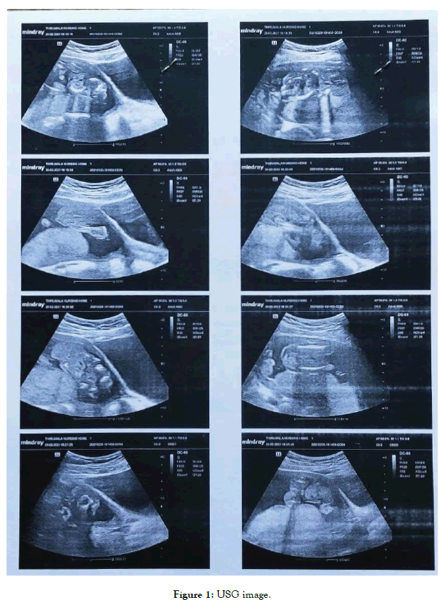
All her blood parameters are with in normal range. Immediately TIFFA scan was performed which showed single, live foetus in cephalic position, placenta in anterior upper segment Grade II umbilical cord showing 2 arteries, 1 vein with gestational age 19 to 20 weeks. But to draw biparietal diameter (BPD) it was noted that foetal skull vault was absent, Frog eye sing seen. Another notable finding is absence of anterior abdominal wall with a mass formed at the site of umbilicus [1-3]. And also noted that spinal card with limbs and rib cage were also absent. Impression of USG is given as multiple fetal anomalies, with Anencephaly and Gastrochisis, Neural tube defects and Polyhydramnios (pol-e-hi-DRAM-nee-os) (Table 1 and Figure 1).
| Test name | Result | Unit | Bio. Ref. Range | Method |
|---|---|---|---|---|
| *CBP (Complete Blood Picture) | ||||
| Hemoglobin | 10.6 | gms % | 12.0 - 15.0 | Cyanmeth Hemoglobin Method |
| HCt | 31.1 | vol % | 36 - 46 | Electronic Pulse & Calculation |
| Total RBC count | 3.7 | mill/cumm | 3.8 - 4.8 | Electrical Impedance |
| Total WBC count | 12400 | cells/cumm | 4000 - 11000 | Electrical Impedance |
| Platelet count | 2.5 | lakhs/cumm | 1.5 - 4.5 | Electrical Impedance |
| Differential Count | ||||
| Neutrophils | 86 | % | 40 - 80 | Electrical Impedance |
| Eosinophils | 2 | % | 1 - 6 | Electrical Impedance |
| Lymphocytes | 10 | % | Female: 36-50; Male: 20-40 | Electrical Impedance |
| Monocytes | 2 | % | 02 - 10 | Electrical Impedance |
| Basophils | 0 | % | 0 - 1 | Electrical Impedance |
| Peripheral Blood Smear | ||||
| RBC | Normocytic, Normochromic | |||
| WBC | Neutrophilic Leucocytosis | |||
| Platelets | Adequate | |||
Table 1: Blood parameters.

Figure 1. USG image.
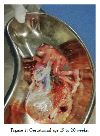
The pros and cons were explained to her and advised her to undergo medical termination of pregnancy. MTP was done, Fetus along with placenta and membranes expelled. Bleeding within normal limits check curettage was done. (Figure 2).

Figure 2. Gestational age 19 to 20 weeks.
Post OP findings uneventful patient improved symptoms and Germodynamically stable. Lab parameters are within normal limits; patient discharged with medication such as antibiotics and antiemetics and advised review after a week. Review scan showed no evidence of retained products of conception. No free fluid in abdomen, no lymphadenopathy [4,5].
Since it is evident that abore case is due to deficiency or womed her about the advantages of intake of folicacid in intake in first trimester and informed about the complication might repeat in future pregnancy if she do not take preconceptional folic acid therapy. Preconceptional usage of folic acid in women of child bearing of reduces rick of neural tube defects. Folic acid supplementation not only prevents NTD but also other ricks such as megatoblastic anemis, abmplio placenta, and intrauternine growth retardation. A dose of 4mg/day is given as prophylaxis in all pregnant women with history of NTD, according to WHO. Government of India is providing folic acid tablets with dose of 500mcg which meets the recommended daily allowance (RDA) of all pregnant women. The ideal prophylactic dose of folic acid is 400mcg which ideally should be given three months before pregnancy but atleast one month before pregnancy and continued till three months after pregnancy. In some countries folic acid fortification is done and made available in foods like pasta, cereals, bread.
Citation: Santhosh K, Naveena P (2021) Importance of Antenatal Folate Supplementation and Neural Tube Defects . J Women's Health Care 10:537. doi:10.35248/2167-0420.21.10.537.
Received: 21-May-2021 Accepted: 04-Jun-2021 Published: 11-Jun-2021 , DOI: 10.35248/2167-0420.21.10.537
Copyright: © 2021 Santhosh K, et al. This is an open access article distributed under the terms of the Creative Commons Attribution License, which permits unrestricted use, distribution and reproduction in any medium, provided the original work is properly cited.