Medicinal & Aromatic Plants
Open Access
ISSN: 2167-0412
ISSN: 2167-0412
Research Article - (2013) Volume 2, Issue 6
Keywords: Nardostachys jatamansi; Roots; Cytotoxicity; Human cancer cell lines; Antitumor
Since age old, plants have been the source of medicines for the treatment of various diseases. Regardless of the availability of a wealth of synthetic drugs, plants remain–even in the 21st century–an integral part of the health care in different countries, especially the developing ones. Efforts are being made to develop safe and cost effective anticancer agents from natural sources. As the age of modern medicine, single pure drugs emerged, and plant-derived active principles, their semisynthetic and synthetic analogs have served as a major route to new pharmaceuticals. Since 1961, plant-derived compounds have been approved for use as anticancer drugs: vinblastine (Velban®), vincristine (Oncovin®), etoposide (VP-16®), taxol (Paclitaxel®), etc [1].
Nardostachys jatamansi DC, (Valirenaceae) commonly known as muskroot, is indigenous to the Himalayan regions of India. Ttraditionally the plants have been used from many years for nervous headache, excitement, menopausal symptoms, flatulence, epilepsy and intestinal colic. The root is of bitter taste and used as aromatic, antispasmodic, diuretic, emmenagogue and nerve sedative in the Indian system of medicine [2]. In Ayurveda, roots and rhizomes of N. jatamansiare used to treat hysteria, epilepsy, and convulsions [3]. The decoction of the drug is also used in neurological disorders, insomnia and disorders of cardiovascular system [4]. The sesquiterpenes (Jatamansic acid, Jatamansone), lignans and neolignans are reported to be present in the roots of this plant [5,6].
Experimentally the plant is reported as anticonvulsant, hepatoprotective and hypolipidemic activities [7-9]. Extracts are reported to improve learning and memory activity in mice [10], antioxidant and lipid peroxidation activity in doxorubicin-induced cardiac damage in rats [11]. Also, the essential oil isolated from the plant had showed the fungicidal activity [12].
The present work was undertaken to evaluate the in vitro and in vivo antitumor potential of Nardostachys jatamansi roots parts.
Plant collection
Roots parts of Nardostachys jatamansi was collected from Himachal Pradesh in the month of October and were authenticated at site by the taxonomist of the Institute i.e., Indian Institute of Integrative Medicine, Jammu, India. A voucher specimen has been deposited at the herbarium of the Institute vide collection no. 17998.
Preparation of the extract and fractions: The dried root part of the plant (200g) was powdered and extracted with 95% alcohol under reflux for 30 min (repeated 3times). The combine solution was filtered and evaporated to dryness using a rotary evaporator under reduced pressure. Then, the initial alcoholic extract (ACE) was desiccated in the vacuum dryer at 400C for about 72 h and the dried extracts (718g) were obtained. The alcoholic extract (200g) was suspended in alcohol: water (1:1) and same was repeated to obtain 50% hydro-alcoholic extract (HAE). The residue was collected and suspended in water and process was repeated to obtain aqueous extract (AQE) respectively. Further, the alcoholic extract (10g) was successively fractionated with n- hexane, chloroform, n-butnol and water to give four fractions (HXF, CHF, BTF and AQF fraction). Fractions were dried with vacuum dryer. The extracts and fractions were kept at 4oC for preparation.
Cell lines and cell cultures: The human cancer cell lines were obtained from National Centre for Cell Science, Pune, India and National Cancer Institute, Frederick, USA. The human lung (A549), prostate (PC-3) and ovary (OVCAR–5) cells were grown and maintained in RPMI-1640 medium (pH 7.4), whereas DMEM was used for liver (HEP-2) cells. The media was supplemented with FCS (10%), penicillin (100units/ml), streptomycin (100μg/ml) and glutamine (2mM).
Preparation of positive controls: Positive controls like Adriamycin, Mitomycin-C and5-fluorouracil were prepared in distilled water. These were further diluted in gentamycin medium to obtain desired concentrations of 1×10⁻⁵ M and
2 ×10⁻⁵ M.
In vitro antiproliferative potential
Test material was subjected to in vitro anticancer activity against various human cancer cell lines [13]. In brief, the cells were grown in tissue culture flasks in growth medium at 37°C in an atmosphere of 5% CO2 and 90% relative humidity in a CO2 incubator (Hera Cell; Heraeus; Asheville, NCI, USA). The cells at sub confluent stage were harvested from the flask by treatment with trypsin (0.05% trypsin in PBS containing 0.02% EDTA) and suspended in growth medium. Cells with more than 97% viability (trypan blue exclusion) were used for determination of cytotoxicity. An aliquot of 100 μl of cells (105 cells/ml) was transferred to a well of 96-well tissue culture plate. The cells were allowed to grow for 24 h. Test material was then added to the wells and cells were further allowed to grow for another 48 h.
The antiproliferative SRB assay [14] was performed to assess growth inhibition which estimates cell number indirectly by staining total cellular protein with the dye SRB. In brief, the cell growth was stopped by gently layering 50 μl of 50% (ice cold) trichloroacetic acid on the top of growth medium in all the wells. The plates were incubated at 4oC for an hour to fix the cells attached to the bottom of the wells. Liquid of all the wells was then gently pipetted out and discarded. The plates were washed five times with distilled water and were air-dried. SRB 100 μl (0.4% in 1% acetic acid) was added to each well and the plates were incubated at room temperature for 30 min. The unbound SRB was quickly removed by washing the cells five times with 1% acetic acid. Plates were air-dried, tris buffer (100 μl, 0.01 M, pH 10.4) was added to all the wells to solubilize the dye and then plates were gently stirred for 5 min on a mechanical stirrer. The optical density was recorded on ELISA reader at 540 nm. Suitable blanks (growth medium and DMSO) and positive controls (prepared in DMSO and distilled water) were also included. Each test was done in triplicate and the values reported were mean values of three experiments.
In vivo studies
Non-inbred Swiss albino mice from an in-house colony were used in the present study. The experimental animals were housed in a standard size polycarbonate cages providing internationally recommended space for each animal. Animals were fed balanced mice feed supplied by M/s Ashirwad Industries, Chandigarh (India) and autoclaved water was available ad libitum. Animals were housed in controlled conditions of temperature (23 ± 2°C), humidity (50-60) and 12:12 of light: dark cycle. The experimental protocol was approved by the Institutional Animal Ethics Committee. Sarcoma 180 model was used to evaluate the samples [15]. Animals of the same sex weighing 20 ± 3 g were injected 1×10⁷ cells collected from the peritoneal cavity of non-in bread Swiss mice, with 8-10 days old ascitic tumor. Cells were then transplanted in the peritoneal cavity for ascites carcinoma and on right thigh for Sarcoma 180 model of fresh non-inbred Swiss mice on Day 0. The next day animals were randomized and divided into test groups with 7 animals each and one control group (15 animals). Test material (0.2 ml) was administered intraperitoneally (i.p) to test groups as suspension in 1% gum acacia (prepared in normal saline) for nine consecutive days. The control group was similarly administered with 1% gum acacia (0.2 ml, i.p) and 5-Flurouracil (22 mg/kg, i.p) was used as positive control. The percent tumor growth inhibition in test groups was calculated on Day 13 for by measuring the dimension of tumor.
Statistical analysis
The values were expressed as mean ± S.D, unless otherwise indicated. Comparisons were made between control and treated groups by one way analysis of variance (ANOVA) and the Student’s t-test. P values < 0.05 were considered significant.
Because of the high number of cancer cases, the interest in alternative therapies using natural products are increasing, especially those derived from plants. Traditional medicine and ethnobotanical information often play an important role in scientific research. Cytotoxicity is one of the main targets by chemicals to produce antitumor activity, number of anticancer drugs possess significant cytotoxic activity [16]. For anticancer activities several plant products have been tested and some of them are now available as the drugs of choice. The vast natural resources of India can provide effective anticancer agents. Whereas, the selection of experimental plant should be based on its traditional uses and testing the selected plants efficacy and safety on the basis of modern science.In order to look for new source of therapeutic agents, many plant extracts and active principle have been studied in vitro and in vivo cancer models, and the correlation of both studies became one of the key steps for the success of this type of research.
In vitro antiproliferative activity
Here in the current study we have used different human cancer cell lines, since different cell lines display various sensitivities towards cytotoxic compounds, the use of more than one cell line was necessary for the detection of these substances. The activity guided fractionation of all the three extracts [95% alcoholic extract (ACE), 50% hydroalcoholic extract (HAE) and aqueous extract (AQE)] of Nardostachy jatamansi, was monitored by in vitro cytotoxic assay in A-549 (lung), HEP-2 (liver), OVCAR-5 (ovary) and PC-3 (prostate) cancer cell lines. After 48-h sulforhodamine B cell viability assay was performed to determine the growth inhibition and cytotoxic properties of ACE and fractions. Here, the growth inhibition in a dose dependent manner was observed against all cell lines by all the extracts. Our results indicated that out of three extracts 95% alcoholic (ACE) extract had exhibited significant and dose-dependent inhibitory effects on all the human cancer cell lines in 48 h of treatment while rest of the extracts showed comparatively lower cancer inhibition (Figure 1).
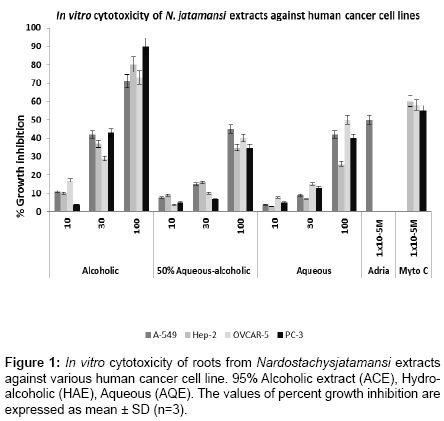
Figure 1: In vitro cytotoxicity of roots from Nardostachysjatamansi extracts against various human cancer cell line. 95% Alcoholic extract (ACE), Hydroalcoholic (HAE), Aqueous (AQE). The values of percent growth inhibition are expressed as mean ± SD (n=3).
The 95% alcoholic extract (ACE) showed percent growth inhibition of 11, 42 and 71 % against lung (A 549), 10, 37 and 80 % against liver (Hep-2), 17, 29 and 73 % ovary (OVCAR-5), 4, 43 and 90 % against prostate (PC-3) human cancer cell lines at 10, 30 and 100 μg/ml respectively (Figure 1). Anti-proliferative activity was observed in dose dependent manner and cell line specific. Percent growth inhibition demonstrated by 50% hydro-alcoholic extract (HAE) and aqueous extract (AQE) was lower than 50% at 100 μg/ml. Based on the result of extracts, 95% alcoholic extract was selected and further sequentially fractionated by various solvents and four fractions were prepared viz., n-hexane fraction (HXF), chloroform fraction (CHF), n-butanol fraction (BTF), aqueous fraction (AQF), respectively. These fractions were again evaluated for in vitro proliferation against same human cancer cell lines. It was observed that among all the tested fractions the n-butanol fraction (BTF) was comparatively more potential then rest of the fractions. n-butanol fraction (BTF) showed percent growth inhibition of 21, 45, 85% against lung (A 549), 28, 50 and 95% against liver (Hep-2), 25, 62 and 96% against ovary (OVCAR-5) and 17,23 and 55% against prostate (PC-3) at 10, 30 and 100 μg/ml followed by n hexane (HXF) fraction and chloroform (CHF) fraction (Figure 2). The hexane fraction showed percent growth inhibition in range of 84-89 % at 100 μg/ml and 40-54% at 30 μg/ml and chloroform fraction demonstrated 62-86 % growth inhibition at 100 μg/ml respectively against all the human cancer cell lines. Whereas, aqueous fraction (AQF) had showed less than 40% growth inhibition at 100 μg/ml against the entire human cancer cell lines (Figure 2). N-Hexane (HXF) fraction showed higher anti-proliferative activity against ovary (OVCAR -5) followed by Hep- 2 (liver)>A-549 (lung)>PC-3 (prostate) human cancer cell lines. The results revealed that extracts of N. jatamansi could decrease the survival rate of four human cancer cell lines in a dose- and time-dependent manner. Cell proliferation was significantly inhibited by ACE and its BTF fraction from the first day of culture.
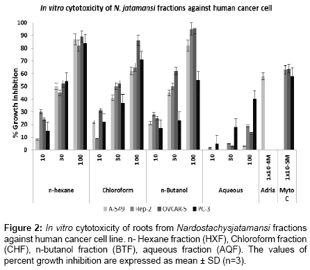
Figure 2: In vitro cytotoxicity of roots from Nardostachysjatamansi fractions against human cancer cell line. n- Hexane fraction (HXF), Chloroform fraction (CHF), n-butanol fraction (BTF), aqueous fraction (AQF). The values of percent growth inhibition are expressed as mean ± SD (n=3).
In vivo anticancer activity
As the extracts exhibit in vitro cytotoxic efficiency, the in vivo studies was also conducted by active n butanol (BTF) fraction of the 95% alcoholic (ACE) extract against Sarcoma-180 solid tumor model at 100 and 200 mg/kg of body weight respectively. The n butanol fraction showed significant 29.53 percent tumor growth inhibition on 9th day at 200 mg/kg of body weight compared to control mice. Positive control 5-fluorouracil was found to be highly significant and not much difference was observed between the different control groups (Table 1).
| Sample | Dose (mg/ kg i.p.) | Animal/Mortality | Body Weight (g) | Tumor Weight (mg) | % Tumor growth inhibition |
|---|---|---|---|---|---|
| Control | NS | 10/0 | 5.25 ± 0.51 | 110.75 ± 2.3 | - |
| BTF | 100 | 7/0 | 20.57 ± 0.64 | 1773.40 ± 71.21 | 10.23 ± 1.29 |
| 200 | 7/0 | 18.57 ± 0.36 | 1268.81 ± 78.51 | 29.53 +1.8* | |
| 5 FU | 22 | 7/0 | 19.11 ± 0.42 | 859.55 ± 77.12 | 55.4 ±1.2* |
Table 1: In vivo anti-proliferative activity of Nardostachys jatamnsi against sarcoma-180 (solid).
Although Nardostachy jatamansi has long been served as traditional medicines very few authentic scientific studies in field of cancer therapy are available. Here, we evaluated the potential anticancer components of Nardostachys jatamansi, extracted with different solvents from nonpolar to polar (n-hexane, chloroform, n-butanol and water), using bioactivity-based assays for evaluating its antitumor activity. The results clearly suggest that non polar alcoholic extract (ACE) and its n-butanol fraction (BTF) has the highest cytotoxic activities among all the three different extracts and four different fractions prepared against four human tumor cell lines. Hence, direct cytotoxic activity was observed by the plant and simultaneously, has significantly reduced the tumor growth when compared to the control group of the Sarcoma 180 tumor model. Meanwhile, some papers reports that roots of the plant contain Jatamansone or valeranone the principal sesquiterpene including other sesquiterpenes nardostachone, dihydrojatamansin, jatamansinol, jatamansic acid etc. [17-19]. Some crude extracts/fractions have compounds that would work synergistically and thus result in weaker anti-carcinogenic activity when separated than when used together in the intact crude extract. Perhaps non polar compounds present in the extracts as well as fraction are responsible for antiproliferative activity shown by the plant. These results are very consistent with others demonstrating the huge potential of the valerian family as a source of new drug development.
Traditionally the plant used widely for the treatment of various ailments, but scientifically few of them was screened out. In conclusion, we proved that roots of Nardostachy jatamansi both extracts and its fractions showed potential in vitro cytotoxicity and antitumor potential against Sarcoma-180 solid tumor model as well. Further, plant can be exploited for the development of the lead molecules for the cancer drug development.