Journal of Clinical and Experimental Ophthalmology
Open Access
ISSN: 2155-9570
ISSN: 2155-9570
Image Article - (2021)Volume 12, Issue 3
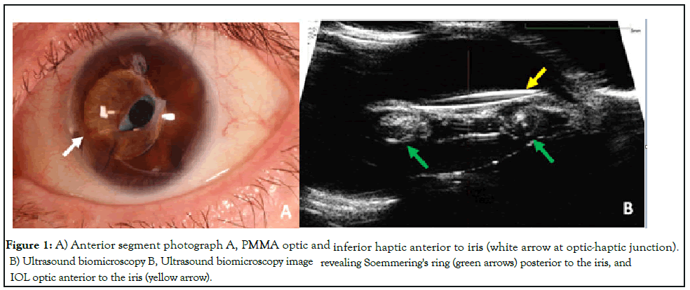
A 58-year-old Caucasian female presented to the clinic with progressive cloudy vision in her left eye. She had an ocular prosthesis in her right eye, and her visual acuity was 20/100 and intra-ocular pressure was 32 mm-Hg in her left eye. Exam revealed a polymethyl methacrylate (PMMA) lens with the optic and inferior haptic anterior to the iris. She had trace pigmented cells and a ring-shaped scar posterior to the iris consistent with a Soemmering's ring.. This was confirmed on ultrasound biomicroscopy and determined to be the causative etiology of her intraocular lens (IOL) dislocation.
Soemmering’s ring formation occurs from proliferation of lens epithelial cells within the peripheral capsule [1]. The formation and thickening of a Soemmering’s ring can cause contact between the intraocular lens (IOL) haptics and the posterior iris leading to iris defects, sphincter damage, as well as mechanical displacement of a sulcus IOL into the anterior chamber [1,2].Ultrasound biomicroscopy can be a useful diagnostic tool to view the anatomic relationship between the IOL haptics, iris and ciliary body[1] (Figure 1).

Figure 1: A) Anterior segment photograph A, PMMA optic and inferior haptic anterior to iris (white arrow at optic-haptic junction). B) Ultrasound biomicroscopy B, Ultrasound biomicroscopy image revealing Soemmering's ring (green arrows) posterior to the iris, and IOL optic anterior to the iris (yellow arrow).
Citation: Soekamto C, Shrestha S, Sohn JH (2021) Intraocular Lens Dislocation Secondary to Soemmering’s Ring. J Clin Exp Ophthalmol.12:878.
Received: 24-May-2021 Accepted: 07-Jun-2021 Published: 14-Jun-2021 , DOI: 10.35248/2155-9570.21.12.878
Copyright: © 2021 Soekamto C, et al. This is an open-access article distributed under the terms of the Creative Commons Attribution License, which permits unrestricted use, distribution, and reproduction in any medium, provided the original author and source are credited.