Anesthesia & Clinical Research
Open Access
ISSN: 2155-6148

ISSN: 2155-6148
Letter - (2023)Volume 14, Issue 3
Double-Chambered Right Ventricle (DCRV) is an uncommon congenital or acquired malformation with anomalous muscle bundles in the Right Ventricle (RV). It is most frequently associated with Peri-membranous Ventricular Septal Defect (PMVSD) [1]. Presentation is usually breathlessness, decreased exercise tolerance, chest pain and syncope or even cyanosis if the muscle bundles are located distal to the VSD (~60%), leading to right-to-left shunting across the VSD [2]. Surgical treatment is suggested in patients with significantly elevated mid ventricular pressure gradient of >40 mm Hg [3]. For the dynamic component of obstruction, β blockers or calcium channel blockers should be started, and intraoperative use of inotropes should be avoided. Hemodynamic goals in such situation will be maintaining right ventricular preload, left ventricular afterload, and a sinus rhythm [4]. We present an interesting example of intraoperative dynamic obstruction due to anomalous RV muscle bundle. An 11 year old female presented with symptoms of progressive shortness of breath and exertional chest pain. She was a known case of Ventricular Septal Defect (VSD) diagnosed in childhood but didn't undergo any intervention. On physical examination, pan systolic murmur (grade IV/VI) was heard at the left sternal border. Chest X ray had RV type of apex in cardiac shadow. Electrocardiogram suggestive of Right Ventricular Hypertrophy (RVH).
Anesthetic agents that cause vasodilation or hypotension should be used with caution, as they may exacerbate dynamic obstruction. Regional anesthesia techniques may be useful in some cases, as they can minimize the need for general anesthesia and reduce the risk of hemodynamic instability. However, they should be used with caution in patients with DCRV, as sympathetic blockade may cause vasodilation and decrease systemic vascular resistance, potentially exacerbating dynamic obstruction.
Transthoracic Echocardiogram (TTE) showed a large perimembranous VSD (PM-VSD) with left to right shunt. In addition, a muscular septation was noted within the RVOT causing obstruction with a peak gradient of 136 mm Hg. The pulmonic valve has doming. Patient planned for intra cardiac repair of PM-VSD and muscle bundle coring to relieve obstruction.
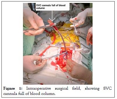
After standard ASA monitors attached, anaesthesia was induced with sevoflurane/oxygen and Fentanyl (2 mcg/kg) followed by rocuronium (0.1 mg/kg) and intubation with 6 mm ID cuffed ETT. Right internal jugular central venous line and right femoral arterial were secured after induction. Blood pressure of 108/66 mmHg, Heart Rate of 88/min NSR, SpO2 of 100% and CVP of 5 cm H2O was noted as baseline. Maintenance was done with sevoflurane/Oxygen/air. After adequate heparinization aortic cannulation was done. To our suprise, on SVC cannulation, blood was overflowing through the cannula, so clamped (Figure 1) and simultaneously ETCO2 fell from 34 to 29 mmHg. It was instantaneously followed by tachycardia (120/min) and fall in BP to 70/32 mmHg. Keeping in mind the muscle bundles in RV leading to probable dynamic obstruction, we started fast IV fluid and phenylephrine 20 mcg. As aortic and svc cannula were already inserted, immediate initiation of CPB was done. We reported this case as dynamic obstruction would have happened at any time in preoperative period or during induction or with sympathetic stimulation during surgery. It warrant knowledge and implication of dynamic obstruction due to muscle bundles and prompt recognition and its management.

Figure 1: Intraoperative surgical field, showing SVC cannula full of blood column.
Double-chambered right ventricle with anomalous muscle bundles is a rare and challenging condition to manage. In this case, the patient presented with symptoms of progressive shortness of breath and exertional chest pain. Physical examination and diagnostic imaging confirmed the presence of a peri-membranous VSD and an anomalous muscle bundle causing obstruction with a peak gradient of 136 mm Hg. During induction, the team encountered an unexpected overflow of blood through the SVC cannula, accompanied by a drop in ETCO2, tachycardia, and hypotension. The immediate suspicion was dynamic obstruction due to muscle bundles, and the team started fast IV fluid and phenylephrine while initiating CPB to manage the hemodynamic instability. This case highlights the importance of recognizing and managing the dynamic obstruction caused by anomalous muscle bundles in patients with double-chambered right ventricle. Preoperative diagnosis and hemodynamic monitoring during surgery are critical in preventing potential complications. It is also important to have a low threshold for initiating CPB to manage any sudden hemodynamic instability in these patients.
[Crossref] [Google Scholar] [PubMed]
[Crossref] [Google Scholar] [PubMed]
[Crossref] [Google Scholar] [PubMed]
[Crossref] [Google Scholar] [PubMed]
Citation: Upadhyay R (2023) Managing Dynamic Obstruction in Double-Chambered Right Ventricle. J Anesth Clin Res. 14:1102.
Received: 14-Apr-2023, Manuscript No. JACR-23-21984; Editor assigned: 18-Apr-2023, Pre QC No. JACR-23-21984 (PQ); Reviewed: 02-May-2023, QC No. JACR-23-21984; Revised: 09-May-2023, Manuscript No. JACR-23-21984 (R); Published: 16-May-2023 , DOI: 10.35248/2155-6148.23.14.1102
Copyright: © 2023 Upadhyay R. This is an open-access article distributed under the terms of the Creative Commons Attribution License, which permits unrestricted use, distribution, and reproduction in any medium, provided the original author and source are credited.