Research Article - (2016) Volume 2, Issue 3
New Natural Medium Using Vitis vinirfera for Siderophore Production from Clinical Isolates of Klebsiella pneumonia
*Corresponding Author: Neihaya H. Zaki, Department of Biology, Collage of science, Al-Mustansiryah University, Baghdad, Iraq Email:
Abstract
Culture media used for isolation and identification of bacteria according to their biochemical and physiological properties, and this new media is cheap and available for use and could be a useful for study the virulence of bacteria and siderophore production. This study included isolation of 50 isolates of K. pneumoniae from different clinical sources from different hospitals in Baghdad city. The number and percentage of isolates according to the sources (urine, blood, sputum, burns, ear swabs, pus, wounds and stool) were 22(44%), 11(22%), 4(8%), 4(8%), 3(6%), 3(6%), 2(4%) and 1(2%) respectively. About 72% (36/50) were indicate as virulence isolates, and 60% (30/50) of isolates produce siderophores on M9 medium, while 70% (35/50) of isolates that produce siderophores when grown on new media. This study aimed to prepare a new natural medium using Vitis vinirfera, and determines the ability of Klebseilla pneumoniae to produce siderophore on it, and relationship between siderophore production and virulence isolates of K. pneumoniae.
Keywords: New natural media; Vitis vinirfera; Klebsiella pneumonia; Siderophore production
Introduction
For any bacterium to propagate for any purpose it is necessary to provide the appropriate biochemical and biophysical environment. The food base that supports the growth of an organism called culture medium; the biochemical (nutritional) environment made available in this culture medium [1]. The food base depending upon the special needs of particular bacteria (as well as particular investigators), so that a large variety and types of culture media have been developed with different purposes and uses. These include sources of organic carbon, nitrogen, phosphorus, sulfur and metal ions including iron. Culture media employed in the isolation and maintenance of pure cultures of bacteria and used for identification of bacteria according to their biochemical and physiological properties [2].
Grapes (Vitis vinifera ) provide many nutrients like carbohydrates (glucose), vitamins, minerals, fibers, phytochemicals and antioxidants. The functional quality of grape fruit characterized by its metabolic compositions. It contains a number of secondary metabolites like flavonols, anthocyanins, proanthocyanidins, stilbene derivatives [3]. The minerals iron, potassium, zinc, manganese, and calcium were present in higher concentrations [4].
Siderophore term is Greek for “iron carrier” and so named because these molecules produced by microorganisms have an extremely high affinity for bind ferric iron and transport it into the bacterial cell. Siderophore is low molecular weight organic molecules [5]. Siderophores have related to virulence mechanisms in microorganisms pathogenic to both animals and plants. In addition, they have clinical applications and are possibly important in agriculture [6]. Siderophores exhibit considerable structural variability and affinity for iron, which determines the growth of a microorganism under competitive conditions when availability is a limiting factor [7].
Klebsiella pneumoniae -siderophore producers- is an opportunistic pathogen responsible for causing a spectrum of hospital communityacquired and nosocomial infection and especially infect patients with indwelling medical devices such as urinary catheters [8]. K. pneumonia is member of Enterobacteriaceae, which is ubiquitously present in the environment such as soil, vegetation, water and from mammalian mucosal surfaces [9].
This study aimed to prepare a new natural medium using Vitis vinirfera , and its low cost medium for siderophore production when Klebseilla pneumoniae were grown on it, and determine if there are any relationship between siderophore production and virulent isolates of Klebseilla pneumoniae .
Material and Methods
The plant collection
Mature and fresh Vitis vinirfera fruits (local name Gooseberry or raisin) collected from markets in Baghdad city. It classified by members in Botany department, college of science, AL-Mustansiriya University. Plant samples carefully washed under running tap water followed by air dried at room temperature (25°C) for five days, crushed into powder using a sterilized electric blender, and stored in airtight bottles.
The Plant extraction and new medium preparation
It done by using cold-water extraction, which is dissolving the powder of plant in cold distilled water using microwave, then the Gooseberry extract is ready for use in media. Composition of the medium was; Gooseberry extract, agar, bile salt, Fe2 (SO4)3, KH2PO4. This medium sterilized by autoclave, cooled and poured in petri dishes to be ready for culturing K. pneumoniae .
Bacterial isolates
Fifty isolates of Klebsiella pneumoniae isolated from three hospitals in Baghdad city (Ibn-El Balady, Al-Kendy teaching and Teaching laboratories in medical city) during period from July/2014 to December/2014. These bacteria isolated from different clinical sources including (urine, blood, sputum, ear swab, burn, stool, wounds and pus).
Identification of bacterial isolates
Initial diagnostic of isolates based on morphological characteristic of the colonies that includes colony shape, colony texture, color, edges studied depending on bacterial growth on MacConkey agar, and blood agar, while microscopic examination exhibited cell shape and arrangement of cells by stained the isolates with Gram stain [10].
Confirmation of bacterial diagnosis done by VITEK-R2 Compact system, which dedicated the identification and susceptibility testing of clinically significant bacteria. The isolates were diagnostic depending on many biochemical tests such as (urease, H2S production, fermentation/ Glucose, lipase, citrate, phosphatase etc).
Detection of virulent K. pneumoniae isolates
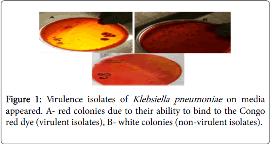
Screening of virulent K. pneumoniae isolates done using Congo red binding assay as described by [11]. Briefly, K. pneumoniae isolates streaked on MacConkey agar and incubated at 37°C for 24 hrs. All the isolates tested for their growth on trypton soy agar (Oxoid, UK) supplemented with 0.015% bile salt and 0.03% Congo red. After 24 hrs of incubation, the cultures left at room temperature for 48 hours to facilitate the annotation of results. Virulent isolates identified by their ability to bind to the Congo red dye and they appeared as red colonies while those that appeared white considered negative.
Siderophores production using M9 media
K. pneumoniae isolates inoculated on M9 media, which prepared according to [12]. It consisted of; solution-1 prepared by dissolving 3 g Na2HPO3, 1.5 g KH2PO4, 0.5 g NH4Cl and 10 g agar in 475 ml of distilled water, then sterilized in autoclave at 121°C. Solution- 2 prepared as followed 2 ml of (1M) MgSO4, 10 ml of (20%) glucose, 0.1 ml of (1M) CaCl2 sterilized by filtration.
Solution 2 was added to solution 1 (after cooling to 50°C), then 0.01562 g of (200) μm of Dipyridyl added. Overnight-activated isolates inoculated to this media and incubated in 37°C for 24 hr, the results based on the presence of growth or not.
Siderophores production using new media
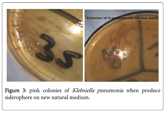
K. pneumoniae isolates inoculated on new media, after activation on brain heart infusion broth at 37°C for 24 hrs. New media prepared to be ready for culturing with K. pneumoniae as mentioned above. Pink large colonies give positive results, while absents of growth give negative results.
Results and discussion
Prevalence of Klebsiella pneumoniae in Clinical specimens exhibited in (Table 1), which show the number and percentage of isolates according to sources were as follow: 22(44%) isolates from urine, 11(22%) blood, 4(8%) sputum, 4(8%) burn patients, 3(6%) ear swab, 3(6%) pus, 2(4%) wounds infection, and 1(2%) stool.
| Type of specimen | Number of isolates | K. pneumoniae isolates (%) |
|---|---|---|
| Urine | 22 | 44 |
| Blood | 11 | 22 |
| Sputum | 4 | 8 |
| Burn | 4 | 8 |
| Ear swab | 3 | 6 |
| Pus | 3 | 6 |
| Wounds | 2 | 4 |
| Stool | 1 | 2 |
| Total | 50 | 100 |
Table 1: prevalence of Klebsiella pneumoniae in Clinical specimens.
The wildly spread of bacteria in hospitals environment in some Baghdad hospitals is the main reasons made this pathogen to multidrug resistant and cause nosocomial infections. K. pneumoniae is an opportunistic pathogen causing serious infections such as pneumonia, urinary tract infection and septicemia. Klebsiella is second only to Escherichia coliin nosocomial Gram-negative bacteremia, as well as in urinary tract infections (UTIs), affecting catheterized patients [13].
The numbers of virulence isolates were (36/50) 72%, when change the color of medium and give a red colonies, while (14/50) 18% of isolates considered a non-virulent isolates according to non-change on color of the medium and give a white colonies (Figure 1).
This result agrees with the results of [11] out of the 33 K. pneumoniae , only 25 (75.8%) virulent isolates showed positive results. Virulence can test by Congo red test due to the presence of a strong correlation between expression of Congo red phenotype and virulence in bacteria. This might be associated with the presence of β-glucan in bacterial cell wall suggesting that Congo red binding can act as a virulence marker [14], while [15] who reported that in vitro pathogenicity testing of E. coli isolates revealed that 46 out of 97 (47.4%) of the isolates were positive for the Congo red binding.
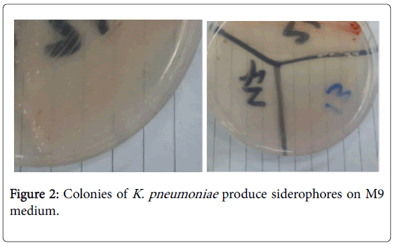
The percentage of K. pneumoniae , which produce siderophore (growth) on M9 medium were 60% (30/50), while 40% (20/50) not produce siderophore (no growth) (Figure 2).
Siderophores implicated as bacterial virulence factors. The role of iron acquisition systems is especially important in light of new findings that siderophores may represent a key front in the interplay between host and pathogen [16]. Subhi and Shaker [17] study the ability of Staphylococcus aureus , and Klebsiella pneumoniae isolated from rhinitis cases to produce siderophores as a virulence factor by using Rogers’s method for extraction of siderophores and then the chemical and biological assay performed to detect siderophore (Figure 3).
The isolates grown in new media to study the different between the siderophore productions in original media with new media, 70% (35/50) only of isolates that produce siderophores grown on new media.
This new media is simple, cheap and available for use and could be a useful media for study the virulence of bacteria and siderophore production. Nutrition content of gooseberry such as Energy 184 kj, Carbohydrates 10.18 g, Dietary fiber 4.3 g, Fat 0.58, Protein 0.88 g, Water 87.87, vitamin C. These nutrients were sufficient to support the growth of gooseberry-pathogenic bacteria [18]. New medium also supported the growth of bacteria (more than M9 medium) even these none or weak grower showed good grow on it indicating that they might require trace elements for growth rather than have sensitivity to some materials in other media.
The relationship between siderophore production and virulence isolates of Klebsiella pneumoniae on new medium revealed in (Table 2), in addition to that clinical source of isolates may play an important role in virulence.
| No. of Isolate | Sources | Male or Female | Virulent or Non-virulence | Siderophore production | media |
|---|---|---|---|---|---|
| 1 | Sputum | Female | Non-virulence | + | - |
| 2 | Urine | Male | Virulence | - | |
| 3 | Urine | Female | Non-virulence | - | |
| 4 | Urine | Female | Virulence | + | + |
| 5 | Ear Swab | Male | Non-virulence | - | |
| 6 | Urine | Female | Virulence | - | |
| 7 | Urine | Female | Virulence | + | _ |
| 8 | Urine | Female | Virulence | + | _ |
| 9 | Urine | Female | Non-virulence | + | _ |
| 10 | Urine | Female | Non-virulence | + | + |
| 11 | Blood | Female | Virulence | + | + |
| 12 | Wounds | Male | Non-virulence | - | |
| 13 | Ear Swab | Male | Non-virulence | + | _ |
| 14 | Burn | Female | Virulence | + | + |
| 15 | Pus | Male | Non-virulence | + | + |
| 16 | Urine | Female | Virulence | - | |
| 17 | Urine | Female | Virulence | + | _ |
| 18 | Urine | Female | Virulence | + | _ |
| 19 | Blood | Male | Non-virulence | - | |
| 20 | Stool | Male | Virulence | + | + |
| 21 | Wounds | Female | Virulence | + | + |
| 22 | Urine | Female | Non-virulence | - | |
| 23 | Urine | Female | Virulence | - | |
| 24 | Urine | Female | Virulence | + | + |
| 25 | Urine | Male | Non-virulence | + | + |
| 26 | Urine | Female | Non-virulence | - | |
| 27 | Urine | Female | Virulence | + | + |
| 28 | Urine | Male | Virulence | - | |
| 29 | Urine | Female | Non-virulence | - | |
| 30 | Urine | Female | Virulence | + | _ |
| 31 | Sputum | Male | Virulence | + | + |
| 32 | Pus | Male | Virulence | + | + |
| 33 | Blood | Female | Virulence | - | |
| 34 | Blood | Male | Virulence | + | + |
| 35 | Blood | Female | Virulence | - | |
| 36 | Blood | Female | Virulence | - | |
| 37 | Blood | Female | Virulence | + | + |
| 38 | Blood | Female | Virulence | - | |
| 39 | Blood | Female | Virulence | - | |
| 40 | Blood | Male | Virulence | + | + |
| 41 | Blood | Female | Virulence | - | |
| 42 | Sputum | Male | Virulence | + | + |
| 43 | Burn | Male | Virulence | + | + |
| 44 | Burn | Male | Virulence | - | |
| 45 | Ear Swab | Female | Virulence | + | + |
| 46 | Burn | Female | Non-virulence | + | _ |
| 47 | Sputum | Male | Virulence | + | + |
| 48 | Urine | Male | Virulence | + | + |
| 49 | Blood | Male | Virulence | - | |
| 50 | Pus | Male | Virulence | + |
Table 2: The relationship between siderophore production and virulence isolates.
Microorganisms require iron for a variety of metabolic processes, so they synthesize and secrete siderophores that actively chelate iron and remove it from eukaryotic iron-binding proteins like lactoferrin & transferring. Thus, iron is a key element of bacterial pathogenesis [19,20]. Vagrali et al., [21] carried out studies that showed siderophores considered as urovirulence markers of uropathogenic E. coli , so siderophore production may be a necessary feature of a virulent bacterium but not a determinant of virulence. Staphylococcus aureus , and Klebsiella pneumoniae produce siderophores and study it as a virulence factor, the results showed ability of all strains to produce siderophores, which confirmed its roles in pathogenesis [17].
References
- Al-Azzauy AAM, Hana DB,Abdalah ME(2011) The use of the water extract of Rosaspp petals as a bacterial growth medium. AJPS 10:84-94.
- Engelkirk PG,Duben-Engelkirk J (2007) Laboratory diagnosis of infectious diseases: Essentials of diagnostic microbiology. LippincottWilliams and Wilkins, USA: 133-134.
- Sindhus S, Radhai SS (2015) Versatile health benefits of active components of grapes (Vitisvinifera). Ind J ApplRes 5:289-291.
- Siham JM (2006) Astudy of the effect of some antigens of Klebsiellapneumoniae on the immune response. PhD thesis. Biology department, College of science, Al-Mustansiriya university.
- Paul A, Dubey R (2015) Characterization of protein involved in nitrogen fixation and estimation of co-factor. Int J Curr Res Biosci Plant Biol 2: 89-97.
- Verma V, Joshi K,Mazumdar B (2012) Study of siderophore formation in nodule-forming bacterialspecies. Res J ChemSci2: 26-29.
- Dimkpa CO, Merten D, Svatoš A, Büchel G, Kothe E (2009) Metal-induced oxidative stress impacting plant growth in contaminated soil is alleviated by microbial siderophores. SoilBiolBiochem 41:154-162.
- Claudia V, Francesca L, Maria PB, Gianfranco D,Pietro EV (2014) Antibiotic resistance related to biofilm formation in Klebsiellapneumoniae. Pathogens 3: 743-758.
- Sharma SK,Mudgal NK, Sharma P,Shrngi BN (2015) Comparison of phenotypic characteristics and virulence traits of Klebsiellapneumoniaeobtained from pneumonic and healthy camels (Camelusdromedarius). AdvAnim Vet Sci 3: 116-122.
- Atlas RM, Parks LC, Brown AE(1995) Experimental microbiology: Laboratory manual.(1st edn.) Mosby, USA.
- Fatma IS,Tarek EB, Ahmed AA, Lamiaa AM (2012) Conjugative plasmid mediating adhesive pili in virulent Klebsiellapneumoniaeisolates. Arch ClinMicrobio 3: 1-9.
- West SA, Buckling A (2003) Cooperation, virulence and siderophore production in bacterial parasites. ProcBiolSci 270: 37-44.
- Niveditha S,Pramodhini S, Umadevi S, Kumar S, Stephen S (2012) The isolation and the biofilm formation of uropathogens in the patients with catheter associated urinary tract infections (UTIs). J ClinDiagn Res 6: 1478-1482.
- Berkhoff HA, Vinal AC (1986) Congo red medium to distinguish between invasive and non-invasiveE. coli pathogenic or poultry. Avian Dis 30: 117-121.
- Shamlal R, Rajarathnam S, Sankaran K, Ramachandran V, Subrahmanyam YV, et al. (1997) Detection of virulent Shigella and enteroinvasiveEscherichia coliby induction of the 43 kDainvasion plasmid antigen, ipaC. FEMS Immunol Med Microbiol17: 73-78.
- Singhai M, Malik A, Shahid M, Malik MA,Goyal R (2012) A study on device related infections with special reference to biofilm production and antibiotic resistance. J Global Infec Dis4:193-198.
- Subhi HK, Shaker GJ (2011) Detection of siderophores from Staphylococcus aureus, Klebsiella pneumonia Isolated from rhinitis cases. Res J BasEduc11: 568-578.
- SathiyaVimalS, Vasantha Raj S, SenthilkumarRP, Jagannathan S (2013) Natural sources of gooseberry component used for Microbial Culture Medium (NSM). J ApplPharmlSci 3: pp. 040-044.
- Pal RB, Gokarn K(2010) Siderophores and pathogenecity of microorganisms. JBiosci Tech 1: 127134.
- BucklingA, Harrison F,Vos M,Brockhurst MA, Gardner A,et al.(2007) Siderophore-mediated cooperationand virulence inPseudomonas aeruginosa. FEMS MicrobiolEcol 62:141.
- Vagarali MA, Karadesai SG, Patil CS, Metgud SC, Mutnal MB (2008)Haemagglutination and siderophore production as the urovirulence markers of uropathogenicEscherichia coli. Ind JMed Microbiol 26:68-70.
Copyright: © 2016 Zaki NH, et al. This is an open-access article distributed under the terms of the Creative Commons Attribution License, which permits unrestricted use, distribution, and reproduction in any medium, provided the original author and source are credited.


