Pediatrics & Therapeutics
Open Access
ISSN: 2161-0665
ISSN: 2161-0665
Case Report - (2024)Volume 14, Issue 3
We are presenting an interesting case of 6 year old with abdominal distension who was symptomatic for 2 years, worked up in the lines of abdominal tuberculosis. He was started antitubercular drugs based on ascitic fluid ADA. As there was no improvement even after 9 months of antitubercular drugs a contrast enhanced CT abdomen was done which showed omental cyst. This case shows that in Tubercular endemic countries like India, we need to look out for other diagnosis as well besides tuberculosis.
Omental cyst; Abdominal; Tuberculosis; Antitubercular drugs
Omental cysts are lymphatic endothelium lined unilocular or multilocular cysts filled with serous fluid arising from obstructed omental lymphatic channels. It can be both congenital and acquired. When small they are asymptomatic and are picked up accidentally on ultrasound. The larger cysts become symptomatic and present with ascites, pain abdomen or a palpable abdominal mass. Common complications which can be seen are infection, torsion and rupture. Cysts or tumors arising from mesentery, peritoneum and retro peritoneum comprise of its differential diagnosis. Treatment involves resection of the cyst [1].
To prevent complications and alleviate symptoms. Surgical removal is typically performed through laparotomy or laparoscopy, depending on the size and location of the cyst. Complete resection is crucial to prevent recurrence. In some cases, if the cyst is adherent to vital structures, partial resection might be performed, but this can increase the risk of recurrence. Postoperative recovery is generally uneventful, but follow-up is necessary to monitor for any signs of recurrence or complications. Omental cysts are relatively rare, and their diagnosis can be challenging due to the non-specific nature of the symptoms and the wide range of differential diagnoses. Imaging studies, such as ultrasound, CT scans, and MRI, play a crucial role in the preoperative assessment, helping to delineate the cyst's size, location, and relationship with adjacent organs. Aspiration of the cyst fluid may be performed for diagnostic purposes but is not recommended as a definitive treatment due to the high likelihood of recurrence [2].
Histopathological examination of the resected cyst confirms the diagnosis and helps rule out other potential causes, such as neoplastic or infectious processes. In children, omental cysts are more often congenital, whereas in adults, they may be associated with trauma, infection, or previous surgeries. Overall, timely recognition and surgical management of symptomatic omental cysts are essential to prevent complications and ensure a good prognosis. Continued research and case studies are necessary to further understand the etiology, optimal management strategies, and long-term outcomes for patients with omental cysts. We are presenting an interesting case of 6 year old male with omental cyst who had been symptomatic for 2 years and had been misdiagnosed as abdominal tuberculosis and had received full course of Antitubercular Drugs (ATD) [3].
A 6 year old male child was admitted with chief complaints of abdominal distention for 2 years and decreased appetite for 1 month. The abdominal swelling had increased gradually over the last 2 years and was not responding to treatment taken by the child over the last 2 years. Fluid tapped from the abdominal swelling 10 months ago was sent for cytology and Adenosine Deaminase (ADA). ADA was 24 u/L was considered positive [4]. Fluid cytology revealed 32 cells of which 20% was neutrophils and 80% was lymphocytes. Sugar was 103 mg/dl, protein 4.13 gm/dl.
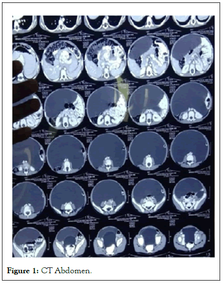
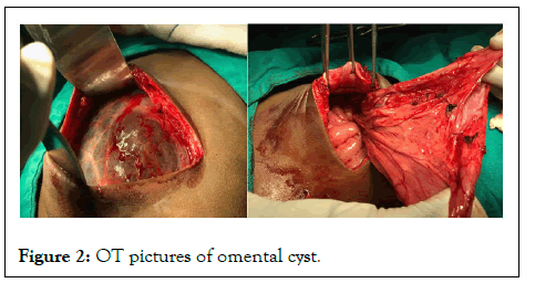
Ultrasound showed gross fluid in abdomen with internal echoes. Based on these tests antitubercular drugs were started. Child took antitubercular drugs for 6 months as there was no response he was taken to another hospital where a peritoneal fluid was again tapped from the abdomen [5]. Cytology of repeat tap showed 120 cells with 35% neutrophils and 65% lymphocytes, glucose was 101 mg/dl and protein was 4.69 gm/dl. This time a contrast enhanced CT abdomen was advised. Child was then admitted in our hospital. On examination he was undernourished. His abdomen was distended but flanks were spared. Rest of the systemic examination was normal. On investigation, Hb was 12.7 gm/dl, Total leucocyte count was 6.094 × 10³/μL (Polymorphs 47%, Lymphocyte 44% Eosinophil; 4% Monocyte 5%) Platelet count was 350 × 103/μL. His liver function test, renal function test and serum electrolytes were normal. Blood coagulation study was also normal [6]. Virology test (HIV, hepatitis B and hepatitis C) were negative. Repeat ultrasound done at our centre revealed a large cystic lesion (19 cm × 13 cm × 16 cm) in the abdominopelvic region displacing the visceral organs and bowel loops likely omental cyst. A contrast enhanced CT abdomen was also done as shown in Figure 1. A huge cystic swelling containing 1.2 litre of serous fluid was resected by lapratomy as shown in Figure 2. The swelling extended superiorly behind inferior border of liver compressing stomach, small bowel upto the pelvis and extended laterally to anterior axillary line compressing the kidneys. Post excision child recovered well, his appetite improved [7].

Figure 1: CT Abdomen.

Figure 2: OT pictures of omental cyst.
It was way back in 1852 when the first report of omental cyst was published omental cysts are fluid filled unilocular or multilocular. They can be simple or multiple and can vary in diameter from few milimeters to more than 40 cm [8]. They usually present with abdominal swelling and need to be differentiated from Intestinal duplication cyst, ovarian cyst in females, choledochal, pancreatic, splenic, or renal cysts, hydronephrosis, cystic teratoma, hydatid cyst and ascites. Good history examination and radiological work up will help to differentiate these. In majority of times when the cyst is small it remains undiagnosed and is accidentally picked up in radiology of abdomen. The problem arises when it is large. Sometimes they can present in emergency as acute abdomen mimicking small-bowel obstruction [9]. There have been occasional case reports where an omental cyst has presented with extra gastrointestinal stromal tumor. Infact there was one case report of acute hemorrhagic anemia in an infant due to giant omental hemorrhagic cyst. No medical treatment works and definitive treatment is excision. In our case the child was symptomatic for 2 years but unfortunately the possibilty of cyst was never kept. He was continuously worked up in line of abdominal tuberculosis [10]. Ascitic fluid tapping was done twice. Radiological work up was done late in the course of treatment. In India tuberculosis is endemic but it can be differentiated from other illness by proper history examination and investigations. The antitubercular treatment was continued for 6 months even when the patient did not respond to treatment and the abdominal distension was persisting.
This case report highlights the importance of proper history taking, examination and adequate work up before starting antitubercular drugs. This policy should be followed even in a countries where tuberculosis is endemic. Further starting antitubercular drugs just on the basis of adenosine deaminase activity is never justified. We must keep the differentials of congenital mesentric ,omental cyst etc in our differential for abdominal swellings particularly in pediatric population.
[Google Scholar] [PubMed]
[Crossref] [Google Scholar] [PubMed]
[Crossref] [Google Scholar] [PubMed]
[Crossref] [Google Scholar] [PubMed]
[Crossref] [Google Scholar] [PubMed]
[Crossref] [Google Scholar] [PubMed]
[Crossref] [Google Scholar] [PubMed]
Citation: Thakur N, Roshan T, Singh S (2024) Omental Cyst a Missed Diagnosis: A Case Report. Pediatr Ther. 14:556.
Received: 15-Jan-2024, Manuscript No. PTCR-24-4741; Editor assigned: 18-Jan-2024, Pre QC No. PTCR-24-4741 (PQ); Reviewed: 01-Feb-2024, QC No. PTCR-24-4741; Revised: 30-May-2024, Manuscript No. PTCR-24-4741 (R); Published: 27-Jun-2024 , DOI: 10.35841/2161-0665.24.14.559
Copyright: © 2024 Thakur N, et al. This is an open-access article distributed under the terms of the Creative Commons Attribution License, which permits unrestricted use, distribution, and reproduction in any medium, provided the original author and source are credited.