Research Article - (2023)Volume 7, Issue 1
Clear aligner treatment is considered to be helpful for periodontitis orthodontics because these aligners are easy to wear and take off. However, studies have shown that the instantaneous force associated with such aligners may affect periodontal tissues. The goal of this study was to identify the optimal orthodontic displacement of clear aligners concerning torque movement using a Three-Dimensional (3D) finite element. We acquired Cone Beam Computed Tomography (CBCT) data from a patient without diabetes and other systemic diseases and who received invisible orthodontics. Based on the different degrees of alveolar bone absorption (1/3 and 1/2 of the root length were reduced horizontally from the alveolar crest along the Z-axis to form alveolar bone models with different degrees of absorption (M1, M2, and M3)), 3D-FEMs of the mandible were established using MIMICS 10.0 and ABAQUS 6.5 software. The 3D stress distribution diagrams and stress values for the teeth (lower incisor, central incisor, lower canine) under three different periodontal conditions (M1, M2, and M3) were then identified by ABAQUS 6.5. The stress values for the root and periodontal ligament increased when the periodontal condition was worse and increased as the level of displacement increased. For patients in which the alveolar bone exhibited 1/3 resorption, we found that when the displacement was 0.18 mm, the stress values of the roots of the anterior teeth (0.591 MPa2.406 MPa) were similar to those of the control group. The stress values of the anterior teeth of the PDL (0.048 MPa-0.216 MPa) were the same as the control group when the displacement was 0.15 mm. For patients in which the alveolar bone exhibited 1/2 resorption, the stress values of the roots of the anterior teeth (0.644-3.969 MPa) were similar to those of the control group; the PDL’s stress values at 0.10 mm (0.056-0.461 MPa) were similar. This experiment showed that when the current tooth is subjected to an additional torque of 5 degrees during lip tilt movement, a clear aligner should be used to reduce the displacement, to avoid greater stress concentration on the root and periodontal ligament. The optimal displacements of M2 and M3 periodontal patients were 0.15 mm and 0.10 mm, respectively.
Clear aligner; Anterior teeth; Periodontal ligament; Displacement
Over recent years, Clear Aligner Treatment (CAT) has been applied extensively and the indications for such treatment have become wider than ever before. There are a series of thermoplastic aligners involved; the teeth can be moved by deforming the aligners. In the early stages, clear aligners were considered as a treatment for simple problems, such as mild crowding and spaces in the arch. However, as this technology developed, clear aligners are becoming increasingly used to treat complicated malocclusions [1]. With the continuous improvement of materials and the study of biomechanics, the CAT is becoming increasingly more reliable.
In the contest to fixed orthodontics, CAT enables teeth to move expectedly to reach a target position via differential displacement between the appliance and the teeth [2]. An increasing body of research has demonstrated that CAT exhibits high efficacy for moving certain types of teeth [3,4]. This efficacy can be further improved by investigating attachment, the steps taken to move teeth, and the setting of tooth-moving stagings [5].
Evidence shows that CAT is increasingly being accepted by adult patients; this is important since the incidence of periodontal diseases among adults can be as high as 76% -92% [6]. Pathological Tooth Migration (PTM) is a common complication of mild to severe periodontitis and manifests as the inclination, elongation, and fan-out of the anterior teeth [7]. When performing orthodontic treatment on patients with periodontitis, we should not only make the tooth in the alveolar bone more stable, and safe and ensure its effective movement, but we must also maintain the health of the periodontal tissue. The application of CAT involves instantaneous high levels of stress with forces that can reach approximately 1.96 N (200 g) [8]. This level of instantaneous stress experienced by the periodontal tissue is 50–500 fold higher than that generated by fixed appliances [9]. These high levels of stress may cause primary periodontal injury in teeth with weak periodontal tolerance. With CAT, it is possible to design the amount of tooth movement per step to reduce stress. However, specific methods to design torque movement have yet to be developed.
Finite element analysis can instantly calculate initial tooth movement after force loading. Since its introduction into oral medicine by thereshertfarah in 1973, it has initiated a new research field and is widely used in many aspects of oral medicine because of its unparalleled superiority in experimental stress analysis methods. In recent years, with the rapid development of electronic computer technology and the development and utilization of various well-characterized large analysis software, the finite element analysis technique can be applied to the mechanical study of various complex problems, and as an effective has exerted potency in the field of biomechanical research in orthodontics. At present, the application of the finite element method in domestic oral research has been quite extensive. This technique has been widely used in biomechanics to analyze the stress and strain response of external forces in residential structures and has been demonstrated to be an effective tool to simulate tooth displacement patterns in orthodontics [10-12]. This method can also be used to calculate the periodontal ligament stress distribution of clear aligners in vitro. Therefore, FEM has become an important method to study the biomechanics characteristics of clear aligners.
In this study, we investigated the effects of torque movement on the anterior teeth in different periodontal conditions (normal periodontal conditions, (M1); 1/3 alveolar bone resorption (M2); 1/2 alveolar bone resorption (M3)) using 3D finite element model (3D-FEM), and explored the optimal displacement of teeth when crown lingual torque was added on the anterior teeth during labial inclination using clear aligners. The specific aim of this research is to improve the efficacy of clinical treatment and reduce the occurrence of complications.
We collated CBCT (Cone Beam CT) data from a patient with normal periodontal conditions who were fitted with clear aligners. Then, we used GEOMAGIC studio (Geomagic Studio 2014, 3D system USA) to build a 3D-FEM model of the mandibular dentition including periodontal ligaments (PDL), and the alveolar bone. Finite element analysis was carried out with Ansys workbench version 19.2 (ANSYS, USA). The outer surface of the mandible teeth was extended outwards by 0.25 mm to generate a preliminary periodontal ligaments (PDL) model. The mandible was also moved inwards by 1.3 mm to generate a bone cortex and cancellous structure, thus generating models for PDL, cortex bone, cancellous bone, the mandible, and mandibular dentition, as shown in Figure 1. The mandibular crown was extended outwards by 0.5 mm to simulate the thickness of the appliance, and each tooth was treated as an independent component; we trimmed along the tooth morphology, and the details of the model were processed to generate a model similar to the invisible appliance, as shown in Figure 2. Then, 1/3 and 1/2 of the root length were reduced horizontally from the alveolar crest along the Z-axis to form alveolar bone models with different degrees of absorption (M1, M2, and M3), as shown in Figure 3.
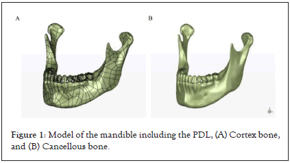
Figure 1: Model of the mandible including the PDL, (A) Cortex bone, and (B) Cancellous bone.
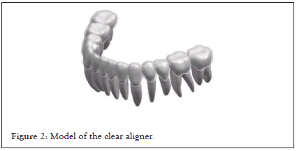
Figure 2: Model of the clear aligner.
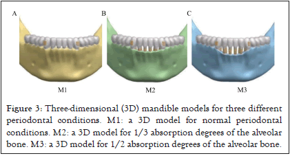
Figure 3: Three-dimensional (3D) mandible models for three different periodontal conditions. M1: a 3D model for normal periodontal conditions. M2: a 3D model for 1/3 absorption degrees of the alveolar bone. M3: a 3D model for 1/2 absorption degrees of the alveolar bone.
The alveolar bone and teeth were set up as a mechanical behavior for linear elastic isotropy and homogeneity [13,14] as shown in Table 1.
| Component | Young’s Modulus (MPa) | Poisson’s Ratio |
|---|---|---|
| Alveolar bone | 1.40 × 10³ MPa | 0.3 |
| Teeth | 1.96 × 10⁴ MPa | 0.3 |
| Clear aligner | 528 MPa | 0.36 |
| Attachment | 12.5 × 10³ MPa | 0.36 |
Table 1: Properties of materials.
A friction condition was established for the contact interfaces between the aligners and the tooth crown surface and the attachments. The contact interfaces on the alveolar bone, PDL, teeth, and the attachment were then bonded. The FEM mesh was then divided into ten tetrahedral nodes, as shown in Table 2.
| Periodontal ligaments | Crown | Root | Alveolar bone | Clear aligner | |
|---|---|---|---|---|---|
| Elements | 120437 | 99906 | 46570 | 141381 | 32193 |
| Nodes | 243104 | 163948 | 78031 | 220076 | 16539 |
Table 2: The number of linear elements and nodes for each material.
Loading method
First, we simulated a moving mode of 5˚ for crown lingual torque. Then, forces of 0.10, 0.15, 0.18, 0.20, and 0.25 mm displacements with labial inclination were applied to the three established alveolar bone models, as shown in Figure 4. The control group featured 0.2 mm displacement under normal periodontal conditions; this was used to analyze and compare the stress generated under different displacements in other periodontal conditions.
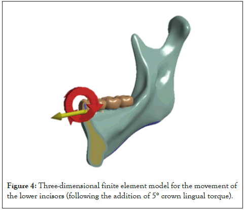
Figure 4: Three-dimensional finite element model for the movement of the lower incisors (following the addition of 5° crown lingual torque).
Force loading method
Preliminary experiments used the established finite element model. We also used ANSYS to calculate the reaction force. The reaction force was defined as the force generated on each tooth when wearing the appliance [15]. In other words, the lower anterior teeth were forced to move and 5° crown lingual torque was applied when the teeth underwent labial inclination to activate the appliance, as shown in Figure 5. This loading method was similar to the clinical force loading. The forces generated by the deformation of the appliance were then calculated by finite element analysis and then loaded back onto the corresponding teeth.
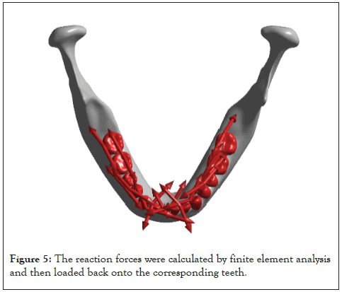
Figure 5: The reaction forces were calculated by finite element analysis and then loaded back onto the corresponding teeth.
Initial tooth displacement
Firstly, we recorded the initial displacement of lower central incisor with and without torque during the force loading (Figure 6).We found after the crown lingual torque added to the lower anterior teeth, the root of the tooth moved slightly to the lip side and the amount of crown lingual movement decreased, indicating the effect of root control. From the analysis of displacement (Table 3), with the decrease of alveolar bone height, the initial displacement of lower central incisor gradually increases, that is to say, when the alveolar bone height decreases, the center of resistance moves down, the worse the torque expression is, the more obvious inclined movement occurs. Combined with Figure 7, it can be seen that the largest displacement occurred in the Y-axis, that is, the buccal-lingual direction of the displacement was large, the inclination trend of the tooth decreased .With the reduction of the alveolar bone height, the inclination movement was more obvious.
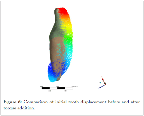
Figure 6: Comparison of initial tooth displacement before and after torque addition.
| X | Y | Z | ||||
|---|---|---|---|---|---|---|
| Maximum | Minimum | Maximum | Minimum | Maximum | Minimum | |
| M1 | 0.0008 | -0.001 | 0.0155 | -0.0433 | 0.0066 | -0.0083 |
| M2 | 0.0009 | -0.0011 | 0.0297 | -0.0967 | 0.0129 | -0.0189 |
| M3 | 0.0006 | -0.0008 | 0.0431 | -0.1873 | 0.021 | -0.0364 |
Table 3: Initial displacement of lower central incisor in 3D direction (0.2 mm displacement as an example).
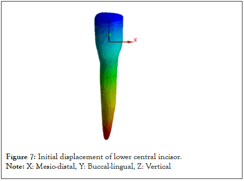
Figure 7: Initial displacement of lower central incisor.
Note: X: Mesio-distal, Y: Buccal-lingual, Z: Vertical
Stress values for the root
When crown lingual torque was added to the lower incisors during labial inclination, we recorded different force systems and behaviors by the anterior teeth. First, we found that the crown moved lingually and there was a small amount of labial movement in the root. Then, the stress values of the lower anterior were calculated: these were 1.193 MPa (central incisor), 1.736 MPa (lateral incisor), and 0.543 MPa (canine), respectively, under normal periodontal conditions and during labial inclination in 0.20 mm displacement, as shown in Figure 8. The stress of the lower central incisors was distributed on the labial side of the root, the lateral incisors on the mesial side, and the canine on the distal side. It means that when we added crown lingual torque to the lower incisors, the areas may have more higher stresses, we should pay attention to the stress to avoid root resorption and increasing alveolar bone resorption, in other words, we should notice the stress distribution of teeth in the treatment of periodontal disease.
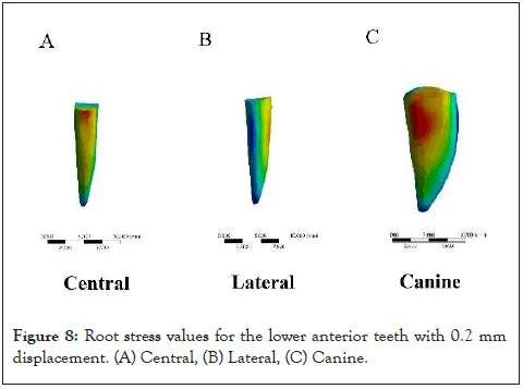
Figure 8: Root stress values for the lower anterior teeth with 0.2 mm displacement. (A) Central, (B) Lateral, (C) Canine.
In normal periodontal condition (M1), the root stress values for the lower anterior teeth increased as the displacement changed; the root stress values for the lower lateral incisor were the greatest, as shown in Figure 9a. Periodontal conditions exhibited a significant influence on root stress. The stress values for the root increased by 24.5% in the model featuring 1/3 absorption degrees (M2: 1.513 MPa (central incisor), 2.406 MPa (lateral incisor), 0.591 MPa (canine)) for the alveolar bone if the displacement of each tooth was 0.2 mm. The stress of the anterior teeth at 0.18 mm displacement was similar to that of the control group (1.296 MPa, 2.061 MPa, and 0.497 MPa). The stress value of the root increased by 92.2% in the model featuring 1/2 absorption degrees (M3: 2.748 MPa, 3.969 MPa, 0.644 MPa) for the alveolar bone if the displacement of each tooth was 0.2 mm; the stress value for the anterior teeth at 0.15 mm displacement was similar to that in the control group (1.792 MPa, 2.589 MPa, 0.419 MPa), as shown in Figure 9b.
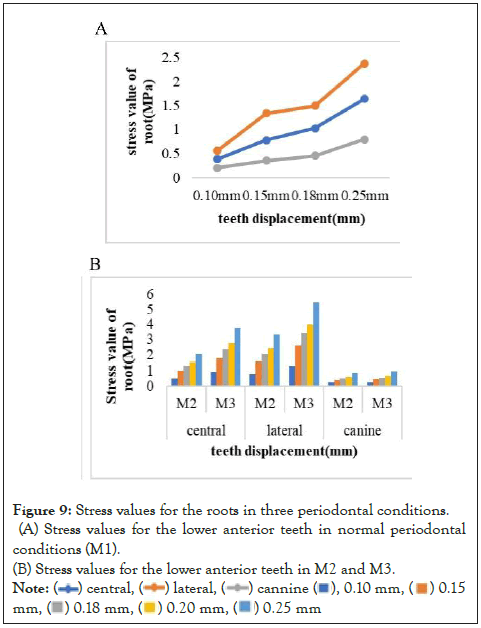
Figure 9: Stress values for the roots in three periodontal conditions.
(A) Stress values for the lower anterior teeth in normal periodontal
conditions (M1). (B) Stress values for the lower anterior teeth in M2 and M3.
Note: ( ) central, (
 ) lateral, (
 ) cannine (
 ), 0.10 mm, (
 ) 0.15
mm, (
 ) 0.18 mm, (
 ) 0.20 mm, (
 ) 0.25 mm
Stress values for the Periodontal Ligament (PDL)
Subsequently, we analyzed the stress of the periodontal ligament under this type of movement. The calculated stress values for the central incisor, lateral incisor, and canine teeth, were 0.111 MPa, 0.106 MPa, and 0.045 MPa respectively under normal periodontal conditions in 0.2 mm displacement. The maximum stress area of the PDL of lower central incisor was in the apex of the tooth, the lateral incisor was in buccal side of the neck, the canine was in the distal side of the neck, as shown in Figure 10. Then we found that the displacement affected the PDL stress values even under normal periodontal conditions, as shown in Figure 11a. In terms of different periodontal conditions and displacements, it exerted a significant influence on PDL stress values. The PDL stress values increased by 152% when the displacement of each tooth was 0.20 mm in the model featuring 1/3 absorption degrees for the alveolar bone (M2: 0.186 MPa, 0.216 MPa, 0.048 MPa); the stress values for the anterior teeth at 0.18 mm (0.159 MPa, 0.185 MPa. 0.041 MPa) was similar to that in the control group. Stress values increased by 214% in the model featuring 1/2 absorption degrees for the alveolar bone (M3:0.425 MPa,0.461 MPa,0.056 MPa), as shown in Figure 11b; under this condition, the stress value for the anterior teeth at 0.10 mm (0.136 MPa,0.149 MPa,0.021 MPa) was similar to that in the control group.
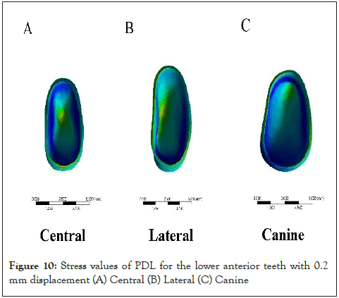
Figure 10: Stress values of PDL for the lower anterior teeth with 0.2 mm displacement (A) Central (B) Lateral (C) Canine
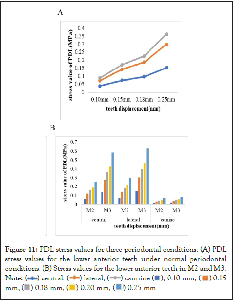
Figure 11: PDL stress values for three periodontal conditions. (A) PDL
stress values for the lower anterior teeth under normal periodontal
conditions. (B) Stress values for the lower anterior teeth in M2 and M3.
Note: ( ) central, (
 ) lateral, (
 ) cannine (
 ), 0.10 mm, (
 ) 0.15
mm, (
 ) 0.18 mm, (
 ) 0.20 mm, (
 ) 0.25 mm
FEM is a widely used and effective method in orthodontic research, especially in clear orthodontic treatment [16]. In a previous study involving the finite element method, Seo et al reported that the thickness of the transparent collimator would affect the stress value of the PDL and change the center of the tooth rotation center [17]. In another study, Nah et al used the finite element method to analyze the stress distribution related to the clear aligner and found that the instantaneous stress was much larger than the stress caused by fixed orthodontics [8]. Nah et al. also used the finite element method to study the stress distribution of the upper incisor during invisible orthodontic treatment and found that the stress distribution was more uniform during labial, lingual, mesial, and distal movement. Tanne et al further show that the stress distribution of parallel motion is more uniform than that of inclined motion, and the non-ideal stress level is significantly lower [18]. In summary, these studies show that the finite element method is very useful for studying the stress behavior related to tooth movement. However, few studies have focused on the biomechanical properties of periodontal transparent locator therapy. In a previous study, we found that periodontal conditions can affect the root stress and the strain of alveolar bone during invasive movement. We should also reduce the displacement to minimize the damage to periodontal tissue [19]. In another study, Wang et al. used the finite element method to analyze the periodontal ligament stress during the indentation of molars with different alveolar bone heights. The analysis showed that the supporting periodontal tissue around the teeth was significantly weakened when the alveolar bone resorption was more than 48% of the root length [20]. Given the application of the finite element method in oral biomechanics and orthodontics, in this study, we used 3D-FEM to study how to optimize orthodontic displacement by increasing torque during labial movement of lower anterior teeth.
Torque is a critical component in an ideal occlusion relationship [21,22]. When performing orthodontic treatment for patients with periodontal disease, it is easy to cause tipping movement by applying orthodontic force; this is due to the disproportionality of the crown and root. This suggests that more attention should be paid to torque, to reduce the periodontal injury caused by tipping movement. The essence of clear aligner treatment is that teeth move under the force generated by the deformation of the appliance [23]. This force is transmitted to the tooth root and periodontal tissues through the inner surface of the appliance, thus causing the tissues to remodel; collectively, these processes result in the movement of the teeth. In a previous study, Djeu et al. found that Invisalign was still inferior to a fixed appliance concerning torque control [24]. Simon et al found the average efficiency of torque movement was about 67%, the control of anterior tooth torque when using clear aligners was also shown to be highly correlated with the amount of buccal and lingual movement. V Magesh et al. carried out a finite element study to show that adding torque during the process of tooth movement could make the body of a tooth move [25]. In the present study, 5 degrees of crown lingual torque was added during the labial inclination of the lower anterior teeth; we found that when using this loading method, the crown was also undergoing inclination. However, in periodontitis, our ability to control torque is much worse; this results in the teeth becoming tilted, thus increasing the risk of bone cracking.
The instantaneous force produced by invisible orthodontics is much larger than that produced by fixed orthodontics [9]; therefore, the stress value of the treatment is very obvious and must be considered. Recent studies have shown that when invisible orthodontics is used, the displacement range of each tooth is 0.2-0.40 mm. [26,27]. More importantly, the default displacement of different brands of transparent aligners is usually within the range of 0.20 mm-0.30 mm per step. Therefore, in this study, the displacement of 0.20 mm under normal periodontal conditions is also regarded as control of other displacements. When the periodontal condition deteriorates, the maximum stress of the anterior teeth should not exceed the stress value when the displacement is 0.20 mm. For alveolar bone resorption, we found that the root stress values of anterior teeth under 0.18 mm displacement ranged from 0.497 to 2.061 MPA, which were similar to those of 0.20 mm under normal periodontal conditions (0.543-1.736 MPa). Similarly, we found that when the alveolar bone resorption was 1/2 under 0.15 mm displacement (0.419-2.589 MPa), the root stress was similar to that of 0.20 mm under normal periodontal conditions. The research results of this experiment are consistent with the reports in the current literature.
In this study, we also determined displacement based on the stress incurred by the PDL. In a previous study, Ren et al. hypothesized that a smaller force can cause the movement of teeth, while a larger force may not cause tooth movement; this theory was mainly related to bone reconstruction [28]. According to Penedo et al., alveolar bone reconstruction begins when the stress value of the PDL exceeds the capillary pressure (0.226 N/mm2) [29]. Oppenheim [30] and Schwarz et al. found that under orthodontic loading, the PDL plays a more important role in alveolar bone reconstruction. Field et al. also reported that high strain in the PDL indicates potential for bone remodeling and that excessive stress can lead to root resorption [31]. In the present study, we analyzed stress values with different displacements in different periodontal conditions; we used 0.20 mm displacement under normal periodontal conditions as a control group while the stress incurred by the lower anterior teeth ranged from 0.045- 0.111 MPa. We found that the PDL stress values for the lower anterior teeth in 0.15 mm displacement ranged from 0.121 - 0.141 MPa; these stress values are similar to those of 0.20 mm’s normal periodontal conditions when the alveolar bone resorption was 1/3. When alveolar bone resorption was 1/2, the PDL stress values for the anterior teeth in 0.10 mm displacement ranged from 0.021-0.149 MPa; we selected 0.10 mm displacement in case the stress value exceeded the control group.
These results implied that the stress incurred by the teeth and the PDL should both be taken into account when pre-setting displacement during clear aligner treatment. So we recommend that the optimal orthodontic displacements are 0.15 mm and 0.10 mm respectively when an alveolar bone is resorption 1/3 or 1/2.
However, there are some limitations to this study that need to be considered. First, this study focused on the biomechanics of clear aligner treatment; many other biological aspects also need to be considered, such as individual and age differences. Second, the properties of the periodontal ligament in FEM are limited; this is a very different scenario than in clinical practice. Third, our FEM findings need to be validated in clinical practice.
In this study, we use FEM to simulate the addition of 5 degrees of crown lingual torque during labial inclination movement in periodontal disease models. Analysis showed that periodontal conditions led to a change in stress values, thus affecting periodontal health. Our findings suggest that we need to reduce such forces to protect periodontal health. During treatment with clear aligners, we should select the optimal orthodontic displacement to minimize the adverse effects of the clear aligner on periodontal tissues. Subsequent research should focus on the specific pattern of tooth movement under periodontal conditions to facilitate clinical treatment.
Ethics approval and consent to participate
This study was approved by the ethics committee of the School of Stomatology, Capital Medical University. (No. CMUSH-IRB-KJPJ- 2020-11) written informed consent was obtained from all subjects.
Consent for publication
Not applicable.
Availability of data and materials
The datasets used and/or analyzed during the current study are available from the corresponding author on reasonable request.
Competing interests
The authors declare that they have no competing interests.
Funding
Youth Clinical Research Fund of Chinese Stomatological Association: Biomechanical analysis of clear aligner in periodontal diseases (CSAMW2021- 07).
Authors’ contributions College Science and Technology Innovation Plan of Shanxi Education Department: Torque compensation of anterior teeth in extractive cases using clear aligners under three periodontal statutes (2021L242).
YNM: Conception, design, and analysis of data, performed the data analyses and wrote the manuscript; BZ: Performed the data analyses;WW: Software; YXC: Analysis of data, performed the data analyses; NZ: Analysis of data; XPW, SL: Contributed to the conception of the study and revised the manuscript; All authors have read and approved the manuscript.
Acknowledgment
Not applicable.
Citation: Wu X, Ma Y, Zhang N, Zhao B, Wang W, Cheng Y, et al. (2023) Optimal Displacement of Teeth by the Torque Movement under Different Periodontal Conditions with Clear Aligners: A Three-Dimensional Finite Element Study. J Odontol. 7: 640.
Received: 29-Nov-2022, Manuscript No. JOY-22-20501; Editor assigned: 06-Dec-2022, Pre QC No. JOY-22-20501 (PQ); Reviewed: 20-Dec-2022, QC No. JOY-22-20501; Revised: 27-Dec-2022, Manuscript No. JOY-22-20501 (R); Published: 03-Jan-2023 , DOI: 10.35248/JOY.23.7.640
Copyright: © 2023 Wu X, et al. This is an open-access article distributed under the terms of the Creative Commons Attribution License, which permits unrestricted use, distribution, and reproduction in any medium, provided the original author and source are credited.