Gynecology & Obstetrics
Open Access
ISSN: 2161-0932
ISSN: 2161-0932
Case Report - (2022)Volume 12, Issue 1
A young woman, health, 33 years old, presented unilateral areola and nipple pruritus and burning about one month ago. She denies smoking or drinking alcoholic beverages. The initial diagnosis was dermatitis and she was treated with topical regimes. She sought further evaluation and care at our institution. Further investigation revealed breast cancer behind the left nipple and areola. She underwent to Breast-Conserving Surgery (BCS) to the left areola and nipple with Sentinel Lymph Node Biopsy (SND) negative. The margins were clear achieved. The surgery for reconstructive breast was Grisotti’s technic. The adjuvant radiotherapy was used to finish the treatment.
Female breast cancer; Breast paget’ disease; Reconstructive surgery; Dermatitis of breast
Paget’s disease of the breast is a rare condition that is associated with underlying breast cancer [1]. It represents 1%-3% of female breast cancer. In situ histology is found in 1/3 cases. Unfortunately, many women were treated with topical steroids because frequently the diagnosis was dermatitis or eczema. However, the true diagnosis remains elusive for long periods [2]. The clinicians must suspect that the first diagnosis can be wrong if the patient did not become better with topical regimens [3]. The case we present is rare because the patient was 33 years old age without any significant breast cancer risk factor and the presenting symptoms were nipple pruritus and burning.
A 33 years old white woman, a refrigerator worker, sought medical attention complaining of itching and burning in her left nipple for more than a month. She underwent a cesarean operation when she was 28 years old. Do not take any medicine for continuous use, only birth control pills. She denies smoking or drinking alcoholic beverages. In her family, there was a case of breast cancer in a paternal aunt at the age of 48 years old.
The breast was small with no visible deformities. When performing the clinical examination of the areola and nipple, thickening of the left areola is observed without injured in the areola. There was neither discharge from the nipples nor mass behind the breast areola. No axillary adenopathy was identified. The diagnosis was dermatitis of the left areola and the patient was treated with topical steroids (Figure 1).
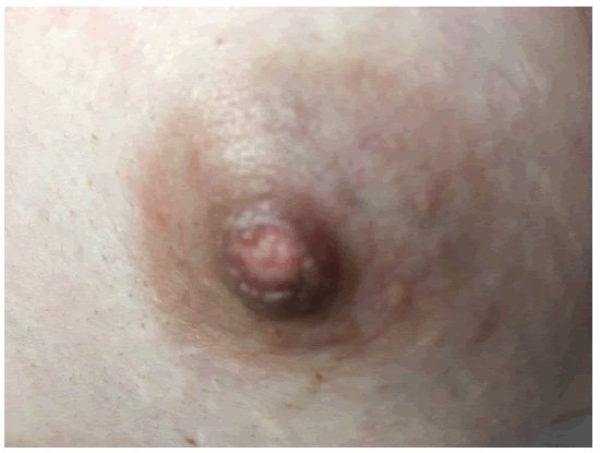
Figure 1: Areola and nipple with normal appearance in the first evaluation.
Her symptoms failed to respond to topical therapy. Twenty days later, she sought further new exams and care at our institution The patient referred a small nodule behind the left areola and she had a few pain too. It was possible to identify a little mass. The patient underwent to ultrasound of the breast and the exam showed the mass behind the areola (Figure 2). It was a new situation and it was necessary to clarify what kind of mass it was.
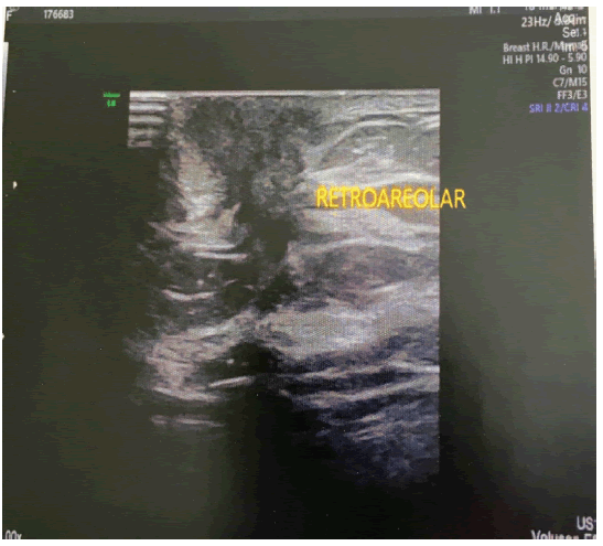
Figure 2: Areola Ultrasound; Node present; Retroareolar means: behind the areola.
The patient underwent to biopsy of the areola and the diagnosis was: Carcinoma Ductal Invasive (CDI). The patient underwent to MRI (Figure 3) before the surgery, because it was necessary to know if she had other tumors or not. The tumor was single and only one lymph node enlarged. It was in the left axilla. The removal of an areola and nipple Breast-Conserving Surgery (BCS) plus reconstructive areola and nipple with Grisotti’s technical and Sentinel Node Biopsy (SNB) was the decision of the physician team.
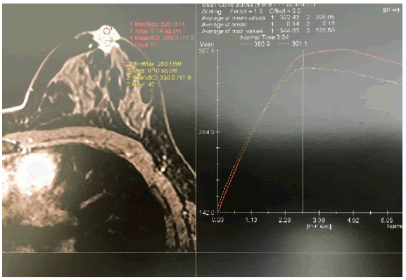
Figure 3: MRI of after the diagnosis of CDI and before tumor removal and reconstructive surgery.
The mass was removed and the surgical margins were clear achieved. It was CDI grade II Nottingham, ranging 1.8 cm. No angiolymphatic invasion was identified. The nipple showed Paget’s disease and SNB was negative. Immunohistochemical tests showed CK7 and CAM 5,2 positive, RE and RP+, Her-2 negative, Ki- 67=10%. The patient recovered well after surgery (Figures 4 and 5). She began radiotherapy on month after the surgery.
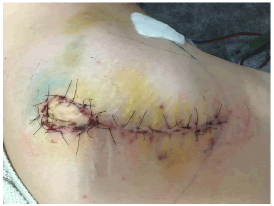
Figure 4: Patient after removal of areola and nipple and reconstruction by Grisotti’s technique-7 days after the surgery.
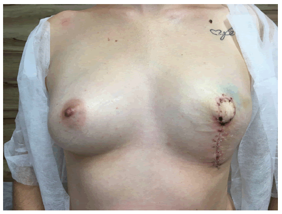
Figure 5: Patient after one month of surgery; Reconstruction using Grisotti.
Paget’s disease of the nipple is a rare malignant condition with a reported incidence among all primary breast cancer of 1%-3% [3]. It is extremely uncommon in young women and there is no evidence at this time that one of the two surgical techniques (conservative or mastectomy) would improve survival [2]. The prognosis depends on the presence of a palpable mass and the invasiveness of cancer [4]. In this case, the patient presented a non-classical clinical indicator of Paget’s disease but she returned to our institution a few days after because she was worse. A new evaluation discovered a mass behind the areola. The diagnosis of breast cancer made our mind to specific treatment and the possibility of reconstructive surgery of the areola. Before the surgery, the patient underwent MRI to detect multifocal or multicentric breast cancer. A recent report recommended MRI because it is more sensitive than Mammography [4 ]. Early and accurate diagnosis of Paget’s disease can be resolved with BCS and SND, especially when the lesion is confined to the epidermis of a nipple, [2]. Breast conservation therapy with lumpectomy and radiation is an effective local treatment strategy for a patient with DCIS and CDI compared with mastectomy [6]. Breast-conserving surgery and sentinel lymph node biopsy are the treatment of choice for most women with early-stage breast cancer. The rarity of Paget’s disease of the breast does not allow randomized trials comparing treatment modalities. The breast-conserving approach has been considered as a treatment option in selected patients [7,8]. Although long-term follow-up will be required, data from previous studies suggest that the type of surgical treatment did not result in differences in DSS and RFS [9].
Paget’s disease is frequently associated with underlying peripheral or multicentric breast cancer. It was clear that Paget’s disease may occur in young women too. Symptoms as itching, burning, or small lesions on the surface in the areola or nipple are not always benign lesions. If initial steroid therapy fails to respond adequately, the clinical investigation should continue to rule out the possibility of breast cancer. MRI may be helpful when considering breast conservation. Breast-conserving surgery and reconstructive of the areola can be done for the selected case of Paget’s disease. Mastectomy is not ever mandatory.
The authors declare that written informed consent was obtained from the patient for the publication of this case report.
None.
Google Scholar Crossref
Citation: Citation: Marinho LAB, Cruz NT. (2021) Paget's Disease of Breast in Young Woman-Unlike But Not Impossible. Gynecol Obstet (Sunnyvale) 11:576.
Received: 08-Dec-2021, Manuscript No. gocr-21-14840 ; Editor assigned: 20-Dec-2021, Pre QC No. gocr-21-14840 ; Reviewed: 13-Jan-2022, QC No. gocr-21-14840 ; Revised: 28-Jan-2022, Manuscript No. gocr-21-14840 ; Accepted: 14-Dec-2021 Published: 15-Feb-2022
Copyright: © 2021 Marinho LAB, et al. This is an open-access article distributed under the terms of the Creative Commons Attribution License, which permits unrestricted use, distribution, and reproduction in any medium, provided the original author and source are credited.