Biochemistry & Pharmacology: Open Access
Open Access
ISSN: 2167-0501
ISSN: 2167-0501
Research Article - (2016) Volume 5, Issue 2
In the present study, two cultivars of pepper (Capsicum annuum and Capsicum frutescens) at two maturity stages (green and red) were evaluated for total phenolic and flavonoid content, organic acids, vitamin C, β-carotene, vitamin E, capsaicin and the antioxidant and anticancer activities of their aqueous extracts. Total phenolic content was found to be ranged from 11.09-26.14 mg GAE/g DW, while total flavonoid content was ranged from 2.7 mg to 5.0 mg QE/g DW. Twenty six phenolic and aromatic compounds, twelve flavonoid compounds and eleven organic acids were identified in all samples by using of HPLC. Vitamin C, β-carotene, vitamin E and capsaicin contents were also estimated by HPLC and detected at high levels which were ranged from 500.0-645.5 mg/100 g DW, 6.56-35.69 mg/100 g DW, 10.44-19.36 mg/100 g DW and 37.46-69.90 mg/100 g DW, respectively. Antioxidant activities of pepper samples were carried out by using of both DPPH•-scavenging activity and total antioxidant capacity (ABTS•+) assays and the extracts exhibited high activities which were ranged from 96.95% to 98.64% and from 77.73% to 93.11%, respectively. Finally, the potential anticancer activity of pepper extracts and capsaicin standard was tested against prostate (PC-3) and breast (MCF-7) carcinoma cell lines in vitro. The results showed that sweet pepper had a higher anticancer activity against PC-3, in contrast, chilli pepper had a higher against MCF-7.
<Keywords: Chilli pepper; Sweet pepper; Capsicum; Phytochemical components; Capsaicin; Antioxidant activities; Anticancer activity
Capsicum is a genus of plants from the family of Solanaceae. Some species of the genus Capsicum are grown for their fruits, which can be consumed fresh (in salads, baked dishes, salsa, pizzas, etc.), cooked, as a dried powder, in a sauce, or processed into oleoresin [1].
Peppers contain phenolics and flavonoids [2], carotenoids [3], vitamin C, vitamin E [4] and alkaloids [5], which play important roles in human health. In other studies, antioxidant activities in peppers were measured by radical-scavenging activity [6,7], inhibition of lipid peroxidation [8] and metal-chelating activity [9]. Capsaicinoids and carotenoids exhibit anticancer [10,11] and antioxidant activities [12-14]. Flavonoids have been shown to act as antioxidants, and they possess anti-inflammatory [15], antiallergic [16], and antibacterial activities [17]. The antioxidant activity of pepper extracts involves bioactive compounds, such as polyphenols, carotenoids, capsaicinoids and ascorbic acid [18-20].
Hot chili peppers that belong to the plant genus Capsicum (family, Solanaceae) are among the most frequently consumed spices throughout the world. The principal pungent ingredient present in hot red pepper (Capsicum annuum L.) and chili pepper (Capsicum frutescence L.) is the phenolic substance named capsaicin (trans-8-methyl-N-vanillyl-6- non-enamide). The capsaicin content of hot peppers varies from 0.1% to 1%. Capsaicin was subjected to extensive investigations with regard to its possible tumorigenicity and genotoxicity [21,22]. However, the compound has recently attracted considerable attention because of its chemoprotective properties against certain carcinogens and mutagens.
Prostate cancer remains the most commonly diagnosed cancer in men living in the Western world [23]. There is increasing evidence that dietary factors play a role in the development and progression of prostate cancer. It is estimated that at least 30% of all prostate cancer patients use complementary and alternative medicine, which includes the consumption of micronutrient supplements [24]. Many dietary agents have been studied for the protective effects on prostate cancer [25,26]. Capsaicin has recently emerged as a potent anti-cancer agent, exhibiting anti-proliferative and pro-apoptotic properties in several different prostate cancer model systems [27]. The use of capsaicin in vitro had been reported to induce apoptosis through the generation of reactive oxygen species (ROS) [28-30] (Figure 1).
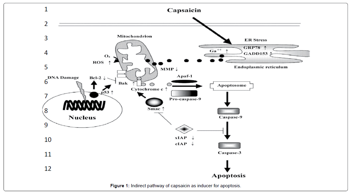
Figure 1: Indirect pathway of capsaicin as inducer for apoptosis.
Breast cancer is the leading cause of death among women worldwide. About 63,300 cases of breast carcinoma in situ are expected to be newly diagnosed in 2012 [31]. Capsaicin or N-vanillyl-8- methyl- 1-nonenamide, the primary pungent and irritating ingredient present in a variety of red peppers of the genus Capsicum [32-34], was reported to selectively inhibit the growth of tumor cells [35]. Despite previous discordant results from studies that determined its potential mutagenic and carcinogenic activity [17], subsequent investigations have shown that capsaicin induces apoptosis in a wide variety of tumor cells [29,36-39]. Additional studies reported that capsaicinoids displayed in vitro and in vivo antitumor activity [40]. In cultured cells, capsaicin blocked the cell migration in breast cancer, while in mice, oral consumption of capsaicin decreased the size of MDAMB 231 breast cancer tumors by 50%, and inhibited the development of pre-neoplastic breast lesions by up to 80%. Also, direct injection of capsaicin led to an 80% reduction in tumor size [39]. Thus, capsaicin can be considered a potential lead against malignant tumors. Capsaicin has been shown to inhibit the growth of ER-positive (MCF-7, T47D, BT-474) and ER-negative (SKBR-3, MDA-MB231) breast cancer cells by causing G0/G1 cellcycle arrest and apoptosis [41].
The aim of the present study was to evaluate the bioactive components, included phenolic and aromatic compounds, flavonoids, organic acids, vitamin C, E, β-carotene and capsaicin in Egyptian dried pepper samples (sweet and chilli) at two ripening stages (green and red). Evaluation was also extended the potential anticancer activity on both prostate and breast carcinoma cell lines in vitro.
Collection of plant materials and treatment
Fruits of hot chilli pepper (Capsium frutescens var. sina) at immature stage (green color) and mature stage (red color) and others of sweet pepper (Capsium annuum var. goduion), green and red, were collected from Vegetable Breeding Research Department, Horticultural Research Institute, Agricultural Research Center in September until December 2012 season and were identified at the same institute.
Fresh fruits were washed by using of tap water, seeds were removed and the edible tissues were cut to small pieces which were oven-dried in Lab Companion oven at 55°C for 48 h.
Chemicals and drugs
All the utilized chemical materials (solvents, mineral salts, etc.) were purchased from El Gomhoryia, El Allamyia, El Nasr and Middle East Pharmaceutical Chemical companies, Egypt and the solvents were purified before using. Chemicals, solvents and all standard materials which were used for fractionation and identification by HPLC, purchased from Sigma/Aldrich Chemical Company, USA.
Carcinoma cell lines
Prostate carcinoma cell line (PC-3) and breast carcinoma cell line (MCF-7) test kits were obtained from Pharmacology Unit, Cancer Biology Department, National Cancer Institute, Egypt.
Preparation of extracts
Ten g of dried pepper samples were extracted with 100 ml distilled water (1:10) (i.e., 10 g/100 ml) to produce the aqueous extracts. The extracts were placed in ultrasonic instrument (BANDELIN SONOREX SUPER RK 514H) for 30 min, left up to 24 h at 15°C and filtered through a Whatman paper No. 1. The resultant extracts were used to determine total phenolic and flavonoid contents and their HPLC fractionation, antioxidant and anticancer activities in vitro.
Determination of total phenolic content
Total phenolic content (TPC) of each sample was determined using a Folin Ciocalteu assay according to the method of Singleton and Rossi [42,43] with slight modification. The reaction mixture contained 1 ml of extract and 0.5 ml of the Folin-Ciocalteu reagent, 1 ml sodium carbonate 7.5% and 7.5 ml of distilled water were added, respectively. After 45 min of reaction at ambient temperature, the absorbance at 765 nm was measured using a UV-visible spectrophotometer (Beckman). A blue color indicated the presence of phenols. A calibration curve was calculated by using of gallic acid standard (0.1 mg/ml). Total phenolic content of samples were determined in triplicates and the results were expressed on dry weight basis (DW) as mg gallic acid equivalents (GAE), per g of each sample.
HPLC analysis of phenolic and aromatic compounds
Phenolic and aromatic compounds were detected by HPLC according to the method of Goupy et al. [44] as follows: the aqueous extracts were centrifuged at 10000 rpm (in ICE Micro-MB Centrifuge/ NARP 64606 instrument) for 10 min and the supernatant was filtrated through a 0.2 μm Millipore membrane filter, then 1-3 ml were collected in a vial for injection into HPLC Agilent (Series 1200) equipped with autosampler injector, solvent degasser, ultraviolet (UV) detector set at 280 nm and quaternary HP pump (Series 1100). The column [Agilent 5HC-C18 (2) 250 × 4.6 mm] temperature was maintained at 35°C. Gradient separation was carried out with methanol and acetonitrile as a mobile phase at flow rate of 1 ml/min. Phenolic acid standards from sigma Co. were dissolved in a mobile phase and injected into HPLC. Retention time and peak area of the tested samples were calibrated against standard solutions of different phenolic and aromatic compounds concentration by the data analysis of HEWLLET Packed (HP) software.
Determination of total flavonoid content
Total flavonoid content was measured by AlCl3 colorimetric assay according to the method of Harborne [45] based on slight modification. Briefly, 500 μl of extract and 2 ml of distilled water, 150 μl of 5% sodium nitrate were added. After 5 min, 150 μl of 10% AlCl3 was added. A total of 2000 μl of sodium hydroxide (1 M) were added after 1 min and followed by 1200 μl of distilled water. The mixture was incubated for 30 min. The absorbance was measured at 510 nm against a prepared blank. A yellow color indicated the presence of flavonoids. A calibration curve was calculated using quercetin standard (0.1 mg/ml). Total flavonoid content of samples was determined in triplicates and the results were expressed on dry weight basis (DW) as mg quercetin equivalents (QE), per g of each sample.
HPLC analysis of flavonoid compounds
Flavonoid fractions were also identified by HPLC according to the method of Mattila et al. [46] as follows: the aqueous extracts were centrifuged at 10000 rpm (in ICE Micro-MB Centrifuge/NARP 64606 instrument) for 10 min and the supernatant was filtrated through a 0.2 μm Millipore membrane filter, then 1-3 ml were collected in a vial for injection into the previous HPLC Agilent (Series 1200) and HP software were used. The ultraviolet (UV) detector was set at 330 nm and the other conditions were set as that previously used in the fractionation of phenolic compounds.
DPPH•-scavenging activity assay
Free radical scavenging activity was determined using the free radical generator DPPH• assay based on slight modifications [47]. One ml of the pepper extract was added to 1 ml of 0.002% methanol solution of DPPH•. The mixture was thoroughly mixed using BioCote/Stuart vortex instrument and kept in the dark for 30 min. The absorbance, using a spectrophotometer, was measured at 517 nm against a blank of methanol without DPPH•.
Total antioxidant capacity assay
Total antioxidant capacity assay was carried out by the improved ABTS•+ method according to the method of Re et al. [48]. ABTS•+ radical cation was generated by reacting 7 mM ABTS•+ and 2.45 mM potassium persulfate after incubation at room temperature (23°C) in the dark for 16 h. The ABTS•+-solution was diluted with 80% ethanol to an absorbance of 0.700 ± 0.005 at 734 nm. 0.1 ml of the tested samples was added to 3.9 ml of ABTS•+ solution and mixed thoroughly. The reactive mixture was allowed to stand at room temperature for 6 min and the absorbance was immediately recorded at 734 nm against a blank of 80% ethanol using a spectrophotometer. The inhibition percent was calculated in both methods (DPPH• and ABTS•+) as follows:
[A control – A extract/A control] × 100
HPLC analysis of organic acids
Organic acid fractionation was conducted according to the method of Wodecki et al. [49]. One g of dried pepper samples were mixed with 50 ml deionized water, placed in the ultrasonic instrument for 30 min and centrifuged at 10000 rpm for 10 min, the supernatant was filtrated through a 0.2 μm Millipore membrane filter then 1 ml was collected in a vial for injection using of HPLC Agilent (Series 1200) equipped with autosampler injector, solvent degasser, ultraviolet (UV) detector (set at 210 nm) and quaternary HP pump (Series 1090). The column [OA-1000 Column S/N: 5927915] temperature was maintained at 55°C. Gradient separation was carried out with methanol and ethanol as a mobile phase.
HPLC analysis of vitamin C: Vitamin C was determined according to the method of Romeu-Nadal et al. [50]. One g of dried pepper samples were mixed with 0.3% metaphosphoric acid solution and centrifuged at 10000 rpm for 10 min and the supernatant was filtrated through a 0.2 μm Millipore membrane filter then 1-3 ml were collected in a vial for injection into HPLC Agilent (Series 1200) equipped with autosampler injector, solvent degasser, ultraviolet (UV) detector (set at 254 nm) and quaternary HP pump (Series 3365). The column [Agilent 5HC-C18 (2) 250 × 4.6 mm] temperature was maintained at 25°C. Ascorbic acid was identified by comparing the retention time of the sample peak with that of the ascorbic standard at 254 nm.
HPLC analysis of β-carotene
β-carotene was determined according to the method of Pupin et al. [51]. Dried (5 g) pepper samples were extracted with ethyl acetate (3 × 50 ml) containing BHT (0.004%). The organic phase was transferred through anhydrous sodium sulfate (50 g) and collected in an ambered round-bottom flask. The dried extract was transferred quantitatively to a 10 ml volumetric flask using portions of 1.5 ml of mobile phase (acetonitrile: methanol: 1,2-dichloromethane, 60:35:5, v/v/v). A vial was injected into HPLC Agilent (Series 1200) equipped with autosampler injector, solvent degasser, ultraviolet (UV) detector (set at 280 nm) and quaternary HP pump (Series 1100). The column [Agilent Hypersil ODS 5 μm 4.0 × 250 mm] temperature was maintained at 35°C.
HPLC analysis of vitamin E
Vitamin E was determined according to the method of Pyka and Sliwiok [52]. Dried (5 g) pepper samples were extracted with hexane (3 × 50 ml) containing BHT (0.004%). The organic phase was transferred through potassium hydroxide (50 g) and collected in an ambered round-bottom flask. The solution was well mixed and further extracted with hexane and petroleum ether (75 and 25 ml, respectively, containing 0.004% BHT). The pooled hexane was evaporated to dryness in a rotary evaporator at 40°C. The extract was transferred quantitatively to a 10 ml methanol. A vial was injected into HPLC Agilent (Series 1200) equipped with autosampler injector, solvent degasser, ultraviolet (UV) detector (set at 290 nm) and quaternary HP pump (Series 1100). Gradient separation was carried out with methanol and water (9:1, v/v) as a mobile phase at a flow rate of 1.5 ml/min. The column [Agilent Hypersil ODS 5 μm 4.0 × 250 mm] temperature was maintained at 35°C. The injection volume was 20 ml of a standard of vitamin E in ethanol.
HPLC analysis of capsaicin
Capsaicin content was determined according to Collins et al. [53]. One g of dried pepper samples was added to 10 ml acetonitrile and placed in 120 ml glass bottles. Bottles were capped, placed in an 80°C water bath for 4 h and then they were swirled manually every hour. After cooling at room temperature, 2 ml of supernatant were taken and filtered (0.45 filter syringe) into a 2 ml glass sample vial, capped, and stored at 5°C until analyzed by HPLC Agilent (Series 1200) equipped with autosampler injector, solvent degasser, ultraviolet (UV) detector set at 280 nm and quaternary HP pump (Series 1100).
The column [Agilent 5HC-C18 (2) 250 × 4.6 mm] temperature was maintained at 35°C. The mobile phase was methanol: water (30%:70%). Standard solution of capsaicin 50% standard was prepared in methanol by dilution of a 2 mg stock solution. Capsaicin was identified by comparing the retention time of the sample peak with that of the CAP standard at 280 nm.
Anticancer activity (Cytotoxicity)
Preparation of the tested samples: Aqueous extracts of dried pepper, which were freeze dried by using of Freeze Dryer Lab Conco USA at -50°C/vacuum, and also capsaicin 50% standard were both tested against prostate (PC-3) and breast (MCF-7) carcinoma cell lines.
In vitro cytotoxicity: The percentage of cell death was estimated by Sulfo-Rhodamine B (SRB) assay. Potential cytotoxicity of both aqueous extracts of dried pepper samples and capsaicin standard (50% capsaicin) was tested using the method of Skehan et al. [54]. Different concentrations of the compounds under test (5, 12.5, 25 and 50 μg/ml DMSO) were added to the cell monolayer triplicate wells which were prepared for each individual dose. Monolayer cells were incubated with the compounds for 48 h at 37°C and in atmosphere of 5% CO2. After 48 h, cells were fixed, washed and stained with Sulfo-Rhodamine-B stain. Color intensity was measured by ELISA reader (TCAL).
Statistical analysis: All results were expressed as means ± standard deviation. Statistical Analysis System SAS 9.1 software package was used to analysis of data and significant differences between mean values were determined by least significant difference (LSD) test at P > 0.05.
Total phenolic content of dried pepper samples and HPLC of their fractionation
Data in Table 1 show that total phenolic content ranged from 19.26- 36.87 mg GAE/g DW and sweet peppers were significantly higher than chilli peppers. The resulted data are in accordance with Aliakbarlu et al. [55] who showed that total phenolic content in red pepper was 34 mg GAE/g DW. But they are higher than those found by Rodríguez- Maturino et al. [56] who found that the Habanero pepper had significantly higher total phenolic content (5.92 mg GAE /g DW) than the Chiltepin pepper (4.85 mg GAE/g DW). The obtained data also show that the total phenolic content was increased with maturation from green to red color. This is in agreement with Howard et al. [57] who found that total phenolics were ranged from 2656-5788 mg/kg FW and total phenols increased with maturation. In contrast, Ghasemnezhad et al. [58] reported that phenolic content decreased with maturity and following ripening. Lin and Tang [59] also observed that total phenols in green, yellow and red pepper were 206.0, 191.2 and 180.3 mg GAE/100g FW, respectively.
| Sample | SGC | SRC | GGS | GRS |
|---|---|---|---|---|
| Total phenolic content (mg GAE/g DW) |
19.26c ± 0.42 | 19.48c ± 0.46 | 29.11b ± 3.82 | 36.87a ± 4.57 |
| HPLC of phenolic and aromatic compounds (mg/100g DW) | ||||
| Gallic acid | 1.31 | 3.00 | 2.18 | 2.12 |
| Pyrogallol | ND | ND | ND | 52.90 |
| 3-Hydroxy tyrosol | 14.00 | 10.00 | 25.54 | 15.62 |
| Benzoic acid | 112.45 | 12.35 | 38.20 | 16.22 |
| 4-Amino benzoic acid | 6.16 | 8.08 | 9.86 | 6.94 |
| Caffeine | 0.54 | 1.77 | ND | 0.77 |
| Protocatchuic acid | 1.28 | 3.11 | 7.03 | 4.42 |
| Catechin | 4.42 | 5.30 | 1.19 | 4.18 |
| Chlorogenic acid | 25.99 | 15.74 | 25.68 | 20.62 |
| Catechol | 12.00 | 8.57 | 4.09 | 9.21 |
| Epicatechin | 5.27 | 7.64 | 15.21 | 7.29 |
| P-hydroxy benzoic acid | 5.73 | 5.52 | 11.48 | 6.97 |
| Caffeic acid | 1.72 | 1.75 | 10.30 | 1.11 |
| Vanillic acid | 2.18 | 3.14 | 3.94 | 3.26 |
| Ferulic acid | 0.30 | 0.66 | 0.70 | 0.49 |
| Isoferulic acid | 0.35 | 0.54 | 0.62 | 0.28 |
| Reverstrol | 1.27 | 0.40 | 0.16 | 0.41 |
| Oleuropein | 86.37 | 5.67 | ND | ND |
| Ellagic acid | ND | ND | 12.24 | ND |
| E-vanillic acid | 15.12 | 20.15 | ND | 28.07 |
| O-coumaric acid | 0.23 | 0.12 | ND | 0.03 |
| 3,4,5-methoxy cinnamic | 0.11 | 0.28 | 0.11 | 0.10 |
| Coumarin | 1.14 | 0.93 | 1.23 | ND |
| Salicylic acid | 2.69 | 1.59 | 1.75 | 0.45 |
| P-coumaric acid | 0.30 | 0.37 | 0.81 | 0.26 |
| Cinnamic acid | 3.21 | 3.06 | 3.43 | 2.14 |
Table 1: Total phenolic content and their HPLC fractionation of dried pepper samples.
Data of the phenolic and aromatic compounds show that 3-hydroxy tyrosol, chlorogenic, catechol, E-vanillic and benzoic acids were detected at higher levels than other phenolic acids (Table 1). Pyrogallol is only detected in GRS sample and ellagic acid only in GGS sample. Coumarin not only found in GRS sample. All of α-coumaric, E-vanillic, caffeine and oleuropein were not found in GGS sample. Other phenolic acids found in all extracts at different contents.
Total flavonoid content of dried pepper samples and HPLC of their fractionation
Data in Table 2 show that total flavonoid content of the dried pepper samples were ranged from 371.7-512.0 mg QE/100 g DW and their contents increased with maturation (red samples had a higher content than green samples). In contrast, Tundis et al. [60] showed that the flavonoids content decreased with the maturity. The results are lower than those of Materska and Perucka [61] who revealed that flavonoids ranged from 16.7-40.7 mg/g DW. But they are higher than those found by Lin and Tang [59] who observed that total flavonoids for green, yellow and red pepper were 7.8, 4.1 and 10.4 mg QE/100 g FW, respectively. Perucka and Materska [62] also found that the low flavonoid contents ranged from 81.2-91.0 mg QE/100 g DW were detected.
| Sample | SGC | SRC | GGS | GRS |
|---|---|---|---|---|
| Total flavonoid content (mg QE/100g DW) |
392.2de ±0.14 | 439.4bc ±0.12 | 371.7def ±0.09 | 512.0a ±0.20 |
| HPLC of flavonoid compounds (mg/100g DW) | ||||
| Naringenin | 3.30 | 1.70 | 4.75 | 7.30 |
| Rutin | 2.65 | 3.85 | 3.76 | 3.75 |
| Hesperidin | 15.35 | 7.18 | 11.37 | 6.67 |
| Rosmarinic | 8.11 | 0.31 | 1.31 | 0.34 |
| Quercetrin | 71.26 | 8.87 | 14.35 | 7.40 |
| Quercetin | 0.18 | 0.32 | 0.17 | 0.09 |
| Naringin | 0.15 | 0.14 | 0.14 | 0.11 |
| Kaempferol | 0.71 | 0.40 | 0.56 | 0.41 |
| Hesperitin | 0.41 | 0.67 | 0.44 | 0.21 |
| Apigenin | 0.21 | 0.18 | 0.29 | 0.12 |
| 7-hydroxy flavone | 0.25 | 0.35 | 0.24 | 0.08 |
| Luteolin | 0.02 | 0.42 | 0.01 | 0.35 |
Table 2: Total flavonoid content and their HPLC fractionation of all dried peppers.
Data of flavonoid fractionation are also observed in Table 2 and they show that quercetrin, hesperidin, naringenin, rosmarinic and rutin are identified at high contents than other compounds. Some of the detected flavonoid compounds increased with maturation and the others were declined (Table 2). These results are in agreement with Ghasemnezhad et al. [58] who reported that the changes in flavonoids (such as quercetin and catachin) were depended on the pepper cultivars.
HPLC of organic acids
Table 3 shows that all pepper samples contain a wide range of organic acids. Lactic, citric, malic and fumaric acids are the major organic acids detected in the different pepper samples at contents which ranged from 29.11-409.79 mg; 14.03-32.08 mg; 109.83-131.62 mg and 3.62-4.34 mg/100 g DW. On contrary, oxalic and maleic acids were not detected in all pepper samples. Finally, the contents of all of the detected organic acids were decreased with the maturation (from green to red color).
| Sample | SGC | SRC | GGS | GRS |
|---|---|---|---|---|
| Acetic | 26.40 | 25.27 | ND | ND |
| Propionic | ND | 58.26 | 187.01 | 33.99 |
| Succinic | 16.46 | ND | ND | 20.76 |
| Formic | ND | ND | 58.02 | 48.41 |
| Butyric | ND | ND | ND | 94.40 |
| Citric | 32.08 | 14.03 | 23.55 | 15.89 |
| Maleic | ND | ND | ND | ND |
| Lactic | 42.57 | 29.11 | 409.79 | 399.30 |
| Malic | 122.63 | 122.40 | 131.62 | 109.83 |
| Fumaric | 4.04 | 4.03 | 4.34 | 3.62 |
| Oxalic | ND | ND | ND | ND |
Table 3: Organic acid contents of all dried pepper samples (mg/100g DW).
These results are in agreement with Serrano et al. [63] who showed that the main organic acid contributing to pepper acidity was citric acid and other organic acids such as succinic, malic, oxalic and fumaric acids were also detected in pepper fruits although at much lower concentrations which ranged from 20-120 mg/100 g FW. In contrast, Matsufuji et al. [64] found that citric acid was ranged from 155-392 mg/100 g FW in different pepper samples.
Antioxidant activities, vitamin C, β-carotene, vitamin E and capsaicin contents of dried pepper samples
The radical scavenging effects and antioxidant activities are demonstrated in Table 4. All of the tested samples were able to reduce the stable free radical 2,2-diphenyl-1-picrylhydrazyl (DPPH) to the yellow-colored diphenylpicrylhydrazine, with inhibition percent ranged from 96.95% to 98.64%. In contrast the previous components, free radical scavenging activity increased in the immature pepper (green color) than mature (red). The free radical scavenging activity on ABTS•+ showed inhibition percent ranged from 77.73% to 93.11% but they increased in the mature pepper (red samples) and sweet pepper had the highest values. The highest antioxidant activities on DPPH• and ABTS•+ radicals were corresponded to the highest values of total phenolic and flavonoid contents. The data are in agreement with Tripathi and Mishra [65] who found that DPPH-radical scavenging activity of powdered red pepper was 96.78%.
| Samples | DPPH• (%) at (100g/L) |
ABTS•+ (%) at (100g/L) |
Vitamin C (mg/100g DW) |
β-carotene (mg/100g DW) |
Vitamin E (mg/100g DW) |
Capsaicin (mg/100g DW) |
|
|---|---|---|---|---|---|---|---|
| SGC | 98.64a ± 0.20 | 77.73d ± 0.07 | 500.00 | 7.28 | 10.44 | 69.90 | |
| SRC | 96.95c ± 0.10 | 84.19c ± 0.17 | 521.21 | 35.69 | 13.95 | 65.98 | |
| GGS | 98.15ab ± 0.11 | 91.52b ± 1.13 | 619.62 | 6.56 | 17.77 | 51.02 | |
| GRS | 97.78bc ± 0.28 | 93.11a ± 0.44 | 645.50 | 35.00 | 19.35 | 37.46 | |
Table 4: Antioxidant activities, vitamin C, β-carotene, vitamin E and capsaicin contents in all pepper samples.
Fresh peppers are the vegetables with the highest vitamin C content. It has been reported that consumption of 100 g FW of peppers provides 100-200% of the RDA (recommended daily administration) of vitamin C [66]. Data in Table 4 show that vitamin C content in all pepper samples ranged from 500.0-645.5 mg/100 g DW. Data also observe that vitamin C contents were higher in sweet peppers than chilli peppers and also increased with the maturation stages. The results are in a good agreement with Howard et al. [57], Mozafar [67], Simonne et al. [68] and Marín et al. [69] who reported an increment in the level of ascorbic acid (AA) during pepper ripening. It might be related to the light intensity and greater levels of glucose, the precursor of ascorbic acid.
β-carotene contents are also shown in Table 4 and ranged from 7.28-35.69 mg/100 g DW in all pepper samples. The resulted data show that red peppers had higher β-carotene content than the green peppers (i.e., β-carotene increased with maturation). The results are in agreement with Perucka and Materska [62] who showed that the contents of β-carotene in red pepper fruits were ranged from 14.0- 39.65 mg/100 g DW in all cultivars. Ozgur et al. [70] found that β-carotene contents were 10.29 and 158.31 mg/kg DW in fresh green and red peppers, respectively and were too increased in dried peppers (173.37 and 2282.45 mg/kg DW, respectively) which disagree with our results of dried peppers.
Results in Table 4 also show that vitamin E contents in dried pepper samples were ranged from 10.44-19.36 mg/100 g DW. These contents increased with maturation and the sweet pepper samples had a higher content than chilli pepper. The data are lower than that found by Perucka and Materska [62] who reported that tocopherole contents in red pepper fruits were ranged from 36.0-68.3 mg/100g DW. Matsufuji et al. [64] also showed that α-tocopherole was ranged from 0.49-5.40 mg/100 g FW. Finally, Isabelle et al. [71] found that α-tocopherole content in Capsicum annuum var. grossum, green and red, was 3.06 and 24.76 μg vitamin E/g FW, respectively, and its content in Capsicum annuum var. longum, green and red chilli, was 8.50 and 56.46 μg vitamin E/g FW, respectively.
Anticancer activity of aqueous extracts of dried pepper samples and capsaicin standard
Tables 5 and 6 and Figures 2 and 3 show the cytotoxicity of freeze dried aqueous extracts of oven-dried pepper samples and capsaicin standard on prostate carcinoma cell line (PC-3) and breast carcinoma cell line (MCF-7). The resulted data observe that sweet peppers had a high inhibition percent on PC-3 than chilli peppers at concentration 50 μg/ml. In contrast, chilli peppers had a higher cytotoxicity on MCF-7. At the same time, capsaicin standard exhibited the highest anticancer activity against PC-3 and MCF-7 that was 71.9% and 78.3%, respectively, at concentration 25 μg/ml for both cell lines.
| Aqueous extract concentrations (µg/ml) | ||||||||
|---|---|---|---|---|---|---|---|---|
| Sample | 5 | 12.5 | 25 | 50 | ||||
| No. | % Inhibition* | Available cells |
% Inhibition | Available cells |
% Inhibition | Available cells |
% Inhibition | Available cells |
| SGC | 13 | 0.87 | 23.9 | 0.761 | 53.3 | 0.467 | 76.8 | 0.232 |
| SRC | 16.7 | 0.833 | 23.9 | 0.761 | 62 | 0.38 | 72.8 | 0.272 |
| GGS | 6.5 | 0.935 | 16.7 | 0.833 | 46.1 | 0.539 | 72.1 | 0.279 |
| GRS | 9.4 | 0.906 | 14.1 | 0.859 | 46.7 | 0.533 | 70.3 | 0.297 |
| CAP | 34.8 | 0.652 | 39.9 | 0.601 | 78.3 | 0.217 | 70.7 | 0.293 |
Table 5: Inhibition percent of aqueous extracts of dried pepper and capsaicin standard on breast carcinoma cell line (MCF-7).
| Aqueous extract concentrations (µg/ml) | ||||||||
|---|---|---|---|---|---|---|---|---|
| Sample | 5 | 12.5 | 25 | 50 | ||||
| No. | % Inhibition | Available cells |
% Inhibition | Available cells |
% Inhibition | Available cells |
% Inhibition | Available cells |
| SGC | 17.1 | 0.829 | 32 | 0.68 | 56.2 | 0.438 | 54.7 | 0.453 |
| SRC | 7.7 | 0.923 | 27.7 | 0.723 | 50 | 0.5 | 56.5 | 0.435 |
| GGS | 3.8 | 0.962 | 26.5 | 0.735 | 64.8 | 0.352 | 67.9 | 0.321 |
| GRS | 16.1 | 0.839 | 24 | 0.76 | 62.5 | 0.375 | 59.3 | 0.407 |
| CAP | 33.5 | 0.665 | 63.3 | 0.367 | 71.9 | 0.281 | 64.8 | 0.352 |
Table 6: Inhibition percent of aqueous extracts of dried pepper and capsaicin standard on prostate carcinoma cell line (PC-3).
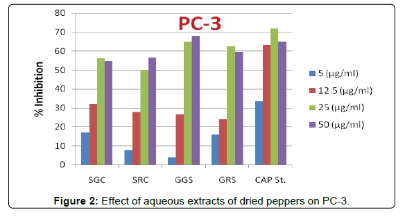
Figure 2: Effect of aqueous extracts of dried peppers on PC-3.

Figure 3: Effect of aqueous extracts of dried peppers on MCF-7.
Quercetin and quercetrin which were detected in these samples with contents ranged from 0.09 to 0.32 and 7.40-71.26 mg/100 g DW, respectively, had been reported to inhibit prostate cancer colony melanoma growth, and act as pro-apoptotic agent [72]. Similarly, ellagic acid, an oxidation product of gallic acid, catechol, kaempferol and its derivatives, which were detected, had undergone different levels of study as possible prostate cancer chemo-preventive agents, with promising results, including potent anti-mutagenesis, antitumor and anti-metastasis properties [73,74], effects that are also relevant to prostate cancer control and chemoprevention (This paragraph is considered a discussion for the results that were found) [75].
Also, all aqueous extracts of dried pepper samples had luteolin contents ranged from 0.01-0.42 mg/100 g DW. This compound had a strong anticancer activity [18,76,77].
The current study showed that aqueous extracts of Egyptian sweet and hot chilli pepper (in the dried form) at two maturity stages (green and red) have antioxidant and anticancer activities. The results observed that these extracts had high total phenolic and flavonoid contents and contain a wide range of phenolic and flavonoid compounds as well as organic acids. Pepper samples were also rich in vitamin C, β- carotene and vitamin E. Capsaicin, the major bioactive compound presented in pepper plants and responsible for the pungency of pepper, was found in these cultivars at high levels. Therefore, these extracts have antioxidant and anticancer activities against prostate and breast carcinoma cell lines. Treatment of the cancer cells with the aqueous extracts of pepper led to their growth inhibition and the induction of the apoptosis in cancer cells. It is apparent from our study that effective drugs produced from the Egyptian sweet and chilli pepper (Capsicum) tend to support a novel therapeutic methods for treatment of human prostate and breast malignancies.