Research Article - (2023)Volume 4, Issue 4
Introduction: Copper is an essential element for the body. It helps in the formation of red blood cells. Nanoparticles have distinctive physicochemical properties like ultra-small sized solid molecules ranging from 1 nm-100 nm, substantial ratio of mass to surface area and increased chemical responsiveness.
Aim: To compare the mechanical, histologic and antimicrobial characteristics of the PRF membrane with and without the addition of Copper Nanoparticles (CuNP).
Materials and methods: 20 volunteers aged 25-35 years were divided in 2 groups. 19 ml blood was collected from each person and PRFs were prepared. Group A comprised of only PRF and group B comprised of PRF with addition of CuNP. The samples were stained with H and E stains to evaluate the density of fibrin meshwork after processing and tensile strength using universal testing machine were evaluated. Zone of inhibition was performed against plaque sample.
Results: Histologically, varying precipitation of CuNPs in the outer layer and homogeneous in the inner core were observed and there was no difference in the density of the fibrin network. The tensile strength was significantly higher in the CuNP+PRF group. The zone of inhibition showed that there was a higher zone of inhibition present around CuNP+PRF compared to the normal PRF.
Conclusion: Incorporation of PRF with CuNP enhanced its mechanical strength and antimicrobial properties of PRF which may help in various periodontal and regenerative procedures.
Antimicrobial; Copper nanoparticles; Platelet rich fibrin; Periodontal pathogens; Tensile strength; Saliva
Dental biofilms which consist of a complex community of bacteria and fungi causes oral infections such as dental caries, periodontitis, and other dental diseases [1]. It has a tendency to accumulate on the surfaces of the teeth, restorations, prosthesis and orthodontic appliances, surgical dressings, and suture materials. The biofilm confers drug resistance and allows microorganisms to escape the host defense mechanism [2].
PRF contains about 97% of platelets in addition to more than 50% of leukocytes in the blood [3]. Macrophages can explicitly stimulates osteogenesis and also possibly helps the process of bone formation by maintaining availability of mesenchymal stromal/progenitor cells [4,5]. Platelets and leukocytes promote bone regeneration by releasing cytokines after activation [6]. The important growth factors in PRF are Bone Morphogenetic Protein-1 (BMP-1), Platelet-Derived Growth Factors (PDGFs), Transforming Growth Factor-1 (TGF-β1), Vascular Endothelial Growth Factor (VEGF), and Insulin-like Growth Factors (IGFs) [7,8]. TGF-β1 promotes new bone formation by stimulating collagen and fibronectin synthesis [9].
According to the American Environmental Protection Agency (EPA), copper is the only metal registered as having antimicrobial properties [10]. No microorganisms can survive on copper surfaces. Bacteria are killed within minutes on copper surfaces or copper alloys containing at least 60% copper, with copper killing capacity of 99.9% for most pathogens within 2 h of the contact period. This killing activity takes place at a rate of at least 7-8 hrs of time interval [11-13]. Copper NPs exhibits a powerful broad antimicrobial spectrum, which is effective against wound related pathogens, such as bacteria, fungi, and viruses [14,15]. The use of Copper (Cu2+) has been described to stimulate blood vessel formation, a key process in tissue regeneration [16].
Nanotechnology has emerged as a promising field for the management of oral diseases due to antimicrobial properties of Nanoparticles (NPs), which have been employed as coatings for implants and suture materials and as root canal irrigants [17-19]. Copper and its derivatives are the oldest known antimicrobial agents used in traditional medicine; it is a trace element in most organisms containing copper proteins, such as tyrosinases, catechol oxidases, and hemocyanins. Thus it is an essential element for the metabolism in animal and plant cells [20].
Nano copper also possesses anti-inflammatory properties. The copper containing coating can be applied to restorative materials, where it kills bacteria, yeasts, and viruses by the process known as “contact killing". The affinity of copper for amines and carboxylic groups in cell walls is thought to explain copper induced damage of the bacterial cell structure. The nanometric dimensions of copper particles enhance its ability to penetrate into the cell, and the oxidation of copper to Cu+ leads to the formation of reactive oxygen species that induces DNA damage.
Copper is a well-established antimicrobial and anti-inflammatory agent with a long history of medicinal applications. The nanosized particles and high surface area to volume ratio allow it to exhibit broad spectrum antibacterial and antiviral activities. Copper nanoparticles may have a similar mode of action as other metal nanoparticles. A few studies were conducted in an attempt to explore the antibacterial mechanism of copper nanoparticles. Mainly, three hypothetical mechanisms were commonly described. First, copper nanoparticles accumulate in the bacterial membrane and change its permeability. They then remove membrane proteins, lipopolysaccharides, and intracellular biomolecules and cause the dissipation of the proton-motive energy around the plasma membrane. Second, reactive oxygen species from nanoparticles (in the form of nanoparticles or ions) process post-oxidative damage in cellular structures. Third, cells’ uptake of ions (generated via nanoparticles) decreases intracellular adenosine triphosphate production and Deoxyribonucleic Acid (DNA) replication. The aim of this study was to compare the histologic, mechanical and antimicrobial characteristics of the PRF membrane with and without the addition of copper nanoparticles.
This in vitro, experimental study (CTRI registration not applicable) was approved by the institutional ethics committee. It was performed with the help of 20 volunteers. The inclusion criteria were healthy subjects ranging from 25-35 years with no history of blood dyscrasias. The exclusion criteria were the history of the known systemic diseases, history of anticoagulant drugs assumption, smoking, use of any medication within the last three months.
Preparing copper nanoparticles suspension
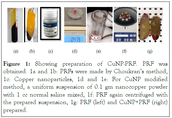
To obtain a uniform suspension, 0.1 grams of nanocopper powder with particles sized <100 nm (Nanoresearch lab, Jharkhand), along with 1 mL normal saline, was poured into the tube and mixed with hand.
Blood samples collection
After obtaining informed consent from the subjects, 19 ml venous whole blood was collected from the subjects who were willing to participate in the study. The blood samples were divided into two silica containing plastic tubes in sterile conditions, one tube containing 10 ml blood for the preparation of the conventional PRF and another containing 9 ml blood in addition with 1 ml copper nanoparticle suspension for the preparation of the CuNP modified PRF, which was then gently mixed with hand to achieve a uniform concentration, similar to the method performed by Ghaznavi, et al.
PRF and CuNP modified PRF preparation
The tubes were immediately centrifuged for 12 minutes at a low speed (2700 rpm, Eppendorf 5702 centrifuge, Hamburg, Germany), according to the protocol provided by Dohan Ehrenfest, et al. The fibrin clot containing platelets were formed in the buffy coat part in middle of the tubes between the red blood cells and plasma. The fibrin clots were removed and placed on a gauze piece, which were pressurized to form membranes (Figure 1).

Figure 1: Showing preparation of CuNP-PRF. PRF was obtained. 1a and 1b: PRFs were made by Choukran’s method, 1c: Copper nanoparticles, 1d and 1e: For CuNP modified method, a uniform suspension of 0.1 gm nanocopper powder with 1 cc normal saline mixed, 1f: PRF again centrifuged with the prepared suspension, 1g: PRF (left) and CuNP+PRF (right) prepared.
Evaluation of histological properties
The whole pieces of the PRF membranes were fixed in 10% formalin for 24 hours and were then embedded in paraffin wax block. The blocks were then subjected to sectioning and H and E staining and with the evaluation by optical microscope (ISKO ISSM-29 triple nose microscope 450X magnification) to appreciate if the embodiment of copper nanoparticles in PRF of exfoliated cells from an epithelial surface. Oral Exfoliative Cytology (OEC) was also attained with H and E staining under optical microscope.
Evaluation of the mechanical properties
The tensile strength of both the groups was performed using the Universal Testing Machine (UTM). The sample was fixed in the UTM device using specialised tongs used for semi-solid or hardgel like samples. The tensile force was applied ranging from 0 MPa-50 MPa until the sample was torn from the thin middle part. The test finished as the sample ruptured.
Evaluation of antimicrobial characteristics
Subgingival plaque samples of subjects who volunteered for blood sample collection were obtained by scraping technique using a sterile gracey curette. These collected samples were immediately transferred in tube containing 5 mL phosphate buffer saline solution and a uniform suspension was obtained by centrifuging these plaque samples at 700 rpm. These samples were then incubated in incubator (bacteriology digital incubator) for 24 hours. After incubation, 0.1 ml of this suspension was then uniformly placed on the blood agar plate which was spread by the L-shaped spreader. A punched piece of normal PRF and CuNP modified membrane of 0.5 cms in diameter was placed on inoculated agar plate. Inoculated blood agar plates were then incubated in an incubator aerobically at 37°C for 48 hours.
Statistical analysis
The obtained data was entered into Microsoft Excel software. The tensile strength of the samples was calculated by dividing the maximum force leading to membrane rupture by their crosssection area. “Paired t-test” was used to compare these values between the two types of membranes and P<0.05 was considered to be statistically significant. For antimicrobial potential, the comparison between zones of inhibition obtained around all the samples was analyzed by one-way ANOVA.
Histological properties
On evaluating the microscopic sections under bright field microscopy, the copper nanoparticles were observed all over the PRF membrane, but in the outer layers they were more densely attached to the fibrin strands compared with the inner layers. Precipitation of the CuNPs was patchy in the outer layers and quite homogeneous in the inner layers. Moreover, the leukocytes were denser in the outer layers than in the inner layers. H and E staining showed the existence of CuNPs in fibrin clot. Bright field microscopy showed small sized dense particles trapped between fibrin strands compared to the PRF alone (Figure 2a and 2b). PAP smear was done to assess successful embodiment of CuNP in PRF. Moreover, the OEC smear confirmed the successful embodiment of copper nanoparticles in PRF.
Figure 2: 2a and 2b: Demonstrating the histologic properties of PRF and CuNP-PRF. Varying precipitation of CuNPs in the outer layers and homogeneous in the inner core were confirmed and there was no difference in the density of the fibrin network, 2c and 2d: PAP smear done to assess successful embodiment of CuNP in PRF.
Tensile strength
The mean tensile strength was 0.154 MPa ± 0.05 MPa in PRF membrane samples and 0.435 MPa ± 0.19 MPa in CuNP-PRF samples modified by CuNPs, being statistically significantly higher in the latter (P=0.01) (Figure 3).
Figure 3: Showing bar graph of CuNP-PRF vs. PRF. The mean tensile strength was significantly higher in the group A at 0.435 MPa ± 0.19 MPa as compared to group B at 0.154 MPa ± 0.05 MPa.
Antimicrobial properties
The antimicrobial efficacy was demonstrated by appearance of a clear zone of inhibition around the samples. Mean zone of inhibition (in cm) around CuNP-PRF was 1.42 ± 0.25 and for the PRF group it was 0.13 ± 0.16. Mean zone of inhibition around CuNP-PRF (P<0.01) and PRF (P<0.05) which was statistically significant. These results indicate that CuNP-PRF has a significant inhibitory effect on growth of oral bacteria in comparison to normal PRF (Figure 4).
Figure 4: Showing a) A clear zone of inhibition of 1.42 ± 0.25 after 48 hours in CuNP-PRF as compared to b) PRF with 0.13 ± 0.16 respectively.
In this study, the tensile strength was significantly higher in the CuNP-PRF membranes than PRF group which signifies that the mechanical properties were improved as a result of adding CuNP-PRF in the fibrin matrix. On microscopic assessment of the membranes CuNP-PRF were more densely mixed with the fibrin strands in the outer layers than in the inner layers. Moreover, their precipitation was patchy in the outer layer and it was observed that it was homogenous in the inner layer. Thus it may signify that the histologically the PRF membrane modified by CuNP did not lose its physical integrity on addition of the nanoparticles. However, further studies by a scanning electron microscope are recommended to confirm its embodiment (Figure 5).
Figure 5: Results indicate that CuNP-PRF has a significant inhibitory effect on growth of oral bacteria in comparison to normal PRF.
During the healing phase, several factors are needed to inhibit the infections and its inflammation and induce cellular proliferation. Platelets are known for their role in the healing of the surgical site due to their growth factors that prevent infection due to antibacterial and fungicidal proteins stored in their granules and blood loss at the site of tissue injury. In the families of platelet concentrates, PRF contains significant concentrations of platelets with a stable dense fibrin matrix and platelet growth factors.
CuNPs are highly biocompatible having antimicrobial properties. They can adhere to the cell membrane of the bacteria and make it porous, which consequently changes the permeability of cell wall and causes cell death. Various studies have shown that the contact between copper nanoparticles and bacteria for free radicals which can damage the bacterial cell membrane and cause bacterial death through increased permeability.
In this study, there was a significant zone of inhibition around the CuNP modified PRF membrane after 48 hours around plaque sample. In vitro studies demonstrate that, at a certain range of concentrations, CuNPs can reduce the viability of cells, depending on the chemical composition, shape, and size of the nanomaterial. Compared to the other metals commonly used in antimicrobial application, copper undergoes oxidation more easily and tends to dissolve in media as ions. According to several studies, CuNP exhibit various actions on the microbes. They are more prone to interact with the membrane of bacteria compromising its integrity. This property of copper nanoparticles can be correlated with our study.
The antibacterial mechanism of copper nanoparticles has been attributed to the fact that Cu2+ ions eluted from nanoparticles are absorbed by bacteria when the nanoparticles concentration is high enough. Copper ions are absorbed onto the bacterial cell surface, imparting damage to the cell membrane by solidifying protein structure or altering enzyme function. Bacterial cells are immobilized and become inactivated by the presence of copper nanoparticles in the growth medium, which results in hampering of their replication process, with subsequent cell death. From our study results, we can determine that binding of copper ions to the bacterial cell surface played an important role in bactericidal activity of the PRF against plaque bacteria.
This study indicated that copper nanoparticles improve the tensile strength and bactericidal activity on addition with PRF, without changing or altering the physical form of PRF. Therefore, it can be incorporated in the bone defects and augmentation. However, further studies are required to assess the mechanism of action related to bone forming cells and periodontal pathogens.
[Google Scholar] [PubMed]
[Crossref] [Google Scholar] [PubMed]
[Crossref] [Google Scholar] [PubMed]
[Crossref] [Google Scholar] [PubMed]
[Crossref] [Google Scholar] [PubMed]
[Crossref] [Google Scholar] [PubMed]
[Crossref] [Google Scholar] [PubMed]
[Crossref] [Google Scholar] [PubMed]
[Crossref] [Google Scholar] [PubMed]
[Crossref] [Google Scholar] [PubMed]
[Crossref] [Google Scholar] [PubMed]
[Google Scholar] [PubMed]
[Crossref] [Google Scholar] [PubMed]
[Crossref] [Google Scholar] [PubMed]
[Crossref] [Google Scholar] [PubMed]
[Crossref] [Google Scholar] [PubMed]
[Crossref] [Google Scholar] [PubMed]
[Crossref] [Google Scholar] [PubMed]
Citation: Bhandare J, Mahale SA, Thosar S (2023) Platelet Rich Fibrins Treated with Copper Nanoparticles Enriches its Physical and Antimicrobial Properties. J Mol Pathol Biochem. 4:159.
Received: 30-May-2023, Manuscript No. JMPB-23-24594; Editor assigned: 01-Jun-2023, Pre QC No. JMPB-23-24594 (PQ); Reviewed: 15-Jun-2023, QC No. JMPB-23-24594; Revised: 02-Aug-2023, Manuscript No. JMPB-23-24594 (R); Published: 09-Aug-2023 , DOI: 10.35248/JMPB.23.4.159
Copyright: © 2023 Bhandare J, et al. This is an open-access article distributed under the terms of the Creative Commons Attribution License, which permits unrestricted use, distribution, and reproduction in any medium, provided the original author and source are credited.