Rheumatology: Current Research
Open Access
ISSN: 2161-1149 (Printed)
ISSN: 2161-1149 (Printed)
Clinical image - (2021)
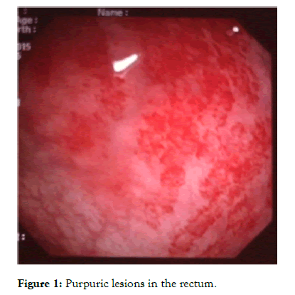
A 27 year-old woman was hospitalized because of abdominal pain and cutaneous purpura on her upper and lower extremities. The laboratory tests showed an inflammatory syndrome. A skin biopsy revealed a Leukocytoclastic Vasculitis (LCV). During her hospital course, she complained of diarrhea. Upper gastrointestinal endoscopic examination did not reveal any abnormality. Rectosigmoidoscpy revealed numerous purpuric lesions in the rectum as shown in Figure 1. Immunofluorescence method was negative in both skin and colic biopsy and positive with presence of IgA in gastric biopsies. The patient was diagnosed with Henoch-Schönlein Purpura (HSP) and was treated with corticotherapy. This resulted in the resolution of skin eruption and digestive symptoms.

Figure 1: Purpuric lesions in the rectum.
HSP is an IgA-associated small-vessel LCV [1] that occurs commonly in children [2,3]. It is characterized by nonthrombocytopenic palpable purpura, arthralgia/arthritis, bowel angina, and hematuria/proteinuria [2,3]. Gastrointestinal (GI) involvement occurs in 50-75% of patients [2] and it includes acute abdominal pain, nausea, vomiting, bloody stools, and upper GI hemorrhage. HSP might present with severe GI involvement, or even life-threatening in the short term [3]. Endoscopic examination is of major importance in detecting the gastrointestinal manifestations of HSP. A large biopsy of purpuric lesions is more likely to detect the vasculitis in the small vessels of the mucosa [4]. Direct immunofluorescence of tissue specimens from skin, GI tract, or kidney may show IgA deposition in both involved and uninvolved tissues [2,4]. In our case, Ig A deposition was observed only in normal gastric mucosa. Although evolution may be spontaneously favorable, The efficacy of corticosteroids on digestive manifestations has been suggested through numerous observations as reported in this case.
Citation: Amira A, Thabet M, Imen A, Ahmed G, Wissal BY, Elhem BJ, et al. (2021) Purpuric Lesions in the Rectum: Clinical Image. Rheumatology (Sunnyvale). 11: 297.
Received: 27-Jul-2021 Accepted: 10-Aug-2021 Published: 17-Aug-2021 , DOI: 10.35248/2161-1149.21.s16.005
Copyright: © 2021 Amira A, et al. This is an open-access article distributed under the terms of the Creative Commons Attribution License, which permits unrestricted use, distribution, and reproduction in any medium, provided the original author and source are credited.