International Journal of Physical Medicine & Rehabilitation
Open Access
ISSN: 2329-9096
ISSN: 2329-9096
Case Report - (2023)Volume 11, Issue 2
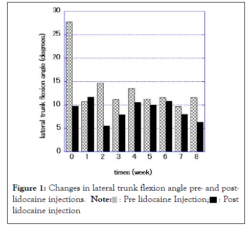
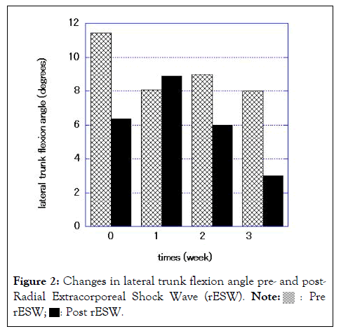
A 58-year-old male with Pisa Syndrome (PS) and Parkinson’s Disease (PD) was first treated with serial lidocaine injections to the External Oblique (EO) muscle of the ipsilateral side to the lateral flexion once time weekly for 8 weeks. The Lateral Trunk Flexion Angle (LFA) was improved from 24 degrees to 6.3 degrees and lumbago resolved. One month later, the LFA was >10 degrees with lumbago, therefore Radial Extracorporeal Shock Wave (rESW) therapy was delivered to the same side of the lateral abdomen in conjunction with the same rehabilitation program once weekly for 4 weeks. The LFA was improved from 11.4 degrees to 6.4 degrees. The LFA was maintained at <10 degrees without lumbago for >2 months. Radial Extracorporeal Shock Wave (rESW) therapy was shown to be effective and safe for PS in a patient with PD.
Pisa syndrome; Parkinson’s disease; Radial extracorporeal shock wave; Lidocaine
PS: Pisa Syndrome; PD: Parkinson’s Disease; rESW: Radial Extracorporeal Shock Wave; EO: External Oblique; LFA: Lateral Trunk Flexion Angle
Pisa Syndrome (PS) in patients with Parkinson’s Disease (PD) is clinically defined as a lateral trunk flexion >10 degrees that can be completely alleviated by passive mobilization or assuming a supine position [1]. Tinzssi et al. estimated the prevalence of PS to be 8.8% in Italian patients with PD. Of note, falls are more frequent in PS patients with PD [2]. Two pathophysiologic hypotheses have been advanced (central and peripheral hypothesis) [1]. Furusawa et al. suggested that dystonia in the External Oblique (EO) muscle is involved in the pathogenesis of upper camptocormia, and then showed that serial lidocaine injection into the EO muscle followed by rehabilitation improved upper camptocormia in 12 PD patients [3]. Moreover, Martino et al. reported improvement of PS in 10 PD patients using the same lidocaine injection protocol into the EO muscle combined with rehabilitation [4]. Radial extracorporeal shock wave (rESW) therapy is a new method for the treatment of dystonia [5,6]. Similar to the effects on tendon disease, Radial Extracorporeal Shock Wave (rESW) has a direct effect on muscle fibrosis and other non-reflex components of muscle hypertonia, which are likely to be present in some dystonic patients [7,8].
The Radial Extracorporeal Shock Wave (rESW) therapy has not been used to treat PS; therefore we used Radial Extracorporeal Shock Wave (rESW) therapy to treat PS in a patient with PD.
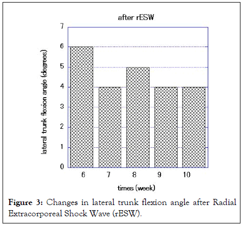
The patient was a 58-year-old Japanese male, with a history of PD for 14 years. His hand began to develop bradykinesia and a resting tremor, therefore levodopa-therapy was started. The symptoms were relieved after taking the drug. When the symptoms gradually worsened, he developed a right deviation of the lumbar spine and right-sided lumbago. The patient underwent rehabilitation for PD in our hospital. We diagnosed PS and two rehabilitation methods were tried (lidocaine injection to the right side of the EO muscle and Radial Extracorporeal Shock Wave (rESW) therapy to the right side of the lateral abdomen). After the first day of lidocaine injection, lumbago resolved. But after 1 month on the last day of the serial lidocaine injections, the LFA was >10 degrees with lumbago, and Radial Extracorporeal Shock Wave (rESW) therapy was then started. After the first Radial Extracorporeal Shock Wave (rESW) therapy session, the LFA decreased from 11.4 degrees to 6.4 degrees and the lumbago resolved. The LFA was maintained at <10 degrees for 3 months after the last day of repeated Radial Extracorporeal Shock Wave (rESW) therapy sessions without lumbago.
The anti-PD medication was not changed during the treatment period for PS. The patient underwent a rehabilitation program that emphasized stretching and gait training with body weight supported during treadmill training for 40 min 2 days per week [9]. LFA was measured on a three-dimensional motion-analysis system (AKIRA®; System Friend, Hiroshima, Japan). Lidocaine (50 mg of 1%xylocaine®, Astrozeneca, Japan) was injected into the right side of the EO muscle under ultrasound guidance. Seral lidocaine injections were administered once weekly for 8 weeks.
Radial extracorporeal shock wave therapy
The Radial Extracorporeal Shock Wave (rESW) pneumatic device (Intellect Mobile 2 RPW, Chattanooga™, Tennessee, USA) was used to provide a single session of shock wave intervention. The pressure pulses were focused on the right side of the abdomen, which was the same site for lidocaine injection to the EO muscle under ultrasound guidance. The treatment protocol for Radial Extracorporeal Shock Wave (rESW) was as follows; 1,500 shots with a pressure of 1.5 bars and wave irradiation of 5 Hz. Serial Radial Extracorporeal Shock Wave (rESW) therapy was administered used once weekly for 4 weeks.
As Figure 1 shows, serial lidocaine injections to the right EO muscle resulted in improvement in truncal lateral flexion. Figure 2 shows, serial Radial Extracorporeal Shock Wave (rESW) therapy to the right lateral abdomen resulted in improvement in truncal lateral flexion. Figure 3 shows, the effect of truncal lateral flexion (<10 degrees) continued for 10 weeks after the last Radial Extracorporeal Shock Wave (rESW) treatment session (Figures 2,3).

Figure 1: Changes in lateral trunk flexion angle pre- and postlidocaine
injections.  lidocaine injection.

Figure 2: Changes in lateral trunk flexion angle pre- and post-
Radial Extracorporeal Shock Wave (rESW). 


Figure 3: Changes in lateral trunk flexion angle after Radial Extracorporeal Shock Wave (rESW).
This is the first case report showing that Radial Extracorporeal Shock Wave (rESW) therapy is effective for PS in a PD patient. The energy generated by the pressure wave of Radial Extracorporeal Shock Wave (rESW) is absorbed into the skin approximately 3 cm deep and spreads a wider beam to a larger target area [10]. Compared to lidocaine injection using ultrasound guidance, Radial Extracorporeal Shock Wave (rESW) therapy is thought to be effective not only EO muscle, but also the Internal Oblique (IO) and Transversus Abdominis (TA) muscles. The main target of Radial Extracorporeal Shock Wave (rESW) therapy might be the EO muscle, because of the equal efficacy of lidocaine injection into the EO muscle. As the long term effect of Radial Extracorporeal Shock Wave (rESW) for post-stroke spasticity and dystonia, in our case, the effect persisted for 10 weeks [5,6,10]. The mechanism underlying the benefit induced by Radial Extracorporeal Shock Wave (rESW) therapy for PS in a patient with PD is uncertain; however, possibilities include inducing Nitric Oxide (NO) production; reducing motor neuron excitability; dysfunction in neuromuscular transmission; and affecting rheologic properties [11].
In this case report, we demonstrated that Radial Extracorporeal Shock Wave (rESW) therapy delivered to the ipsilateral side of the abdomen was equally effective for PS in a patient with PD as lidocaine injection into the EO muscle. Given the safety, efficacy and long-lasting effect, Radial Extracorporeal Shock Wave (rESW) therapy may be useful for PS in patients with PD; however, further trials are warranted before making recommendations.
This study did not receive any funding support. The authors declare no conflicts of interest.
Not applicable.
[CrossRef] [Google Scholar] [PubMed]
[CrossRef] [Google Scholar] [PubMed]
[CrossRef] [Google Scholar] [PubMed]
[CrossRef] [Google Scholar] [PubMed]
[CrossRef] [Google Scholar] [PubMed]
[CrossRef] [Google Scholar] [PubMed]
[CrossRef] [Google Scholar] [PubMed]
[CrossRef] [Google Scholar] [PubMed]
[Google Scholar] [PubMed]
[CrossRef] [Google Scholar] [PubMed]
Citation: Inobe J, Otsuka T, Shibuta Y, Yamazaki R, Kato T (2023) Radial Extracorporeal Shock Wave is Effective for Pisa Syndrome in a Patient with Parkinson’s disease: A Case Report. Int J Phys Med Rehabil. 11:659.
Received: 24-Dec-2022, Manuscript No. JPMR-22-21153; Editor assigned: 28-Dec-2022, Pre QC No. JPMR-22-21153 (PQ); Reviewed: 13-Jan-2023, QC No. JPMR-22-21153; Revised: 20-Jan-2023, Manuscript No. JPMR-22-21153 (R); Published: 27-Jan-2023 , DOI: 10.35248/2329-9096.23.11.659
Copyright: © 2023 Inobe J, et al. This is an open-access article distributed under the terms of the Creative Commons Attribution License, which permits unrestricted use, distribution, and reproduction in any medium, provided the original author and source are credited.