Journal of Clinical Toxicology
Open Access
ISSN: 2161-0495

ISSN: 2161-0495
Case Report - (2020)Volume 10, Issue 6
Objective: Rattlesnake envenomation is a pathophysiologically complex process with a range of local effects including pain, erythema, local edema resembling compartment syndrome, ecchymosis that extends beyond the bitten extremity, as well as the development of blisters, bullae and tissue necrosis in severe cases. Previous animal studies have suggested that lymphatic flow is a crucial part of dissemination of venom after initial injection to produce systemic effects. However, the effects of baseline lymphatic obstruction on local injury in humans have yet to be described clinically.
Methods: Three cases of patients that sustained rattlesnake bites with a history of lymphatic disruptions were reviewed, two with histories of mastectomies and one with chronic lower extremity lymphedema. All three patients experienced bites on the limb affected by lymphatic obstruction, yet their presentation of local symptoms and treatment regimens varied.
Results: The first case describes a woman with a history of bilateral mastectomy and complete lymph node dissection who was bitten on her upper extremity and developed severe local cytotoxicity and tissue necrosis despite standard antivenom administration protocols. Providers in the second case treated the patient, who also had a history of mastectomy and complete lymph node dissection, more aggressively with repeat loading doses rather than maintenance dosing based on standard criteria of swelling progression greater than an inch an hour. In this instance, the patient had a favorable outcome with complete recovery of the local tissue and no evidence of necrosis. The final case, which involved a bite on an extremity with chronic lymphedema, resulted in delayed necrotic skin breakdown requiring two surgical debridements after initial standard antivenom dosing.
Conclusion: The clinical outcomes of these cases revealed a more severe local injury with lymphatic disruption, suggesting that either venom is unable to travel systemically and concentrates at the site of injection or that antivenom therapy is unable to reach the site of the bite. Future studies are needed to better understand this relationship as this case series suggests that lymphatic obstruction may be a risk factor for more severe local injury.
Rattlesnake; Lymphatics; Antivenom
Rattlesnake envenomation is a pathophysiologically complex process with a range of local effects including pain, erythema, local edema resembling compartment syndrome, ecchymosis that extends beyond the bitten extremity, tissue damage from the fang puncture as well as the development of blisters, bullae and tissue necrosis in severe cases. Local effects of edema and erythema are likely due to damage of the vascular endothelial cells allowing for extravasation of erythrocytes and plasma [1], while more severe effects including skin necrosis are thought to be due to cytotoxicity through disruption of the lipid bilayer [2]. Animal studies have supported the role of the lymphatic system in the development of local effects. In a study with rabbit subjects, neurotoxic tiger snake venom was injected into the limbs of animals with ligated lymphatic channels. Survival was prolonged to 60 minutes in the group with ligated lymphatic vessels compared to 10 minutes in the control group [3]. An additional study used a cooling technique to slow lymphatic flow in rat limbs and found that the time to peak toxin concentrations were prolonged in the group with slowed lymphatics [4]. One study cannulated and drained the thoracic ducts of sheep’s and found that compared to the control group, these sheep had lower steady state concentrations suggesting the importance of lymphatic flow in the maintenance of steady state levels [5]. Though animal studies are promising, the effects of baseline lymphatic obstruction on local injury in humans have yet to be described clinically. Here, we present a series of cases in which patients were bitten by a rattlesnake in an extremity with disrupted lymphatic flow and detail their clinical presentation, management, and outcomes.
Case 1
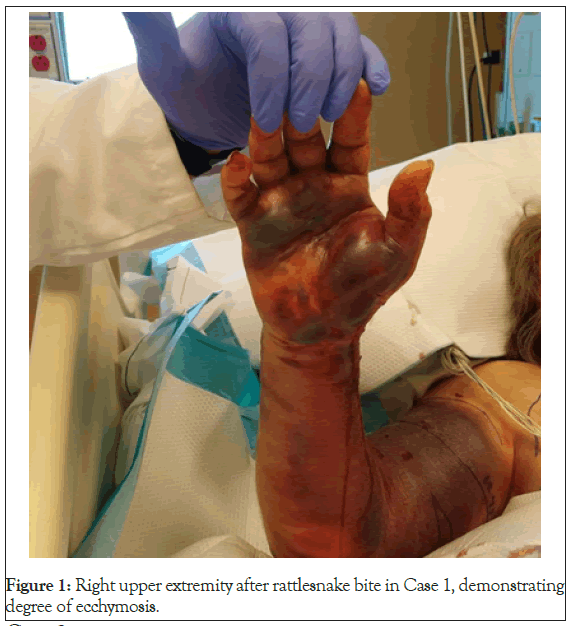
An 80-year-old woman with a history of bilateral mastectomy and lymph node dissection sustained a snake bite to her right wrist while outside gardening. When EMS was initially contacted, the swelling was localized to the wrist, however upon their arrival it had quickly extended to the elbow. On initial evaluation in the emergency department, the edema extended to the bicep. Initial laboratory evaluation revealed a thrombocytopenia with a platelet count of 82,000, but other labs were without abnormalities. She received a 6 vial loading dose of Crotalidae Polyvalent Immune Fab (Ovine), brand name CroFab®, approximately one hour after the initial injury. As she was transferred to a tertiary center, she received another 6 vials of AV with notable swelling and ecchymosis into the axilla. Over the next few hours, she developed several hemorrhagic bullae over the volar wristand palm, which were aspirated. Following this, she received an additional eight vials leading into the following day.
On the second day of her hospitalization, an examination of the leading edge of edema showed progression greater than an inch per hour, and extension into the mid-chest was noted along with scattered ecchymosis. Platelet count recovered to 229,000 and other coagulation studies remained normal by 3 hours post antivenom administration (Table 1).
| Time (Hours post administration) | Dose (Vials) |
|---|---|
| 1 | 6 |
| 4 | 6 |
| 6 | 4 |
| 11 | 4 |
| 21 | 2 |
| 27 | 2 |
| 33 | 2 |
| 47 | 4 |
Table 1: Antivenom administration in Case 1.
After obtaining initial control, the patient was switched to maintenance dosing. The patient experienced several episodes of hypotension. Further workup demonstrated a lactic acidosis and a bedside toxicology examination was performed. Pulse oxygenation was normal, and ultrasound of the right upper extremity did not show any evidence of a DVT. No further ecchymosis or edema was noted on the exam. A CBC revealed a drop-in hemoglobin from 12.6 to 10.2 which was attributed to be dilutional secondary to IV fluids or related to the consistent oozing of serosanguineous fluid from her previous bulla. During this time, she was receiving an additional four vials of CroFab®.
The following morning, coagulation studies revealed an H/H of 7.3/22.2, which had been slowly downtrending. While considering transfusion and continuation of anti-venom, the patient developed paroxysmal atrial fibrillation with signs of hypervolemia with pulmonary edema. Further treatment of her snake bite was terminated to focus on pressing medical issues. Her physical examination showed severe ecchymosis with no further edema. She was discharged after management of her CHF exacerbation (Figure 1).

Figure 1: Right upper extremity after rattlesnake bite in Case 1, demonstrating degree of ecchymosis.
Case 2
An 84-year-old woman with a history of bilateral mastectomy with 17 lymph node resection and dementia sustained a snake bite to the right thumb while performing yard work. Initially, the patient and her husband noted localized swelling and violaceous skin changes with streaking erythema to the right forearm. On presentation to the emergency department, vitals were stable, coagulation studies within normal limits, and loading dose of 5 vials of Crofab was administered. After serial stable examinations of swelling the decision to move to maintenance dosing was made.
Before the first maintenance dose was actually administered, the leading edge was noted to have progressed to the triceps area, and the decision was made to deliver an additional 4 vial loading dose instead of the planned 2 vial maintenance dose. During the second loading dose, the leading edge progressed an additional 2” to the mid-upper arm. Given the previous mastectomy case outcome, more aggressive management was chosen, and the patient received a third loading dose of 4 vials. The edematous extremity began to improve after the 3rd loading dose and the decision to proceed to maintenance dosing was made. After the standard 3 maintenance doses, her swelling receded to the elbow and she was stable for discharge with no further complications on follow up (Table 2).
| Time (Hours post administration) | Dose (Vials) |
|---|---|
| 1 | 5 |
| 9 | 4 |
| 14 | 4 |
| 24 | 2 |
| 30 | 2 |
| 36 | 2 |
Table 2: Antivenom administration in Case 2.
Case 3
A 64-year-old male presented to the emergency department after he sustained a rattlesnake bite to the left heel. His past medical history was noncontributory other than chronic left lower extremity swelling, which was presumed to be secondary to lymphedema. On initial evaluation, the left lower extremity was noted to have localized ecchymosis and was edematous, but difficult to approximate acute change from baseline.
He was administered 10 vials of AnaVip (Crotalinae Equine Immune F(ab)2 Antivenom) and IV analgesia with initial coagulation studies showing a mild thrombocytopenia with a platelet count of 87,000 but otherwise normal. Two hours after administration of the antivenom, the patient’s platelet count normalized to 152,000 and other studies were table (Table 3).
| Time (Hours post administration) | Dose (Vials) |
|---|---|
| 1 | 10 |
Table 3: Antivenom administration in Case 3.
The following day, the patient’s swelling had not progressed, he was transitioned from IV to PO analgesia, evaluated by physical therapy and determined suitable for discharge. Over the next two weeks, the patient was seen several times by his primary care physician for follow up of his hospitalization and other medical issues. Approximately 12 days after discharge, the patient reported increased redness and bruising over the lateral ankle. His provider started to express concern for a post-snakebite infection, though these are typically rare. Due to clinical presentation and leukocytosis on CBC, he received an intramuscular dose of Ceftriaxone and a 10-day oral antibiotic course. Despite compliance with this therapy, the patient clinically worsened in terms of reported pain and erythema on examination. The patient received an outpatient ultrasound, which revealed no evidence of abscess or thrombosis. While objectively afebrile, the patient felt a subjective fever and was subsequently sent back to the emergency room 19 days after the envenomation.
Examination in the emergency department revealed a developing area of necrotic tissue, and he was admitted for broad-spectrum antibiotics and debridement. The patient’s case resulted in two debridements of necrotic tissue with subsequent skin grafting and he continues to experience increased swelling and pain as a result.
Rattlesnake venom is complex and composed of various proteins, peptides, enzymes and other macromolecules. Specific venom composition can vary based on snake species, geographical location, prey, age and size of the animal [6], making its study a difficult task. Although some components have been identified and used medicinally, there are ongoing studies to determine the underlying mechanism of many snake venom components.
Rattlesnake envenomation has a variety of clinical presentations, which can include edema, ecchymosis, erythema, and coagulopathy. Necrosis, which is a more severe and less commonly seen effect, was an outcome in two of the three cases presented. These necrotic skin outcomes in patients with a history of lymphatic disruption may indicate that the lymphatic system is a crucial part of dissemination of the venom after initial injection in humans.
When considering pathology involving the lymphatic system, lymphatic filariasis can serve as a model of lymphatic obstruction and flow disruption. Using mosquitoes as an intermediate host, these parasites produce millions of larvae that initially enter the lymphatic system before migrating into the vascular system [7]. The above mechanism may explain the initial or subsequent accumulation of venom in the lymphatics of extremity before entering the vascular system. This could potentially explain significant localized tissue toxicity seen in patients with lymphatic disruptions; the venom is not able to leave the local tissue and distribute in the local vasculature. This potential accumulation of venom may be associated with increased local tissue damage and lower incidence of systemic effects as the venom is inhibited from accessing the bloodstream. While animal models of western diamondback envenomation have shown that applying pressure to disrupt lymphatic flow prolongs survival, a unified treatment algorithm discouraged the use of pressure immobilization or lymphatic constricting bands due to the unknown effects on local injury [8]. In support of these guidelines, the cases presented above demonstrate the severe localized effects of snake envenomation on a limb with lymphatic obstruction in the absence of significant systemic effects.
Predictors of severe localized effects at the envenomation site include cyanosis, ecchymosis, and history of alcohol use [9]. This case series suggests that another indicator of local severity is lymphatic obstruction of the affected limb. Each patient presented with erythema and edema at the bite site and two patients had persistent venom effects. In the second case, given the treatment center’s prior experience with treating a patient with a history of mastectomy, the positive outcome may have been related to more aggressive initial treatment due to perceived increased risk.
If a patient displays signs of envenomation, standard management for a rattlesnake bite usually involves an initial loading dose that is repeated until initial control is determined to have been achieved. Depending on the antivenom being used and local practices, maintenance doses of antivenom may then be scheduled. If lymphatic disruption can worsen outcomes, current management strategies may have to adapt. If the patient is positive for this risk factor, alternative dosing strategies with a total larger scheduled dose regimen may need to be developed. Choice of anti-venom is also an important consideration. Crofab, a singular Fab fragment of IgG, has a relatively small molecular size and would theoretically be able to penetrate affected tissues effectively. By comparison, Anavip is a recently FDA approved Fab 2 fragment that due to its larger molecular mass may theoretically be unable to reach affected tissues as effectively as a singular Fab fragment. Additional studies are needed to evaluate the minimum effective dose and individual therapeutic benefits of these antivenoms in the setting of impaired lymphatic flow.
The clinical outcomes of these cases revealed a more severe local injury with lymphatic disruption, suggesting that either venom is unable to travel systemically and concentrates at the site of injection or that antivenom therapy is unable to reach the site of the bite. Future studies are needed to better understand this relationship as this case series suggests that lymphatic obstruction may be a risk factor for more severe local injury.
This work was supported by the Arizona Poison & Drug Information Center.
We would like to thank the Arizona Poison & Drug Information Center for supporting this project.
Citation: Brady M, Junak M, Smelski G, Shirazi FM (2020) Rattlesnake Envenomation in the Setting of Disrupted Lymphatic Flow: A Case Series. J Clin Toxicol. 10:455. DOI: 10.35248/2161-0495.20.10.455
Received: 17-Sep-2020 Accepted: 01-Oct-2020 Published: 08-Oct-2020 , DOI: 10.35248/2161-0495.20.10.455
Copyright: © 2020 Brady M, et al. This is an open-access article distributed under the terms of the Creative Commons Attribution License, which permits unrestricted use, distribution, and reproduction in any medium, provided the original author and source are credited.