Journal of Clinical & Experimental Dermatology Research
Open Access
ISSN: 2155-9554
ISSN: 2155-9554
Clinical image - (2019)Volume 10, Issue 2
Sarcoidosis is a systemic granulomatous disease which is frequently revealed by cutaneous lesions. Involvement of the scalp with cicatricial alopecia is exceptional. We present an observation of a patient who presented with scalp sarcoidosis as the only cutaneous manifestation of the disease. This clinical form must be known in order to make the diagnosis and allow early treatment before the installation of cicatricial alopecia.
Sarcoidosis; Scalp alopecia; Cicatricial; Dermoscopy
Sarcoidosis is a systemic granulomatous disease which is frequently revealed by cutaneous lesions. Involvement of the scalp with cicatricial alopecia is exceptional.
Dermoscopic features of scalp sarcoidosis had been rarely reported.
We present a 67-year-old woman presented with asymptomatic patchy alopecia evolving since 8 months. Personal and family history of the patient was otherwise unremarkable.
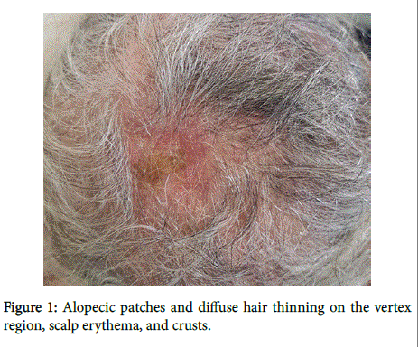
At clinical examination, she presented with alopecic patches and diffuse hair thinning on the vertex region, scalp erythema, and crusts (Figure 1).

Figure 1: Alopecic patches and diffuse hair thinning on the vertex region, scalp erythema, and crusts.
Systemic examination revealed no abnormalities in any other organ.
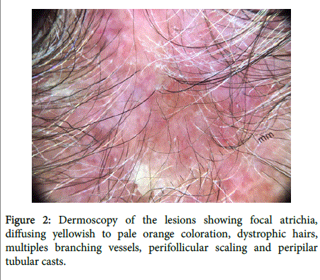
Dermoscopy of the lesions showed focal atrichia, diffuse yellowish to pale orange coloration, dystrophic hairs, multiples branching vessels, perifollicular scaling and Peripilar tubular casts (Figure 2).

Figure 2. Pre-operative and post-operative photo progression of Patient #2 with acne keloidalis nuchae.
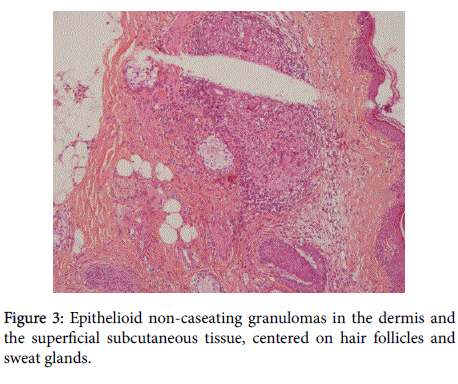
A 6 mm punch biopsy specimen obtained from the margin of the affected area for histopathology revealed an epithelioid non-caseating granulomas in the dermis and the superficial subcutaneous tissue, centered on hair follicles and sweat glands (Figure 3). The special colors were negative. The diagnosis of sarcoidosis was mentioned.

Figure 3: Epithelioid non-caseating granulomas in the dermis and the superficial subcutaneous tissue, centered on hair follicles and sweat glands.
Routine laboratory data, including erythrocyte sedimentation rate, CRP, serum calcium levels, calcium concentration in 24 hour urine, were all normal or negative. Except converting enzyme (ACE) level that was elevated 76 U/L. Chest X-ray and spirometry revealed normal lung function. Ophthalmological and nasal examination of the patient was unremarkable.
Kveim test was not realized in our patient.
Diagnosis of cutaneous sarcoidosis with no other organ involvement was made and the patient was given oral hydroxychloroquine 200 mg twice daily.
This clinical form must be known in order to make the diagnosis and allow early treatment before the installation of cicatricial alopecia.
Citation: El Anzi O, Sqalli A, Maouni S, Mai S, Meziane M, Ismaili N, et al. (2019) Scalp Sarcoidosis: A Clinical and Dermoscopic Features. J Clin Exp Dermatol Res 10: 488. doi:10.4172/2155-9554.1000488
Received: 03-Feb-2019 Accepted: 07-Mar-2019 Published: 14-Mar-2019
Copyright: © 2019 El Anzi O, et al. This is an open-access article distributed under the terms of the Creative Commons Attribution License, which permits unrestricted use, distribution, and reproduction in any medium, provided the original author and source are credited.