Journal of Clinical and Experimental Ophthalmology
Open Access
ISSN: 2155-9570
ISSN: 2155-9570
Clinical image - (2023)Volume 14, Issue 3
Neuroretinitis is a focal inflammation of the optic nerve and peripapillary retina or macula. It can be either infectious or idiopathic and is characterized by acute unilateral vision loss [1,2]. The pathophysiology of neuroretinitis is characterized by an inflammation of the optic disc vasculature with exudation of fluid into the peripapillary retina. Gass [3] established optic disc leakage by fluorescein angiography and suggested the term neuroretinitis.
It has been reported that visual prognosis in patients with idiopathic neuroretinitis is excellent, with or without interventions. Albeit no well-defined treatment plan exist, the use of corticosteroids has proven beneficial in some reported studies.
60-year old female presented with sudden onset painless loss of vision in left eye. On examination, visual acuity was 5/60 no improvement on pin hole test. Colour vision was abnormal and contrast sensitivity reduced. Pupillary light reaction showed relative afferent pupillary defect. On dilated fundus examination, there was optic disc edema and superotemporal retinal patch with macular star suggesting neuroretinitis [4].
60 year old female patient came to ophthalmology Outpatient Department (OPD) with chief complaint of diminution of vision in left eye since 3 days. She had past history of fever 15 days back. On ocular examination, she had visual acuity of counting finger 1 meter not improved on pinhole test, colour vision was abnormal and contrast sensitivity reduced in left eye. On indirect ophthalmomoscopic examination, disc edema, macular star and single whitish lesion superior temporal to the optic disc was seen. Fundus was suggestive of neuroretinitis in left eye. On examination right eye was within normal limits.
Further investigating for QuantiFERON TB gold test was found to be positive. Afterwards, she was started with anti-tubercular drug therapy for 6 months and oral corticosteroids were weekly tapered for 6 weeks.
She was followed for day 7th, day 15th, day 30th and 3 months and 6 months respectively. After completing above treatment visual acuity, colour vision, contrast sensitivity was improved.
And on fundus examination there was complete resolution of neuroretinitis (Figures 1-5).
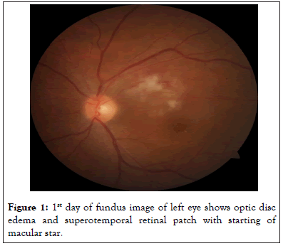
Figure 1: 1st day of fundus image of left eye shows optic disc edema and superotemporal retinal patch with starting of macular star.
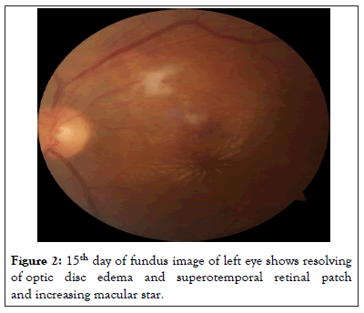
Figure 2: 15th day of fundus image of left eye shows resolving of optic disc edema and superotemporal retinal patch and increasing macular star.
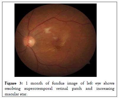
Figure 3: 1 month of fundus image of left eye shows resolving superotemporal retinal patch and increasing macular star.
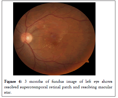
Figure 4: 3 months of fundus image of left eye shows resolved superotemporal retinal patch and resolving macular star.
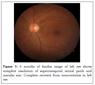
Figure 5: 6 months of fundus image of left eye shows complete resolution of superotemporal retinal patch and macular star. Complete recovery from neuroretinitis in left eye.
[Crossref] [Google Scholar] [PubMed]
[Google Scholar] [PubMed]
Citation: Selukar KV, Daigavane SV, Mathurkar S, Thool A (2023) Serial Images of Post Infectious Neuroretinitis Complete Recovery. J Clin Exp Ophthalmol. 14:947
Received: 01-May-2023, Manuscript No. JCEO-23-22304; Editor assigned: 03-May-2023, Pre QC No. JCEO-23-22304 (PQ); Reviewed: 17-May-2023, QC No. JCEO-23-22304; Revised: 24-May-2023, Manuscript No. JCEO-23-22304 (R); Published: 02-Jun-2023 , DOI: 10.35248/2155-9570.23.14.947
Copyright: © 2023 Selukar KV, et al. This is an open-access article distributed under the terms of the Creative Commons Attribution License, which permits unrestricted use, distribution, and reproduction in any medium, provided the original author and source are credited.